Asia Pacific
allergy
Asia Pacific
allergy
pISSN 2233-8276 · eISSN 2233-8268
Copyright © 2017. Asia Pacific Association of Allergy, Asthma and Clinical Immunology.
The asthma and chronic obstructive pulmonary disease overlap syndrome in tertiary care setting Thailand
Theerasuk Kawamatawong1,2,*, Sanruethai Charoenniwassakul1, and Ticha Rerkpattanapipat1,3
1Department of Medicine, Faculty of Medicine, Ramathibodi Hospital, Mahidol University, Bangkok 10400, Thailand
2Division of Pulmonary and Critical Care Medicine, Department of Medicine, Faculty of Medicine, Ramathibodi Hospital, Mahidol University, Bangkok 10400, Thailand 3Division of Allergy Immunology and Rheumatology, Department of Medicine, Faculty of Medicine, Ramathibodi Hospital, Mahidol University, Bangkok 10400, Thailand
Background: Asthma and chronic obstructive pulmonary disease (COPD) overlap syndrome (ACOS) is an increasingly recognized clinical entity. ACOS significantly impacts on patient outcome compared to isolated asthma or COPD. However, ACOS definition and diagnosis criteria have not been well standardized. ACOS prevalence and clinical features in Thailand has never been studied.
Objective: To investigate the prevalence and clinical features of ACOS compared to isolated asthma or COPD among patients with clinician-diagnosis of obstructive airway diseases.
Methods: Spirometry, skin prick test (SPT) and allergens specific IgE (sIgE) were done. Serum total IgE, exhaled nitric oxide (FeNO) and blood eosinophils were measured. High resolution computed tomography (HRCT) was performed. Smoking history, pollution, biomass exposure and symptoms (Asthma Control Test [ACT], COPD assessment test [CAT], Modified Medical Research Council Dyspnea Scale [MMCR]) were assessed. Patients were classified to isolated asthma, COPD or ACOS according to predefined definitions for this study.
Results: A total 92 patients were enrolled: 58 patients with clinician-diagnosed of late onset asthma and 34 with clinician-diagnosed COPD.
The mean age was 67.4 years. Thirty-four asthma patients (58.6%) were considered to have ACOS with postbronchodilator forced expiratory volume in 1 second (FEV1)/forced vital capacity ratio <0.7 and/or presence of emphysema on HRCT. In addition, 10 COPD patients (28.6%) were classified as ACOS if they had bronchodilator reversibility (FEV1 ≥ 12% and ≥ 200 mL) and positive SPT or sIgE. Hence, total of 44 from 92 patients (47.8%) with obstructive airway diseases were found to have ACOS, while isolated asthma and COPD were found in 24 patients equally. No difference in symptoms assessed by CAT, ACT, or MMRC was found between 3 groups of patients. Neither serum total IgE nor blood eosinophils counts distinguished ACOS from asthma and COPD (p = 0.83 and p = 0.40). FeNO was higher in pure COPD than ACOS and asthma (p = 0.03).
Conclusion: ACOS is prevalent in late-onset asthma or clinician-diagnosed COPD who were treated in tertiary care clinic. However, we found no difference in symptoms, blood eosinophils or serum total IgE between groups.
Key words: Asthma and chronic obstructive pulmonary disease overlap syndrome; Prevalence; Tertiary care centers; Clinical features;
Thailand
*Correspondence: Theerasuk Kawamatawong
Division of Pulmonary and Critical Care Medicine, Department of Medicine, Faculty of Medicine , Ramathibodi Hospital, Mahidol University Bangkok 10400, Thailand
Tel: + 66-2-201-1619, Fax +66-2-201-1629 E-mail: [email protected] Received: March 31, 2017 Accepted: October 12, 2017
This is an Open Access article distributed under the terms of the Creative Commons Attribution. Non-Commercial License (http://creativecommons.
org/licenses/by-nc/4.0/) which permits unrestricted non-commercial use, distribution, and reproduction in any medium, provided the original work is properly cited.
https://doi.org/10.5415/apallergy.2017.7.4.227 Asia Pac Allergy 2017;7:227-233
Hypothesis & Experience
Original Article
INTRODUCTION
Asthma and chronic obstructive pulmonary disease (COPD) are common obstructive pulmonary diseases in clinical practice.
Although asthma and COPD frequently represent with different clinical characteristics, significant overlap features between 2 diseases are observed. The observational study has initially shown that prevalence of asthma-COPD overlap syndrome (ACOS) range from 15% to 20% in a tertiary care severe asthma clinic and triggered worldwide controversy [1, 2]. ACOS was proposed by Global Initiative for Asthma (GINA) and Global Initiative for Chronic Obstructive Lung Disease (GOLD) [3, 4]. However, definition of ACOS has never been completely standardized. ACOS is commonly defined as either the diagnosis of COPD in a patient with previously diagnosed asthma, or incompletely reversible airway obstruction in asthmatic patients. In addition, ACOS diagnosis depends on the patient’s presentations and laboratory tests [5, 6]. Moreover, ACOS patients experience uncontrolled symptoms despite medical treatments, more frequent exacerbations in the 6th decade of life, and poorer prognosis when compared to COPD or asthma alone. Furthermore, ACOS was associated with higher risks for exacerbations, increased and worse global health status compared with those with COPD alone [7-9]. The prevalence of ACOS in Thailand is not known but may represent an unaddressed public healthcare disparity. The aim of this study was to investigate the prevalence and characteristics of ACOS among COPD and high-risk asthma patients in a tertiary healthcare setting in Thailand.
MATERIALS AND METHODS
Cross-sectional study was conducted in Ramathibodi Hospital, Bangkok Thailand from August 2014 to October 2015. The clinician-diagnosed asthma and COPD patients were recruited at outpatient clinic. The study was approved by the Committee on Human Right Related to Research Involving Human Subjects, Faculty of Medicine Ramathibodi Hospital, Mahidol University (ID 08-57-06). All patients gave the informed consents before participating in the clinical study.
Patient inclusion criteria were as followings: (1) Enrolled asthmatic or COPD patients were diagnose by physician with age ≥40 years. (2) The clinician diagnosed asthma patients were identified and diagnosed following GINA [4]. (3) Clinician diagnosed COPD patients were identified and diagnosed
according to GOLD [3].
The exclusion criteria were as followings: any severe illness that limit the capability to perform pulmonary function testing.
Methods
All patients completed a questionnaire including onset of chronic airway diseases, smoking status, pack-years of tobacco use and history of biomass exposure. The current patient’s symptoms using COPD Assessment Test (CAT, GlaxoSmithKline group, Middlesex, United Kingdom), Asthma Control Test (ACT, GlaxoSmithKline group) and Modified Medical Research Council Dyspnea Scale (MMRC) in Thai language version. Medical information and patients’ characteristics including age, age onset of disease, and body mass index (BMI) were obtained. Spirometry and bronchodilator reversibility tests were performed according to the American Thoracic Society (ATS)/European Respiratory Society (ERS) standardization [10]. Fractional exhaled nitric oxide (FeNO) was measured. Total serum IgE and serum specific IgE (sIgE) for aeroallergens (Dermatophagoides pteronyssinus, Dermatophagoides farinae, and Aspergillus fumigatus) were measured. Eosinophils were counted. Skin prick tests for aeroallergens were performed. High resolution computed tomography (HRCT) of chest was performed for detecting emphysema and other radiologic findings.
Definition of ACOS
Patients were defined ACOS according to the following: (1) The clinician-diagnosed COPD patients were considered to have ACOS if there were evidence of atopy by positive skin prick test and/or sIgE and postbronchodilator FEV1 reversibility after 400- µg salbutamol inhalation ≥ 12% or ≥ 200 mL. (2) The clinician- diagnosed asthma patients were considered to have ACOS if they had a history of smoking or biomass exposure > 10 years AND their postbronchodilator FEV1/forced vital capaticy (FVC) ratio was less than 0.7 OR presence of emphysematous change by HRCT of lung. (3) This sentence could be deleted (I think it is repetitive with the criteria (1) mentioned earlier).
All patient had spirometry and bronchodilator reversibility challenge performed as part of routine care at each clinic visit;
only the results within 1 year were included. All patients were clinically stable without previous respiratory infections for the past 3 months. The procedures were performed according to the ATS/European Respiratory Society guidelines [10]. Spirometry parameters including FVC, FEV1, and FEV1/FVC ratio performed
The ACOS in tertiary care setting Thailand
pre- and post-bronchodilator testing using 400 mcg salbutamol were recorded. The volumes of spirometry were expressed in litre and percent of predicted values.
FeNO measurements
FeNO was measured by using electrochemical technique (NOBREATH, Bedfont Scientific Ltd., Kent, United Kingdom). FeNO was measured and reported in part per billion (ppb) according to the standard procedures recommended by the manufacturer.
FeNO was acquired in a clinically stable condition without previous respiratory infections the past 3 months prior and before performing spirometry maneuver [11, 12].
Serum total IgE and serum allergen sIgE measurements Serum total IgE was measured using enzyme-linked immunosorbent assay and data were expressed in IU/mL. Serum sIgE measurement was performed by means of Pharmacia CAP- System using fluoroenzyme immunosorbent assay (CAP-System- FEIA, Pharmacia Diagnostic Co., Uppsala, Sweden). The positivity of sIgE was determined by using level ≥ 0.35 KUA/L [13].
Skin prick test to common aeroallergens
Skin prick test was done in COPD patients under a stable condition. Positive results were defined as wheal > 3 mm at immediately posttest within 15 minutes. Atopy was defined as positive result to at least 1 common aeroallergens that were pollens (Bermuda, Timothy, Johnson grass, Careless weed, or Acacia), mold (A. fumigatus), animal dander (cat, dog), house dust mite, and cockroach.
High-resolution computed tomography of chest HRCT of chest was done by using thin collimation (1- mm thickness) technique and bone algorithm. Centrilobular emphysema was diagnosed in the presence of low attenuation lung area (less than -950 Hounsfield unit). The report of HRCT was done by radiologist independent of clinical diagnosis.
Statistical analysis
The clinical characteristics between asthma, COPD and ACOS patients including age, BMI, pack-years of tobacco use and years of biomass exposure were expressed as mean and range of results, and data from CAT, ACT score, and MMRC scale were expressed as mean and standard deviation (SD). The result of investigations (FeNO, total IgE, sIgE, and eosinophil count)
were compared between two or more independent groups by using the chi-square, Fisher exact tests, and Kruskal-Wallis as appropriate. All statistical analyses were performed using Stata ver. 12 (StataCorp LP., College Station, TX, USA).
RESULTS
From 100 patients recruited from outpatient clinic, 92 were included in the analysis. There were 58 clinician-diagnosed asthma patients and 34 clinician-diagnosed COPD patients (Fig. 1). Characteristics of clinician-diagnosed asthma and COPD were compared in Table1. Patients’ age, spirometry parameters, symptom score (CAT, ACT, and MMRC), serum total IgE proportion of patients with atopy, and mean of eosinophil counts were similar between clinician-diagnosed COPD and asthma. However, clinician-diagnosed COPD patients were older, later onset of disease shorter duration of symptoms, more smoking pack-years and having higher proportion of male patients.
Thirty-four of 58 clinician (58.6%) diagnosed asthma patients were considered to have ACOS due to persistent airflow limitation from spirometry findings and/or HRCT detected pulmonary emphysema. By using the investigation definition, 30 ACOS patients presented with postbronchodilator FEV1/FVC < 0.70 and
100 Asthma COPD Patients
92 Patients included analysis
58 Clinician diagnosed
asthma 34 Clinician-diagnosed
COPD
3 No history df smoking
or biomass exposue 55 History of smoking
or biomass exposue 4 Post-BD reversibility FEV1
≥ 12% or 200ml with atopy 6 Post-BD reversibility FEV1 12% and 200ml
24 (26.0%) Pure asthma according to study definition
21 No emphysema 31 Emphysema 21 FEV1/FVC ≥ 0.7 31 FEV1/FVC < 0.7
24 (26.0%) Pure asthma according to study definition 8 Spirometry measurements
were not available
44 (47.8%) ACOS (34 from asthma and 10 from COPD)
Fig. 1. Diagram classifying patients with isolated (pure) asthma, isolated (pure) COPD and ACOS according to study definition. COPD, chronic obstructive pulmonary disease; ACOS, asthma and COPD overlap syndrome; BD, bronchodilator; FEV1, forced expiratory volume in 1 second; FVC, forced vital capacity.
4 ACOS patients had emphysematous change from HRCT. Ten of 34 clinician (29.4%) diagnosed COPD patients were considered to having ACOS. These patients had postbronchodilator FEV1 reversibility ≥ 12% and ≥ 200 mL and had atopy by either positive skin prick test or positive aeroallergen sIgE.
Hence, total 44 (47.8%) from both clinician diagnosed asthma and COPD were classified ACOS. While 24 were finally diagnosed as isolated COPD and 24 with isolated asthma. The patient classification (ACOS, pure asthma, and pure COPD) was shown in Fig. 1.
Comparison of ACOS, isolated asthma, and isolated COPD COPD patients were older and associated with the longer tobacco smoking (pack-years) than asthma and ACOS. No significant difference in duration of biomass exposure and symptoms score between each groups. ACOS patients have more reversibility of FEV1 than others. More ACOS patients had
reversibility than asthma and COPD groups and the magnitude of the reversibility in FEV1 was greater than those with pure asthma and pure COPD. Asthma patients had the higher blood eosinophil count than other groups. In addition, the higher serum allergen sIgE for aeroallergen were noted in asthma and ACOS, particularly, D. farinae. The comparison between 3 groups of obstructive airway disease was shown in Table 2.
The highest FeNO was noted in COPD in comparison to asthma and ACOS patients (p = 0.04). The FeNO level in atopic COPD was statistically significantly different from those without atopy (mean 66.6 ppb vs. 46.8 ppb in nonatopy vs. atopy patients, respectively p < 0.005). Higher serum total IgE was noted in COPD with atopy than COPD without atopy (825.9 IU/mL vs. 227.3 IU/mL, p < 0.005).
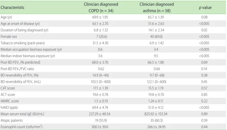
Table 1. Patient characteristic of patients with diagnosed asthma and COPD
Characteristic Clinician diagnosed Clinician diagnosed
p value
COPD (n = 34) asthma (n = 58)
Age (yr) 69.9 ± 1.95 65.7 ± 1.39 0.08
Age at onset of disease (yr) 63.1 ± 2.70 51.6 ± 2.63 <0.005
Duration of being diagnosed (yr) 6.8 ± 1.32 14.1 ± 2.34 0.02
Female sex 7 (20.6) 40 (69.0) <0.005
Tobacco smoking (pack-years) 31.5 ± 4.30 6.9 ± 1.42 <0.005
Median occupation biomass exposure (yr) 0.6 3.4 <0.005
Median indoor biomass exposure (yr) 3.6 9.5 <0.005
Post-BD FEV1 (% predicted) 68.0 ± 3.70 66.5 ± 1.80 0.69
Post-BD FEV1/FVC ratio 0.62 0.66 0.14
BD reversibility of FEV1 (%) 14.9 (0–40) 9.7 (0–68) 0.38
BD reversibility of FEV1 (mL) 103.5 (0–400) 122.1 (0–600) 0.45
CAT score 17.1 ± 1.39 15.5 ± 1.19 0.57
ACT score 19.6 ± 0.78 19.8 ± 0.70 0.85
MMRC score 1.5 ± 0.19 1.24 ± 0.11 0.22
FeNO (ppb) 69.4 ± 4.74 51.0 ± 4.12 <0.005
Mean serum total IgE ((IU/mL) 227.29 ± 48.54 825.92 ± 153.34 0.89
Atopic patients 19 (55.9) 35 (60.3) 0.59
Eosinophil count (cells/mm3) 300.1± 39.0 266.1± 24.95 0.44
Values are presented as mean ± standard deviation, number (%), or median (range).
COPD, chronic obstructive pulmonary disease; FEV1, forced expiratory volume in 1 second; FVC, forced vital capacity; BD, bronchodilator; CAT, COPD assessment test; ACT, Asthma Control Test; MMCR, Modified Medical Research Council Dyspnea Scale; FeNO, fractional exhaled nitric oxide; ppb, part per billion.
p < 0.05, statistically significance.
The ACOS in tertiary care setting Thailand
DISCUSSION
Prevalence of ACOS was 47.8% among obstructive airway disease patients treated in a tertiary care clinic in Thailand. In comparison with previous observation, the prevalence of ACOS in United States (US) population, there was 15.8%–23.4% of obstructive airway disease patients in general clinic and severe asthma clinic [1]. However, recent studies show that prevalence of ACOS from both asthma and COPD cohort in the different region varies from 14.6%–56% [7, 14, 15]. Prevalence of ACOS in Thai chronic airway disease patients was similar to Australia as well as United Kingdom. The US cohort found that half of patients with diagnosed obstructive lung disease were ACOS [15, 16].
Thirteen percent of COPD patients in COPDgene cohort and
17.4% of Spanish COPD cohort were labelled ACOS according to history of previous physician-diagnosed asthma [8, 17]. In contrast to our study, one-third (29.4%) of clinician-diagnosed Thai COPD was classified ACOS according bronchodilator reversibility and presence of atopy. Apart from ACOS definition or description, patient characteristics, clinical severity, and ethnicities contributed to the difference between ACOS prevalence in COPD.
For ACOS from the asthma aspect, one-fifth (22.8%) of Latin American cohort with fixed airway obstruction were reported a prior diagnosis of asthma [18]. The prevalence of Scandinavian ACOS in asthmatics in primary care clinic was 27.4% [19]. Since we enrolled more severe asthma in specialist clinic, selection bias is associated with the higher ACOS prevalence in our study. Age of Thai ACOS is comparable with isolated asthma and isolated COPD Table 2. Patients’ characteristics according to final diagnosis and investigator definition
Characteristic Patients with pure Patients with pure Patients with ACOS
p value
COPD (n = 24) asthma (n = 24) (n = 44)
Age (yr) 75.0 (49–91) 61.3 (40–78) 66.6 (48–93) 0.0001
Tobacco smoking (pack-years) 31 (0–120) 6.7 (0–30) 13 (0–70) 0.0001
Body mass index (kg/m²) 23.55 (13.38–30.60) 27.50 (20.60–37.42) 25.13 (17.26–41.64) 0.062
Occupational biomass (yr) 0.83 (0–12) 3.58 (0–22) 2.59 (0–25) 0.24
Indoor biomass (yr) 4 (0–30) 9.13 (0–30) 8.14 (0–30) 0.05
Females sex 6 (25.0) 20 (83.3) 21 (47.7) 0.0001
CAT score 15.75 ± 7.31 12.58 ± 9.42 12.20 ± 6.57 0.12
ACT score 19.88 ± 4.35 20.5 ± 4.18 19.91 ± 4.5 0.85
MMRC scale 1.42 ± 0.88 1.21 ± 1.18 1.16 ± 0.57 0.42
FeNO (ppb) 72.22 ± 30.21 53.94 ± 23.51 52.54 ± 29.04 0.04
Post-BD FEV1 (% predicted) 65.6 ± 20.2 68.3 ± 11.8 67.2 ± 18.0 0.86
Patients with BD reversibility 0 (0) 1 (4.2) 12 (27.3) 0.002
BD reversibility of FEV1 number to
BD reversibility of FEV1 (%) 4.8 ± 4.3 8 ± 5.9 12.5 ± 14.7 0.009
BD reversibility of FEV1 (mL) 63.6 115.8 149.8 0.009
Total serum IgE (IU/mL) 471.6 ± 641.6 442.4 ± 432.36 747.23 ± 1,243.6 0.84
Serum sIgE to Dp (kUA/L) 1.05 ± 3.38 6.70 ± 21.64 6.11 ± 17.96 0.06
Serum sIgE to Df (kUA/L) 2.00 ± 7.70 5.40 ± 16.42 5.93 ± 17.70 0.03
Serum sIgE to Af (kUA/L) 0.42 ± 1.39 0.09 ± 0.12 0.96 ± 3.90 0.08
Eosinophil counts (cells/mm3) 255.48 ± 240.0 311.38 ± 199.17 271.38 ± 159.48 0.40
Atopy (+SPT/ sIgE) 11 (45.8) 15 (62.5) 29 (65.9) 0.26
Values are presented as median (range), mean ± standard deviation, or number (%).
COPD, chronic obstructive pulmonary disease; ACOS, asthma and COPD overlap syndrome; CAT, COPD assessment test; ACT, Asthma Control Test; MMCR, Modified Medical Research Council Dyspnea Scale; FeNO, fractional exhaled nitric oxide; BD, bronchodilator; FEV1, forced expiratory volume in 1 second;
ppb, part per billion; sIgE, specific IgE; Df, Dermatophagoides farina; Dp, Dermatophagoides pteronyssinus; Af, Aspergillus fumigatus; SPT, skin prick test.
p < 0.05, statistically significance.
that were similar to previous report [8].
Tobacco smoking is the most important risk factor of COPD around the world including Asian region [3]. However, duration of tobacco smoking in Thai ACOS did not reach GOLD definition of COPD. Previous Asian studies has supported that cigarette smoking was related to the development of fixed airway obstruction in asthma which is a criterion in the definition of ACOS [20].
No difference in symptoms assessed by CAT, ACT, or MMRC was found between 3 groups in the present study. Our study compared symptoms using composite score including COPD (CAT) and asthma (ACT) in all obstructive airway patients. The composite score could not differentiate severity among there clusters of airway diseases. Generally poor quality of life is noted in ACOS compared to isolated asthma and isolated COPD.
The limitation of our study is lack of measuring quality of life by using specific questionnaire either asthma-related quality of life questionnaire or St. George Respiratory Questionnaire.
Neither serum total IgE nor blood eosinophils count distinguishes ACOS from asthma and COPD in Thai cohort. This finding is different from previous study of biomarker study in ACOS which shown role of these biomarkers [5, 21]. However, FeNO was significant higher in isolated COPD than ACOS and isolated asthma. The increased FeNO in our COPD group may relate to tobacco smoke inhibits nitric oxide synthase and the presence of Th2 inflammation in COPD with atopy may lead to increased FeNO. For these, reason, FeNO cannot be recommended for differentiating asthma from COPD [12]. Moreover, we found that atopic status could be a confounding variable for high FeNO level in Thai COPD. Since allergen sIgE is recommended for defining atopy and it was used with FeNO for diagnosed ACOS in Japanese cohort [21]. Nevertheless, Thai ACOS had the higher serum total IgE and FENO in comparison with Japanese population. Different biomarkers may reflect the different racial basis and atopic background. For this reason the current biomarkers including lung function bronchodilator reversibility, total IgE, and FeNO are limited in terms of ACOS diagnosis across the different ethnicities and their role needs to be further investigated.
In conclusion, prevalence of ACOS is common in severe late-onset adult asthma and COPD who were treated in a Thai tertiary care clinic. However, there is no difference in symptoms score, lung functions and biomarkers of atopy and systemic inflammation were found. Atopy is common in our Thai COPD cohort that has never been previously reported. Further studies
are needed for characterizing ACOS whether it is a part of the spectrum of asthma or COPD or another entirely different entity.
REFERENCES
1. Zeki AA, Schivo M, Chan A, Albertson TE, Louie S. The asthma-COPD overlap syndrome: a common clinical problem in the elderly. J Allergy (Cairo) 2011;2011:861926.
2. Louie S, Zeki AA, Schivo M, Chan AL, Yoneda KY, Avdalovic M, Morrissey BM, Albertson TE. The asthma-chronic obstructive pulmonary disease overlap syndrome: pharmacotherapeutic considerations. Expert Rev Clin Pharmacol 2013;6:197-219.
3. Global Initiative for Chronic Obstructive Lung Disease (GOLD). Global strategy for the diagnosis, management, and prevention of chronic obstructive lung disease [Internet]. [place unknown]: Global Initiative for Chronic Obstructive Lung Disease; 2014 [cited 2014 Sep 1]. Available from: http://goldcopd.org/global-strategy-diagnosis-management- prevention-copd.
4. The Global Initiative for Asthma. GINA Report, Global strategy for asthma management and prevention. Revised 2014 [Internet]. The Global Initiative for Asthma; c2014 [cited 2014 Jan 1]. Available from:
http://ginasthma.org/archived-reports/
5. Cosio BG, Soriano JB, López-Campos JL, Calle-Rubio M, Soler-Cataluna JJ, de-Torres JP, Marín JM, Martínez-Gonzalez C, de Lucas P, Mir I, Peces- Barba G, Feu-Collado N, Solanes I, Alfageme I, Casanova C; CHAIN Study.
Defining the asthma-COPD overlap syndrome in a COPD cohort. Chest 2016;149:45-52.
6. Sin DD, Miravitlles M, Mannino DM, Soriano JB, Price D, Celli BR, Leung JM, Nakano Y, Park HY, Wark PA, Wechsler ME. What is asthma-COPD overlap syndrome? Towards a consensus definition from a round table discussion. Eur Respir J 2016;48:664-73.
7. Kauppi P, Kupiainen H, Lindqvist A, Tammilehto L, Kilpeläinen M, Kinnula VL, Haahtela T, Laitinen T. Overlap syndrome of asthma and COPD predicts low quality of life. J Asthma 2011;48:279-85.
8. Hardin M, Silverman EK, Barr RG, Hansel NN, Schroeder JD, Make BJ, Crapo JD, Hersh CP; COPDGene Investigators. The clinical features of the overlap between COPD and asthma. Respir Res 2011;12:127.
9. Menezes AMB, Montes de Oca M, Pérez-Padilla R, Nadeau G, Wehrmeister FC, Lopez-Varela MV, Muiño A, Jardim JRB, Valdivia G, Tálamo C; PLATINO Team. Increased risk of exacerbation and hospitalization in subjects with an overlap phenotype: COPD-asthma.
Chest 2014;145:297-304.
10. Standardization of Spirometry, 1994 Update. American Thoracic Society.
The ACOS in tertiary care setting Thailand
Am J Respir Crit Care Med 1995;152:1107-36.
11. Pisi R, Aiello M, Tzani P, Marangio E, Olivieri D, Chetta A. Measurement of fractional exhaled nitric oxide by a new portable device: comparison with the standard technique. J Asthma 2010;47:805-9.
12. Dweik RA, Boggs PB, Erzurum SC, Irvin CG, Leigh MW, Lundberg JO, Olin AC, Plummer AL, Taylor DR; American Thoracic Society Committee on Interpretation of Exhaled Nitric Oxide Levels (FENO) for Clinical Applications. An official ATS clinical practice guideline: interpretation of exhaled nitric oxide levels (FENO) for clinical applications. Am J Respir Crit Care Med 2011;184:602-15.
13. Chiriac AM, Bousquet J, Demoly P. In vivo methods for the study and diagnosis of allergy. In: Adkinson NF, Bochner BS, Burks AW, Busse WW, Holgate ST, Lemanske RF Jr, O'Hehir RE, editors. Middleton's allergy:
principles & practice. 8th ed. Philadelphia (PA): Elsevier Saunders; 2014.
p. 1119-32.
14. Andersén H, Lampela P, Nevanlinna A, Säynäjäkangas O, Keistinen T.
High hospital burden in overlap syndrome of asthma and COPD. Clin Respir J 2013;7:342-6.
15. Fu JJ, Gibson PG, Simpson JL, McDonald VM. Longitudinal changes in clinical outcomes in older patients with asthma, COPD and asthma- COPD overlap syndrome. Respiration 2014;87:63-74.
16. Soriano JB, Davis KJ, Coleman B, Visick G, Mannino D, Pride NB.
The proportional Venn diagram of obstructive lung disease: two approximations from the United States and the United Kingdom. Chest 2003;124:474-81.
17. Miravitlles M, Soriano JB, Ancochea J, Muñoz L, Duran-Tauleria E, Sánchez G, Sobradillo V, García-Río F. Characterisation of the overlap COPD-asthma phenotype. Focus on physical activity and health status.
Respir Med 2013;107:1053-60.
18. Tálamo C, de Oca MM, Halbert R, Perez-Padilla R, Jardim JR, Muiño A, Lopez MV, Valdivia G, Pertuzé J, Moreno D, Menezes AM; PLATINO team. Diagnostic labeling of COPD in five Latin American cities. Chest 2007;131:60-7.
19. Kiljander T, Helin T, Venho K, Jaakkola A, Lehtimäki L. Prevalence of asthma-COPD overlap syndrome among primary care asthmatics with a smoking history: a cross-sectional study. NPJ Prim Care Respir Med 2015;25:15047.
20. Lee HY, Kang JY, Yoon HK, Lee SY, Kwon SS, Kim YK, Rhee CK. Clinical characteristics of asthma combined with COPD feature. Yonsei Med J 2014;55:980-6.
21. Tamada T, Sugiura H, Takahashi T, Matsunaga K, Kimura K, Katsumata U, Takekoshi D, Kikuchi T, Ohta K, Ichinose M. Biomarker-based detection of asthma-COPD overlap syndrome in COPD populations. Int J Chron Obstruct Pulmon Dis 2015;10:2169-76.