The Chewing Efficiency of Occlusal Stabilization Appliances by Anatomy of the Occlusal Surface
Yeong-Gwan Im
1, D.D.S.,M.S.D., Choong-Ho Choi
2*, D.D.S.,M.S.D.,Ph.D., Jae-Hyeong Kim
1*, D.D.S.,M.S.D.,Ph.D., Chong-Ouk Rhee
3, Ph.D.,
Byung-Gook Kim
1*, D.D.S.,M.S.D.,Ph.D.
Dept. of Oral Medicine, School of Dentistry, Chonnam National University
1Dept. of Preventive Dentistry, School of Dentistry, Chonnam National University
2Dental Science Research Institute*, Dept. of Food Science & Technology, Chonnam National University
3Occlusal appliance therapy has been proven to be very useful and effective in reducing signs and symptoms of patients with TMD. However, there are no reports about the masticatory efficiency of the occlusal appliance.
The purpose of this study was, first, to investigate the masticatory efficiency of the conventional stabilization appliance experimentally in normal healthy subjects, by comparing it with that of their natural dentition; and, second, to develop a modified stabilization appliance as an attempt to increase masticatory efficiency.
Eleven subjects (mean age 25.3 years, range from 23 to 33) participated in this study. Six were men and five were women. They were healthy and had complete or near―complete natural dentition, and did not present with signs or symptoms of TMD.
Two kinds of occlusal appliances―the conventional flat maxillary stabilization appliance (i.e., FSA) and a modified maxillary stabilization appliance with additional anatomic structures on its occlusal surface (i.e., ASA)
―were made for every subject. Subjects chewed peanuts that were selected as a food to test the three masticatory conditions of the natural dentition, the ASA, and the FSA. The number of chewing strokes was counted during each 1-minute chewing period. Chewed peanut boluses were recovered and their hardness was measured by texture analysis. Statistical tests were performed. The following results were obtained.
1. The masticatory efficiency of the FSA was 38.6 percent that of the natural dentition. The efficiency of the ASA was 78.2 percent that of the natural dentition.
2. The number of chewing strokes in the natural dentition condition was measured to be 1.5 strokes per second.
It decreased to 90 percent in the ASA and FSA conditions.
These results indicate that the ASA could serve an improved masticatory capacity as well as its therapeutic effects in TMD. A clinical application of the ASA should be considered to extend the management of TMD patients.
Key words : Masticatory efficiency, Occlusal stabilization appliance, Texture analysis, Occlusal surface
Corresponding Author : Prof. Byung-Gook Kim
Department of Oral Medicine, School of Dentistry, Dental Research Institute Chonnam National University 8 Hak-1-Dong Dong-Gu Gwangju 501-190, Korea
E-mail: [email protected] received: 2005-06-26
accepted: 2005-08-10
Ⅰ. INTODUCTION
Temporomandibular Disorder (TMD) is a condition that encompasses various functional disturbances in the masticatory system. TMD presents its clinical signs and symptoms on the major masticatory system components (i.e., masticatory muscles, joints and teeth). Patients suffering TMD frequently show decreased or impaired masticatory function as a result.
Helkimo
1)and Molin
2)reported that patients with TMJ pain or dysfunction symptoms have been found to have a reduced bite force. Christensen
3)suggested that the masticatory muscles of TMD patients may be more susceptible to fatigue when compared to normal subjects. Mongini
4)compared the chewing strokes between normal and diseased persons and observed that normal persons masticate with chewing strokes that are well rounded, whereas the chewing strokes of persons with TMJ pain were a repeated pattern. TMD has been considered as one of factors influencing masticatory efficiency, which is generally defined as the capacity to reduce food during mastication. Agerberg
5)showed that chewing ability was closely correlated to symptoms of mandibular dysfunction. Tzakis
6)concluded that the masticatory function of TMD patients seems to be compromised, and the treatment of TMD has a positive effect on the masticatory function, because both masticatory efficiency and occlusal force endurance improved after treatment.
Among the wide spectrum of therapeutic modalities, occlusal appliance therapy has been proven to be very useful and effective in reducing signs and symptoms of the patients with TMD
7,8). An occlusal appliance is a removable device, altering the mandibular position and contact pattern of the teeth. This provides a therapeutic mandibular position and occlusion that fits the criteria for optimum occlusal relationships of the jaw. When the appliance is being worn, an occlusal contact pattern is established in harmony with the optimum condyle-disc-fossa relationship for the patient, providing orthopedic stability
9). The stabilization
appliance uses specifically this condylar position or centric relation as a therapeutic jaw position. The stabilization appliance induces an optimum occlusal condition that reorganizes the neuromuscular reflex activity, which in turn reduces abnormal muscle activity (i.e., parafunctional activity).
Clinical usage of an occlusal appliance is varied according to the disorder that is to be treated.
Intracapsular disorders respond well with continuous use of an occlusal appliance. When a patient has a myogenous pain disorder, nighttime use is usually effective. Nighttime usage is also effective in the management of bruxism and the symptoms closely related to nocturnal parafunc- tional habits
10,11).
After proper insertion of the stabilization appliance, patients are told to close their jaw gently to touch the acrylic occlusal surface of the appliance slightly with the teeth of their opposite dentition during most of the non-functioning resting period.
There are no consistent criteria about whether patients should wear and chew with an occlusal appliance at meals or take it off to chew with their natural teeth. Additionally, there are no reports about the masticatory efficiency of the occlusal appliance. However, patients are usually instructed to remove the appliance and have a meal using their own dentition. The reason for this may be attributed to the expectation that wearing an occlusal appliance will disturb normal masticatory function and deteriorate chewing efficiency.
It is mostly at chewing time when heavy functional forces are generated from masticatory muscles and applied to teeth, periodontium and temporomandibular joints. Therefore, if a patient has a compromised masticatory system or an inappropriate occlusal condition as an etiologic agent of TMD, chewing without removing the stabilization appliance during mealtime might help continue to retain an optimized jaw relation and occlusal condition over the mastication period. This might further promote the resolution of signs and symptoms of TMD.
In other words, it is possible that wearing the
stabilization appliance even at mealtime as well as other times can improve the treatment effect in patients that need it. Then, the masticatory capacity of the appliance plays an important role in mastication and dietary absorption. Up to now, there are no data available about the chewing efficiency of occlusal appliances. The first step to verify the above assumption is to investigate the chewing efficiency of the stabilization appliance itself. Then, any modification of the appliance can be made to increase the chewing efficiency. Ultimately, the appliances could be clinically tested to evaluate their therapeutic effect.
The purpose of this study was, first, to reveal the masticatory efficiency of the conventional stabi- lization appliance experimentally in normal healthy subjects, by comparing it with that of their natural dentition; and second, to develop a modified stabilization appliance as an attempt to increase masticatory efficiency.
Ⅱ. MATERIALS AND METHOD
The criteria used for patient selection were the following: ⑴ all subjects had no notable pain or dysfunction of TMD at present and no such past history; ⑵ there was no considerable malocclusion or occlusal interference in their dentition; ⑶ they had complete or near-complete natural dentition without any missing teeth except the third molars;
⑷ in all subjects no significant facial asymmetry was found. Of the students in the dental college of Chonnam National University, 11 met these criteria after clinical examination. Six were men and five were women. Their mean age was 25.3 years, with a range from 23 to 33 years of age. The mean number of teeth was 28.5, with a range from 28 to 32; and that of restored teeth was 5.0 in each subject. They were all fully informed and all consented to their participation in this study.
Two types of occlusal appliances were included for studying chewing efficiency. One was a conventional maxillary stabilization appliance with a flat occlusal surface and canine rises, or the flat
stabilization appliance (FSA). Okeson presents a full description of this occlusal appliance
11). The other was a modified maxillary stabilization appliance, which has additional anatomic structures such as cusps, grooves and interdental spaces on the acrylic occlusal surface, mimicking natural tooth anatomy.
This occlusal appliance was accordingly named the anatomic stabilization appliance (ASA).
Both an ASA and an FSA for each subject were constructed following the same clinical and laboratory procedures, except the step for formation of anatomic occlusal structures on the ASA. A Lucia jig was made on the upper central and lateral incisors of an acrylic resin (Orthodontic Resin
®, Dentsply, USA). Its thickness was adjusted to make 2-3 mm interocclusal spaces at the first molars bilaterally when the jaw was closed. Centric relation (CR) of the jaw was guided by this Lucia jig.
Mandible was further guided by the bimanual technique of Dawson to ensure the accurate CR position. The CR interocclusal relation was then recorded using a silicone impression material (Futar D Occlusion
®, Kettenbach, Germany). Irreversible alginate impressions of the maxillary and mandibular arches were taken. The working casts, produced from those impressions, were mounted on an articulator (Twin Hoby
®, Shioda, Japan) with the CR interocclusal record inserted between the upper and lower casts. The articulator components that control incisal and condylar movement were set to
"Condition 2" for construction of the incisor and canine occlusal area of the appliances, and
"Condition 1" for that of the premolar and molar occlusal area
12)(Table 1).
Acrylic bases for the two appliances were
prepared prior to formation of occlusion. An
eccentric guiding ramp at the anterior occlusal area
was built up the same way on the two occlusal
appliances, but the anatomic tooth morphology of
cuspal ridges, fossae, grooves and interdental spaces
at the posterior occlusal area was formed only on
ASAs. Occlusal contacts in centric and eccentric
mandibular positions were checked and adjusted
further in the mouth after completion of the
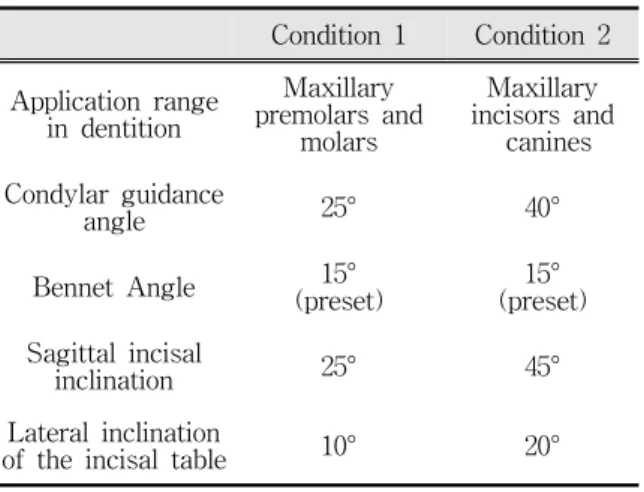
Condition 1 Condition 2
Application range in dentition
Maxillary premolars and
molars
Maxillary incisors and
canines Condylar guidance
angle 25° 40°
Bennet Angle 15°
(preset) 15°
(preset) Sagittal incisal
inclination 25° 45°
Lateral inclination
of the incisal table 10° 20°
Table 1. Articulator control settings for the construction of the occlusal surfaces of two kinds of stabilization appliances
Fig. 1. An anatomic stabilization appliance (ASA) with an occlusal view (left) and a close-up view of the posterior occlusal area (right).
Fig. 2. A flat stabilization appliance (FSA) with an occlusal view (left) and a close-up view of the posterior occlusal area (right).
appliances in the laboratory (Fig. 1 and 2).
Roasted peanuts were selected for the test food.
They were obtained from a market in Gwangju, Korea. They had been harvested and processed in 2004, and stored in a cool, dry place before use. Ten grams of peanuts was dispensed in each paper cup in a sufficient number.
Subjects were instructed to chew the dispensed
ten grams of peanuts as they usually did for 1
minute without swallowing. Chewing strokes during
the 1-minute chewing period were counted and
recorded by an examiner. The subjects were
directed to stop chewing after 1 minute and to spit
out the masticated peanut bolus into a disposable
glass bottle. Their mouth was washed out with water. This chewing scheme was repeated more than 5 times in the same condition. Next, the subjects inserted a prefabricated ASA in their mouth and made themselves accustomed to the appliance for more than 10 minutes. After the period of adaptation, they performed the chewing trial as described above using their natural dentition. This chewing scheme was repeated more than 5 times.
Finally, the subjects changed an ASA into an FSA and executed the chewing scheme another 5 times.
Every bolus of chewed peanuts in glass bottles was transferred to a Petri dish (50 φ × 12 mm). Petri dishes were filled up evenly with peanut bolus particles (Fig 3).
Uniaxial compression tests were performed with a texture analyzer (TA-XT
®, Stable Micro-Systems Survey, England) (Fig. 4). A 5.00 kg load compression cell was mounted in the moving crosshead. Chewed peanut bolus samples for tests were placed on the stainless steel bottom plate, and the stainless steel plunger―cylindrical in shape, 20 mm in diameter―was attached to the load cell on the upper movable crosshead. Each chewed peanut
Fig. 3. Chewed peanut bolus samples in Petri dishes.
Different sizes of comminuted peanut particles in the three samples are distinguishable with the naked eye (A, a sample from the condition of natural dentition; B, from an ASA; and C, from a FSA).
bolus sample placed on the stationary plate was compressed once to determine peak compression force. Crosshead speed was set at 1.0 mm/s.
Deformation was set at 80 percent of the original height of each sample was applied. Peak compression force was used as the measure for hardness.
Every hardness value obtained as a result of hardness measurements was paired with the number of chewing strokes counted during a 1-minute chewing period. Means and standard deviations of hardness values and chewing stroke numbers in each three chewing condition of all subjects were computed. One or two extreme hardness values were excluded with their counterparts (i.e., chewing stroke numbers) in order to ensure that standard deviations of hardness values fell within 3.0 and, if possible, 2.0. Revised means and standard deviations of hardness values and chewing stroke numbers were obtained as a result of this adjustment. Only mean values were used for further analysis.
For statistical analysis, nonparametric tests were selected because of the small number of subjects and the results of normality tests. The Friedman
Fig. 4. The texture analyzer used to measure the hardness of chewed peanut bolus samples (TA-XT®, Stable Micro-Systems Survey, England).
analysis of variance by ranks was used to determine from these revised means whether the three masticatory conditions were different in hardness and chewing stroke numbers. The Wilcoxon signed-rank test was used to distinguish the pairs of difference at the 0.05 level of significance. The Mann-Whitney test was used to see whether there is a difference between two genders at the 0.05 level of significance. All computing procedures were processed by a statistical analysis computer software application (SPSS 12.0 for Windows, SPSS Inc., USA).
Ⅲ. RESULTS
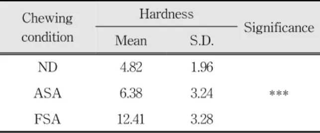
The total mean of the hardness values of 11 subjects was computed from the revised means of chewed peanut samples in the three masticatory conditions. The total mean hardness of chewed peanut samples was 4.82 kgf in the chewing condition of the natural dentition. That was 6.38 kgf in the ASA condition, which was about 1.3 times greater than that of the natural dentition. The total mean hardness in the FSA condition was even greater (about 2.6 times) and was measured to be 12.41 kgf (Table 2).
By means of the Friedman test, it was concluded that hardness values were not completely equal among the three chewing conditions (P < 0.001 at the 0.05 level of significance). The Wilcoxon signed-rank test showed that all three conditions were significantly different from one another. The differences in mean hardness between natural dentition and ASA, between natural dentition and FSA, and between ASA and FSA were all significant (all P < 0.01) (Fig 5). No significant difference was found between the two genders (Table 3).
These results clearly showed that hardness in the natural dentition condition is the lowest among the
Masticatory efficiency
(Occlusal appliance) = Hardness (Natural dentition)
× 100 (%) Hardness (Occlusal appliance)
Chewing condition
Hardness
Significance
Mean S.D.
ND 4.82 1.96
ASA 6.38 3.24 ***
FSA 12.41 3.28
The Friedman analysis of variance by ranks was used at the 0.05 level of significance.
ND: natural dentition; ASA: anatomic stabilization appliance; FSA: flat stabilization appliance; *** : P <
0.001
Table 2. Total means and standard deviations of the hardness of chewed peanut samples in the three masticatory conditions.
(unit: kgf)
Chewing condition
Mean for 6 men
Mean for 5
women Significance
ND 4.69 4.98 NS
ASA 5.89 6.97 NS
FSA 12.04 12.85 NS
The Mann-Whitney test was used at the 0.05 level of significance (NS: not significant).
ND: natural dentition; ASA: anatomic stabilization appliance; FSA: flat stabilization appliance
Table 3. Means of the hardness values of the two genders and significance in their difference in three masticatory conditions.
(unit: kgf)
three conditions and that hardness in the ASA
condition is closer to that in the natural dentition
than that in the FSA. Greater hardness of chewed
peanut samples results from less comminuted
peanuts and thus reflects less efficiency in
masticatory function. It was concluded from the
ND ASA FSA Hardness of chewed peanut samples (kgf)
0 2 4 6 8 10 12
14 Men
Women Total
**
**
**
Fig. 5. Hardness of chewed peanut samples in the three masticatory conditions. The Wilcoxon signed-rank test was used at the 0.05 level of significance. All three conditions were significantly different from one another (** : P < 0.01).
ND: natural dentition; ASA: anatomic stabi- lization appliance; FSA: flat stabilization appliance
above results that chewing with an occlusal appliance is not as efficient as chewing with the natural dentition, and that chewing with the ASA is more efficient than with the FSA.
Masticatory efficiency is related to the capacity to chew and comminute a food material easy for swallowing and digestion. Hardness of comminuted test food was supposed to be inversely associated with masticatory efficiency. Therefore, the
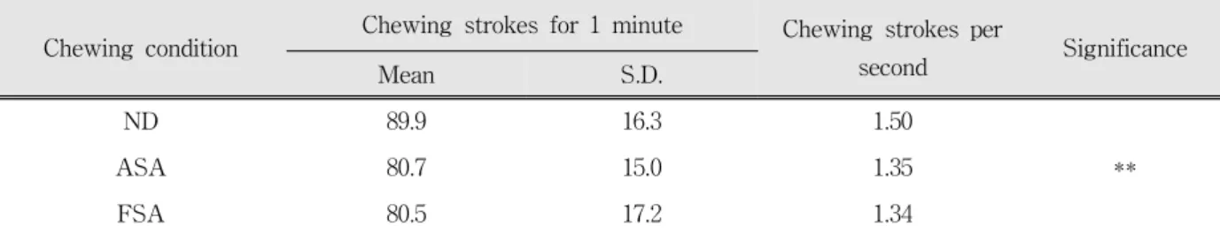
Chewing condition Chewing strokes for 1 minute Chewing strokes per
second Significance
Mean S.D.
ND 89.9 16.3 1.50
ASA 80.7 15.0 1.35 **
FSA 80.5 17.2 1.34
The Friedman analysis of variance by ranks was used at the 0.05 level of significance(** : P < 0.01).
ND: natural dentition; ASA: anatomic stabilization appliance; FSA: flat stabilization appliance
Table 4. Total means of chewing stroke numbers for chewing 10g peanuts for 1 minute in the three masticatory conditions.
ND ASA FSA
The number of chewing strokes
0 20 40 60 80 100
120 Men
Women Total
Fig. 6. Chewing stroke numbers for chewing 10 g of peanuts for 1 minute in the three masticatory conditions
ND: natural dentition; ASA: anatomic stabili- zation appliance; FSA: flat stabilization appliance
efficiency of an occlusal appliance relative to that of the natural dentition could be calculated from the ratio of their hardness values of chewed peanut samples. The masticatory efficiency of a certain type of an occlusal appliance was defined as follows.
The masticatory efficiency of the natural dentition
is 100 percent by this definition. The efficiency of
the ASA of each subject was calculated according
to this equation. All values were averaged to
compute the mean efficiency. The mean masticatory
efficiency of the ASA of all subjects was 78.2
percent; and that of the FSA was 38.6 percent
(Table 4). Thus the ASA was about two times more efficient in chewing than the FSA.
The same statistical tests were performed to compare chewing stroke numbers in the three chewing conditions. Total means of chewing stroke numbers of 11 subjects were computed from the revised chewing stroke numbers in the three masticatory conditions. The mean of the chewing stroke numbers in the condition of the natural dentition was 89.9 times per 1 minute (1.5 strokes/sec). That of the ASA was 80.7; and that of the FSA was 80.5 (Table 4) (Fig 6). The chewing stroke numbers of the ASA and the FSA were reduced to about 90 percent of that of natural dentition (P < 0.01).
Ⅳ. DISCUSSION
Mastication of food is the initial stage in the process of digestion. Large pieces of food are reduced for swallowing. The food is broken apart, and the surface area is increased for the efficient action of digestive enzymes and to facilitate solubilization of food substances in saliva to stimulate taste receptors
13). Efficient chewing implies the breakdown of food with the minimum effort and maximum rate of particle-size reduction.
The number of chews, or the time spent chewing before voluntary swallowing may be a reflection of the efficiency of the chewing process
14). Masticatory efficiency is usually defined as the capacity to reduce food during mastication. It is often given as index values obtained in specific tests
15).
Masticatory function can be assessed in several ways. Fractional sieving
16-26)is the oldest and most widely used method. It measures the capacity to pulverize certain test materials using a standardized sieve system. Questionnaires
26,27)have been used to evaluate the subjective chewing ability of an individual. Bite force measurements
25,28-32)have been considered as a reliable way to measure biome- chanical aspects of the masticatory system and also chewing efficiency. Electromyography (EMG)
33-37)basically measures muscle function and has been
successfully used as a masticatory function test.
Texture analysis describes the mechanical properties of test food. Changes in texture of a certain test material following mastication can implicate the functional capacity of the masticatory system. Horio
38)measured hardness, cohesiveness, gumminess, flexibility, adhesiveness and stickiness of five different foods, and suggested that hardness of food and shear force are the major factors controlling the pattern of chewing movements.
Mioche
39)found that measuring the shear force of the bolus may be a relevant method to assess chewing efficiency of a cohesive, fibrous food.
Texture of hard food like peanuts can be understood properly in the aspect of brittleness because hard food usually responds to mechanical stress by fracturing. Peanuts, which were chosen as a test food in this study, undergo a substantial change during the process of mastication through breakdown into smaller particles, incorporation of saliva, agglomeration and shaping of the mixture into a cohesive bolus. Consequently, hardness is the appropriate texture to measure rather than brittleness for the resultant chewed peanut bolus.
Because comminution of peanuts is under the influence of masticatory function, hardness of chewed peanut bolus reflects the functional capacity of the masticatory system: less hardness means more efficient mastication. The equation used to calculate the efficiency of an occlusal appliance is based on the assumption that masticatory efficiency is inversely associated with the hardness of comminuted test food.
The masticatory efficiency of dentures and various conditions of natural teeth has been studied.
Manly
16)investigated the masticatory performance and efficiency of complete dentitions and dentures.
The masticatory performance and efficiency were defined separately. The average performance of complete dentitions was 88 percent; dentitions lacking the third molar averaged 78 percent;
dentitions that possess two premolars and one molar in occlusion had a mean performance of 55 percent;
and denture cases averaged 35 percent. The
efficiencies of the four groups were calculated to be 166, 100, 44 and 23 percent, respectively.
In another study, the chewing efficiency of the denture wearer was reported as less than one-sixth that of the subject with a dentition
19). In the masticatory performance tests by Rissin
21), the patients with natural teeth had the highest score (90
%), followed by the overdenture patients (79 %), and the complete denture patients (59 %). In one study,
40)the substantial functional improvement was reported after insertion of a bridge mounted on osseointegrated oral implants in the lower jaw of edentulous patients. The results indicated the superiority of fixed prostheses over removable dentures.
The masticatory function in patients with myogenous TMD was evaluated by Tzakis
25). Masticatory efficiency increased from 54 percent before treatment to 65 percent after treatment, respectively. However, in healthy individuals pro- longed intense chewing decreased the masticatory efficiency, and chewing training for 1 month did not influence the masticatory efficiency
24). Therefore, it was suggested that healthy individuals, having a complete natural dentition and already a satisfactory chewing performance, may not improve their masticatory efficiency despite chewing training.
The masticatory efficiency of occlusal appliances used in the treatment of patients with TMD is not known. In this study, the masticatory efficiency of the conventional flat stabilization appliances or FSA was revealed to be 38.6 percent of that of the natural dentition in healthy subjects when peanuts were used as a test food. Meanwhile, the efficiency of ASA was 78.2 percent and about twice greater than that of FSA.
The only difference in the two stabilization appliances was whether the occlusal surface had anatomic tooth form. Modification of FSA with anatomic occlusal morphology resulted in increased masticatory efficiency. This fact implies that anatomic occlusal morphology is a critical factor associated with increased efficiency.
Masticatory efficiency has been shown to be
affected by many factors. Age, the state of dentition including number of teeth, quality and type of prosthetic appliances, occlusal morphology, food size, food texture, the tongue and other oral soft tissues, chewing habits, and dysfunction of the masticatory system were suggested.
Several authors have investigated tooth related factors. Occluding pairs of teeth was closely correlated with chewing efficiency, and use of the number of occluding pairs of teeth was proposed as a relatively reliable measure of chewing efficiency
20). The rate of comminution with increasing numbers of chews was most highly correlated with the occlusal area of the postcanine teeth
22). In another study, masticatory efficiency was well correlated with food platform area
18). The food platform area and the size of molars were the most important determinants for chewing ability among adults with a normal or defective natural dentition. Similarly, chewing performance was shown to vary directly with potential contact area
41). A reduction in the size of the potential contact area of artificial posterior teeth caused a loss in chewing effectiveness. It was also found that broad areas of intermediate occlusal contact appeared to contribute to chewing efficiency more than a few tight contact points
42). Bourdiol
43)further discriminated functional surface areas from static interarch contacts and found functional surface areas were positively correlated with occlusal areas.
These studies present sufficient explanation for understanding the difference in the efficiency of the two appliances. According to the above studies, the occlusal contact area, specifically the functional surface area, is the key for controlling masticatory efficiency in the occlusal appliance. Anatomic occlusal form built on the occlusal area of the ASA increased the occlusal contact area in the posterior occlusal surface of the appliance and improved efficiency remarkably.
Mechanisms of mastication are extremely
complex and highly flexible. A considerable
variation in chewing cycle characteristics is
observed even among healthy subjects. In this
experiment, the number of chewing strokes in the natural dentition was measured at 1.5 strokes per second. This result is in agreement with other studies
37,38). In one study
37), the number of cycles per second in subjects with complete or near complete dentition varied considerably from 0.6 to 1.6 cycles per second. In another study
45), the number of cycles ranged from 1.4 to 1.9 cycles per second among subjects with complete dentition; and 1.3 to 1.4 cycles per second among subjects with reduced periodontal support.
However, the number of chewing strokes was decreased to 90 percent in the presence of occlusal appliances. This means that an occlusal appliance worn in the mouth affects the normal masticatory process by slowing down the rate at which subjects chew. It is interesting to note that there was no difference in the number of chewing strokes between ASA and FSA, whereas the masticatory efficiency of ASA was about twice that of FSA.
Therefore, the occlusal anatomy of the occlusal appliance itself did not seem to be associated with a reduced number of chewing strokes.
In this study, there was no significant difference between the two genders in both hardness of chewed test food and the number of chewing strokes in all the three chewing conditions. This result coincides with other studies
17,20). Helkimo
20)reported no significant difference in chewing efficiency (the number of chewing strokes between sexes).
Manly
17)also concluded that there was no indication that the sex of the patient was a factor of efficiency.
On the contrary, Mioche
39)found that bolus properties and chewing variables were affected by gender. In his study females chewed for a shorter duration and developed less muscle activity than males. When ready for swallowing, the bolus texture was less comminuted and less homogenous.
There are some limitations in this study. A small number of subjects participated in this experiment.
Possibly they did not have enough time to adjust themselves to the occlusal appliances before chewing test food. Two or more kinds of test food other than peanuts could have been tested for better
assessment of masticatory efficiency. Masticatory function could be sufficiently evaluated in a multidimensional approach using more than one method―for example, texture analysis of chewed food; a questionnaire; and electromyography of masticatory muscles. For best results, these methods could be used all together in one experiment. Thus, studies should be extended further to include many subjects and patients using various examination methods.
Nonetheless, this study elucidated the masticatory efficiency of the FSA which is very effective in the management of pain and dysfunction of the masticatory system. In addition, the ASA was proved to have superior efficiency than the FSA.
The ASA shares all the features with the FSA in common except the anatomic occlusal surface morphology. These facts implicate the possibility that the ASA could serve improved masticatory capacity as well as its therapeutic effects in TMD.
Extended use―including at mealtime―of the ASA is expected to increase treatment effects in relevant TMD patients, though the effects should be proven in the future studies. A clinical application of the ASA should be considered to extend the management of TMD patients.
Ⅴ. CONCLUSIONS
The masticatory efficiency of two stabilization appliances (FSA and ASA) was investigated experimentally in healthy subjects by texture analysis of chewed peanut samples.
1. The masticatory efficiency of the FSA was 38.6 percent that of the natural dentition. The efficiency of the ASA was 78.2 percent that of the natural dentition.
2. The number of chewing strokes in the natural
dentition condition was measured to be 1.5
strokes per second. It decreased to 90 percent in
the ASA and FSA conditions.
REFERENCES
1. Helkimo E, Carlsson GE, and Carmeli Y. Bite force in patients with functional disturbances of the masticatory system. J Oral Rehabil 1975;2:397-406 2. Molin C. Vertical isometric muscle forces of the
mandibule. A comparative study of subjects with and without manifest mandibular pain dysfunction syndrome. Acta Odontol Scand 1972;30:485-499.
3. Christensen LV. Jaw muscles fatigue and pains induced by experimental tooth clenching: a review. J Oral Rehabil 1981;8:27-36.
4. Mongini F and Tempia-Valenta G. A graphic and statistical analysis of the chewing movements in function and dysfunction. Cranio 1984;2:125-134.
5. Agerberg G and Carlsson GE. Chewing ability in relation to dental and general health: Analyses of data obtained from a questionnaire. Acta Odontol Scand 1981;39:147-153.
6. Tzakis MG, Dahlstrom L and Haraldson T. Evaluation of masticatory function before and after treatment in patients with craniomandibular disorders. J Cranio- mandib Disord 1992;6:267-272.
7. Carraro JJ and Caffesse RG. Effect of occlusal splints on TMJ symptomatology. J Prosthet Dent 1978;40:
563-566.
8. Okeson JP, Kemper JT and Moody PM. A study of the use of occlusion splints in the treatment of acute and chronic patients with craniomandibular disorders.
J Prosthet Dent 1982;48:708-712.
9. Okeson JP. Management of temporomandibular disorders and occlusion. 5th ed., Philadelphia, 2003, Mosby, p.373
10. Wilkinson T, Hansson TL, McNeill C and Marcel T.
A comparison of the success of 24-hour occlusal splint therapy versus nocturnal occlusal splint therapy in reducing craniomandibular disorders. J Craniomandib Disord 1992;6:64.
11. Okeson JP. Management of temporomandibular disorders and occlusion. 5th ed., Philadelphia, 2003, Mosby, p.519
12. Jo Y, Hobo S, and Takayama H. Occlusion. Seoul, 1996, Koon Ja publishing Inc., pp.30-37
13. Bradly RM. Essentials of oral physiology. St. Louis, 1995, Mosby Inc., p.188
14. Wilding RJC. The association between chewing efficiency and occlusal contact area in man. Arch Oral Biol 1993;38:589-596.
15. Carlsson GE. Masticatory efficiency: the effect of age,
the loss of teeth and prosthetic rehabilitation. Int Dent J 1984;34:93-97.
16. Manly RS and Braley LC. Masticatory performance and efficiency. J Dent Res 1950;29:448-462.
17. Manly RS and Vinton P. A survey of the chewing ability of denture wearers J Dent Res 1951;30:
314-321.
18. Manly RS. Factors affecting masticatory performance and efficiency among young adults. J Dent Res 1951;30:874-882.
19. Kapur KK and Soman SD. Masticatory performance and efficiency in denture wearers. J Prosthet Dent 1964;14:687-694.
20. Helkimo E, Carlsson GE and Helkimo M. Chewing efficiency and state of dentition. Acta Odontol Scand 1978;36:33-41.
21. Rissin L, House JE, Manly RS and Kapur KK. Clinical comparison of masticatory performance and electro- myographic activity of patients with complete dentures, overdentures, and natural teeth. J Prosthet Dent 1978;39: 508-511.
22. Luke DA and Lucas PW. Chewing efficiency in relation to occlusal and other variations in the natural human dentition. Br Dent J 1985;159:401-403.
23. Lucas PW, Luke DA, Voon FCT, et al. Food breakdown patterns produced by human subjects possessing artificial and natural teeth. J Oral Rehabil 1986;13:205-214.
24. Tzakis MG, Kiliaridis S and Carlsson GE. Effect of chewing training on masticatory efficiency. Acta Odontol Scand 1989;47:355-360.
25. Tzakis MG, Dahlstrom L and Haraldson T. Evaluation of masticatory function before and after treatment in patients with craniomandibular disorders. J Cranio- mandib Disord 1992;6:267-272.
26. Hirai T, Ishijima T, Koshino H and Anzai T.
Age-related change of masticatory function in complete denture wearers: evaluation by a sieving method with peanuts and a food intake questionnaire method. Int J Prosthodont 1994;7:454-460.
27. Agerberg G and Carlsson GE. Chewing ability in relation to dental and general health: Analysis of data obtained from a questionnaire. Acta Odontol Scand 1981;39:147-153.
28. Helkimo E, Carlsson GE and Helkimo M. Bite force and state of dentition. Acta Odontol Scand 1977;35:
297-303.
29. Haraldson T, Karlsson U and Carlsson GE. Bite force and oral function in complete denture wearers. J Oral
Rehabil 1979;6:41-
30. Gibbs CH, Mahan PE, Lundeen HC, etc. Occlusal forces during chewing-Influences of biting strength and food consistency. J Prosthet Dent 1981;46:
561-567.
31. Heath MR. The effect of maximum biting force and bone loss upon masticatory function and bone loss upon masticatory function and dietary selection of the elderly. Int Dent J 1982;32:345-356.
32. Mioche L and Peyron MA. Bite force displayed during assessment of hardness in various texture contexts.
Arch Oral Biol 1995;40:415-423.
33. Rissin L, House JE, Manly RS and Kapur KK. Clinical comparison of masticatory performance and electromyographic activity of patients with complete dentures, overdentures, and natural teeth. J Prosthet Dent 1978;39: 508-511.
34. Tallgren A, Holden S, Lang BR et al. Jaw muscle activity in complete denture wearers-a longitudinal electromyographic study. J Prosthet Dent 1980;44:
123-.
35. Tallgren A, Holden S, Lang BR et al. Correlations between EMG jaw muscle activity and facial morphology in complete denture wearers. J Oral Rehabil 1983;10:105-120.
36. Karkazis HC and Kossioni AE. Re-examination of the surface EMG activity of the masseter muscle in young adults during chewing of two test foods. J Oral Rehabil 1997;24:216-223.
국문요약
교합안정장치 교합면의 모양에 따른 저작효율
전남대학교 치의학전문대학원 구강내과학교실
1, 예방치과학교실
2, 전남대학교 치의학연구소
*전남대학교 농업생명과학대학 식품공학과
3임영관
1․최충호
2*․김재형
1*․이종욱
3․김병국
1*교합장치요법은 측두하악장애 환자의 증상과 징후를 감소시키는데 매우 효과적인 방법으로 밝혀져 있다. 교합 장치의 저작효율에 관한 보고는 아직 없는 실정이다. 이 연구의 목적은 정상인에서 실험적으로 자연치열과 비교 한 교합안정장치의 저작 효율을 조사하고, 개선된 저작 효율을 가진 변형된 교합안정장치를 개발하는 것이었다.
열한명의 연구대상자(평균 연령 25.3세, 연령 범위 23-33세)가 실험에 참여했다. 이 중 남자는 6명, 여자는 5명 이었다. 이들은 완전하거나 거의 완전한 자연치열을 가지고 있고 측두하악장애의 증상과 징후가 없는 건강한 사람들이었다. 기존의 편평한 교합면이 있는 상악 교합안정장치(FSA) 및 교합면에 부가적으로 해부학적인 구조
37. Rilo B, Silva JL, Gude F and Santana U. Myoelectric activity during unilateral chewing in healthy subjects:
Cycle duration and order of muscle activation. J Prosthet Dent 1998;80:462-466.
38. Horio T and Kawamura Y. Effects of texture of food on chewing patterns in the human subject. J Oral Rehabil 1989;16:177-183.
39. Mioche L, Bourdiol P and Monier S. Chewing behaviour and bolus formation during mastication of meat with different textures. Arch Oral Biol 2003;48:193-200.
40. Lindquist LW and Carlsson GE. Changes in masticatory function in complete denture wearers after insertion of bridges on osseointegrated implants in the lower jaw. Adv Biomaterials 1982;4:151.
41. Lambrecht JR. The influence of occlusal contact area on chewing performance. J Prosthet Dent 1965;15:
444-450.
42. Wilding RJC. The association between chewing efficiency and occlusal contact area in man. Archs Oral Biol 1993;38:589-596.
43. Bourdiol P and Mioche L. Correlations between functional and occlusal tooth-surface areas and food texture during natural chewing sequences in humans.
Arch Oral Biol 2000;45:691-699.
45. Fernandes CP, Psarras V, Freitas LB and Ahlgren J.
Jaw-closing muscles: electromyographic activity of human subjects with reduced periodontal support. J Oral Rehabil 1994;21:165-175.