The First Successful Heart-Lung Transplant in a Korean Child with Humidifier Disinfectant-Associated Interstitial Lung Disease
From 2006 to 2011, an outbreak of a particular type of childhood interstitial lung disease occurred in Korea. The condition was intractable and progressed to severe respiratory failure, with a high mortality rate. Moreover, in several familial cases, the disease affected young women and children simultaneously. Epidemiologic, animal, and post-
interventional studies identified the cause as inhalation of humidifier disinfectants. Here, we report a 4-year-old girl who suffered from severe progressive respiratory failure. She could survive by 100 days of extracorporeal membrane oxygenation support and finally, underwent heart-lung transplantation. This is the first successful pediatric heart-lung transplantation carried out in Korea.
Keywords: Heart-Lung Transplantation; Extracorporeal Membrane Oxygenation;
Interstitial Lung Disease; Inhalation Exposure Won Kyoung Jhang,1 Seong Jong Park,1
Eun Lee,1 Song I Yang,1 Soo Jong Hong,1 Ju-Hee Seo,2 Hyung-Young Kim,3 Jeong-Jun Park,4 Tae-Jin Yun,4 Hyeong Ryul Kim,4 Yong-Hee Kim,4 Dong Kwan Kim,4 Seung-Il Park,4 Sang-Oh Lee,5 Sang-Bum Hong,6 Tae-Sun Shim,6 In-Cheol Choi,7 and Jinho Yu1
1Department of Pediatrics, Asan Medical Center Children’s Hospital, College of Medicine, University of Ulsan, Seoul, Korea; 2Department of Pediatrics, Korea Cancer Center Hospital, Seoul, Korea;
3Department of Pediatrics, Pusan National University Yangsan Hospital, Yangsan, Korea; 4Department of Thoracic and Cardiovascular Surgery, University of Ulsan College of Medicine, Asan Medical Center, Seoul, Korea; 5Division of Infectious Disease, Department of Internal Medicine, University of Ulsan College of Medicine, Asan Medical Center, Seoul, Korea; 6Division of Pulmonary and Critical Care Medicine, Department of Internal Medicine, University of Ulsan College of Medicine, Asan Medical Center, Seoul, Korea; 7Department of Anesthesiology and Pain Medicine, University of Ulsan College of Medicine, Asan Medical Center, Seoul, Korea
Received: 24 November 2014 Accepted: 30 April 2015 Address for Correspondence:
Jinho Yu, MD
Department of Pediatrics, University of Ulsan College of Medicine, Asan Medical Center Children’s Hospital, 88 Olympic-ro 43-gil, Songpa-gu, Seoul 05505, Korea E-mail: [email protected]
http://dx.doi.org/10.3346/jkms.2016.31.5.817 • J Korean Med Sci 2016; 31: 817-821
INTRODUCTION
Children’s interstitial lung disease (chILD) comprises a group of heterogeneous lung conditions characterized by diffuse lung pathology and disordered gas exchange, re- sulting in high morbidity and mortality (1-3). Recently, we reported 16 cases of a par- ticular type of chILD that occurred between 2006 and 2011 in Korea, initially present- ing with mild symptoms suggestive of a respiratory infection but rapidly progressing to respiratory failure with a high mortality rate (44%) (4). In addition, two familial cases with the same clinical manifestations were reported (5). Pathologic findings of centri- lobular bronchiolar destruction and alveolar damage with fibrosis suggested intersti- tial lung disease (ILD), possibly associated with inhalation toxicity (6). Epidemiologic and animal studies identified humidifier disinfectants (HD) as a likely cause of this fa- tal lung disease (7-9). Indeed, no new cases were identified during the 2 years after hu- midifier disinfectants were removed from markets (6).
Here, we report a child with irreversible respiratory failure caused by HD-associated ILD who underwent heart-lung transplantation after a long period of extracorporeal membrane oxygenation (ECMO) support. This is the first documented case of pediat- ric heart-lung transplantation in Korea.
CASE DESCRIPTION
A 4-year-old girl was transferred to our hospital with a 2-month history of progressive respiratory problems. Initially, she developed a mild dry cough with no underlying lung disease. However, she developed dyspnea and tachypnea about 2 weeks after ini- tial symptom onset, which became progressively worse and necessitated admission to a tertiary hospital to which her mother and 1-year-old sister had been admitted several days earlier suffering from similar symptoms. The patient was initially diagnosed with chILD of unknown origin and was treated with methyl prednisolone, hydroxychloro- quine, and cyclophosphamide. However, the clinical symptoms and radiologic find- ings worsened (Fig. 1), which brought her to our hospital at June 11th, 2011. Unfortu-
nately, her younger sister could not be transferred and expired one week after her transfer. The patient and her family mem- bers had used HD which contained polyhexamethylene-guani- dine (PHMG) as one of components.
Her initial respiratory rate was 77 breaths per minute, her pulse rate was 136 beats per minute, and her blood pressure was 113/81 mmHg. Because the patient had already developed air leak syndrome involving subcutaneous emphysema and pneumomediastinum, we tried to avoid intubation and main- tained oxygenation as efficiently as possible. Thus, the patient received 100% oxygen via a 15 L reservoir mask bag and was lightly sedated to prevent further air leak. She also received in- travenous immunoglobulin, azathioprine, and montelukast.
However, her condition continued to worsen. She was intubat- ed on Day 8 post-hospitalization. Despite high-frequency oscil- latory ventilator care with nitric oxide, desaturation and hypox- emia became worse (saturation < 80% and the PaO2 ≤ 40 mmHg).
For respiratory support, we applied veno-venous ECMO via right internal jugular vein and right femoral vein surgical cannula- tion on Day 12 post-hospitalization. ECMO initially improved the hypoxemia, and her condition appeared to be stabilized;
however, respiratory failure progressed to the point that it was considered irreversible. Therefore, she was registered to lung transplantation. During ECMO, progressive pulmonary hyper- tension was observed by serial echocardiography follow-up.
She was switched from veno-venous ECMO to veno-arterial ECMO with right common carotid arterial annulation to pro- vide more effective systemic perfusion with cardiac and pul- monary support after 83 days of ECMO support. Eventually, on Day 100 of ECMO support, she received heart and lung from 11-year-old girl with brain death. The donor to recipient body weight ratio was 1.3 (23.1/17 kg). The ratios of preoperatively measured lung size (donor/recipient) were as follows: T1 (aor- tic arch level to transverse dimension) 17.6/11.8, T2 (diaphragm A
D E
B C
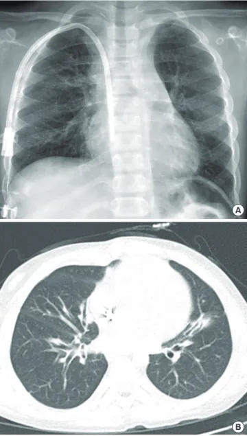
Fig. 1. Serial follow-up of radiologic examination before transfer to our hospital. (A) Initial chest x-ray showed normal chest radiography. (B) Chest radiograph taken one month after symptom develop revealed diffuse haziness in both lung field. (C) Diffuse reticulo-nodular opacities in both lung fields were aggravated and pneumomediastinum along the bilateral mediastinum were developed after two months. (D) A high-resolution CT scan performed 2 weeks after symptom onset revealed peribronchial and subpelural consoli- dation, especially in dependent portion of both lower lobes. (E) A follow-up CT scan performed 2 months later showed increased diffuse ground glass opacities with traction bronchiectasis and cystic lesions in both lungs. Pulmonary interstitial emphysema in the left lower lobe and pneumomediastinum also detected.
level to transverse dimension) 19.8/17, L1 (apex of right lung to diaphragm level) 15.9/11.3 and L2 (apex of left lung to diaphragm level) 15.8/12.3. Due to size mismatch and overinflation of the lung, the sternum was left open. However, we could perform sternal closure without further procedure on Day 2 post trans- plant. Histological examination of the explanted lung showed interstitial organizing fibrosis and multiple microabscesses (Fig.
2). The patient was extubated on Day 11 post-transplant with- out acute complication; however, she suffered several morbidi- ties due to prolonged ECMO support including chronic renal failure requiring hemodialysis and pancreatic pseudocyst. She was transferred to a general ward on Day 83 post-transplant. Six months after transplantation, a follow-up abdominal CT acci- dentally detected lesions in the liver and kidney, which were diagnosed as monomorphic B-cell post-transplant lymphopro- liferative disease. She received chemotherapy with a monoclo- nal anti-CD20 antibody (rituximab®), and the condition resolved.
The latest pulmonary function test (performed at 3 years post- transplant) showed a forced expiratory volume in 1 second of 0.95 L and a forced vital capacity of 1.01 L, which were 76.3%
and 74.2% of predicted values, respectively. Echocardiography
found no evidence of pulmonary hypertension with good func- tion (ejection fraction 62% and fractional shortening 32%). Rou- tine follow-up imaging study revealed no evidence of transplant- associated complications such as bronchiolitis obliterans (BO) (Fig. 3).
DISCUSSION
chILD comprises a diverse group of rare lung diseases, which often makes it difficult to identify a specific underlying cause.
From 2006, an outbreak of a particular type of chILD had oc- curred in Korea. First nationwide survey was performed in 2008, which reported 78 cases with as high as 46.1% of mortality, but could not identify the cause (10). Although progressive clinical Fig. 2. Explanted lung of recipient. (A) Gross finding of explanted lung tissue showed
diffuse parenchymal consolidation and multiple mucus plugs in the bronchus. (B) His- tologic examination of explanted lung biopsy revealed interstitial organizing fibrosis and multiple microabscesses.
A
B
Fig. 3. Follow up imaging studies at three years post-heart-lung transplantation. (A) Simple chest x-ray showed no active lung lesion. (B) Chest CT scans showed subseg- mental atelectasis and diffuse bronchial wall thickening.
A
B
course and fatal outcome of the disease are similar to those of acute interstitial pneumonitis, this disease is characterized by unique pathologic patterns of fibrosis in the bronchioles that are not observed in other chILD.
This disease gained much attention in 2011 when it was first reported in adults, mainly because the outcome was often fatal in pregnant and postpartum women. Finally, epidemiologic and animal studies including a nationwide investigation by the Korean Centers for Disease control and Prevention were un- dertaken, which identified toxic inhalation of HD as the cause (7-9). It contains PHMG, oligo (2-[2-ethoxy] ethoxyethyl) gua- nidium chrolide, 5-chloro-2-methylisothiazol-3(2H)-one/2- methyl isothiazol-3-one, and didecyldimethylammonium chlo- ride. The annual incidence of the disease dropped to zero after sales of humidifier disinfectant were suspended in 2012 (6).
Recently, lung transplantation was considered as a possible therapeutic option for patients with end-stage lung disease. Al- though median 5 years survival rates after lung transplantation are still around 50%, any patient with a pulmonary disease as- sociated with a life expectancy of less than 2 years (e.g., cystic fi- brosis, primary or secondary pulmonary hypertension, ILD with pulmonary fibrosis, bronchiolitis obliterans, and surfactant pro- tein b deficiency in children) is a potential candidate (11-14).
According to the official registry report by the International So- ciety for Heart and Lung Transplantation (ISHLT), lung and/or heart-lung transplantation was first performed in adults and children in 1963 and 1986, respectively. As of 2012, a total of 43,428 lung and 3,703 heart-lung transplantations have been performed in adults, and 1,875 and 667, respectively, have been performed in children, worldwide according to ISHLT registry (14,15). However, long-term results are somewhat disappoint- ing, with a median survival of 5.4 years in adults and 4.9 years in children. The 5 years survival rate for children undergoing a lung transplant is about 50%, and BO is responsible for 48% of deaths in those that survive more than 5 years post-transplant (13). The first lung transplant in Korea was performed in an adult patient in 1996. Prior to the present report, no such procedure had been performed in a child in Korea.
Many barriers to pediatric lung and/or heart-lung transplan- tation remain; for example, the scarcity of the available donors, size mismatches, and the more technically demanding surgical procedures required for small children. These constraints often extend the waiting time for organ allocation, which may neces- sitate longer periods of mechanical ventilator or ECMO sup- port. As in the current case, these therapeutic interventions may increase the rate of treatment-related complications post- transplant. In our case, the patient was on ECMO for 100 days before a donor organ became available (by comparison, her mother received ECMO for only 5 days). Her mother suffered no sequelae, however, the patient suffered from multiple organ failure including the heart, kidney, gastrointestinal tract, and
pancreas due to prolonged ECMO support. A combined heart- lung transplant was required due to severe pulmonary hyper- tension and related both ventricular failure. The decision to per- form lung or heart-lung transplantation is generally dictated by the presence/absence of irreversible left ventricular failure, al- though the preference of individual surgical teams will ultimate- ly determine the options available (16). Airway complications such as bronchial anastomotic dehiscence, bronchial stenosis, or malacia are the major causes of morbidity and mortality af- ter lung transplantation (11,17); these complications may be more common in pediatric cases due to the smaller airways.
Thus, surgical preference and the size of the recipient may de- termine the best treatment option. However, recent improve- ments in surgical technique (such as living donor lobar trans- plantation) and size reduction procedures (such as lobectomy, wedge resection, and split partitioning to the donor lung) all helped to overcome the aforementioned constraints (18-20).
Considering the improved life expectancy of pediatric patients after surgery, we suggest that lung transplantation be consid- ered more actively as a life-saving therapeutic option in Korea.
In summary, we report a case of pediatric heart-lung trans- plantation in Korea. The patient was diagnosed with a particu- lar type of chILD caused by toxic inhalation of humidifier disin- fectant. Although she suffered post-transplant comorbidities, her life was saved by the heart-lung transplant.
DISCLOSURE
The authors have no potential conflicts of interest to disclose.
AUTHOR CONTRIBUTION
Conception and coordination of the study: Jhang WK, Yu J. Ac- quisition of data: Jhang WK, Lee E, Yu J. Manuscript prepara- tion: Jhang WK, Park SJ, Lee E, Yang SI, Hong SJ, Seo JH, Kim H, Park J, Yun T, Kim HR, Kim Y, Kim DK, Park S, Lee S, Hong S, Shim T, Choi I, Yu J. Writing the first draft of the manuscript:
Jhang WK. Review and revising the manuscript: Jhang WK, Yu J.
Manuscript approval: all authors.
ORCID
Won Kyoung Jhang http://orcid.org/0000-0003-2309-0494 Seong Jong Park http://orcid.org/0000-0003-0250-2381 Eun Lee http://orcid.org/0000-0002-7462-0144 Song I Yang http://orcid.org/0000-0002-9648-4585 Soo Jong Hong http://orcid.org/0000-0003-1409-2113 Ju-Hee Seo http://orcid.org/0000-0001-6783-2942 Hyung-Young Kim http://orcid.org/0000-0001-8591-1783 Jeong-Jun Park http://orcid.org/0000-0003-0212-5475 Tae-Jin Yun http://orcid.org/0000-0001-8441-4574
Hyeong Ryul Kim http://orcid.org/0000-0002-6691-7693 Dong Kwan Kim http://orcid.org/0000-0003-1984-0352 Seung-Il Park http://orcid.org/0000-0002-8729-0498 Sang-Oh Lee http://orcid.org/0000-0003-1381-8787 Tae-Sun Shim http://orcid.org/0000-0001-6653-816X In-Cheol Choi http://orcid.org/0000-0002-7386-5043 Jinho Yu http://orcid.org/0000-0002-1226-8077 REFERENCES
1. Fan LL, Deterding RR, Langston C. Pediatric interstitial lung disease re- visited. Pediatr Pulmonol 2004; 38: 369-78.
2. Dinwiddie R, Wallis C. Paediatric interstitial lung disease (PILD)—an up- date. Curr Paediatr 2006; 16: 230-6.
3. Clement A, Eber E. Interstitial lung diseases in infants and children. Eur Respir J 2008; 31: 658-66.
4. Lee E, Seo JH, Kim HY, Yu J, Jhang WK, Park SJ, Kwon JW, Kim BJ, Do KH, Cho YA, et al. Toxic inhalational injury-associated interstitial lung disease in children. J Korean Med Sci 2013; 28: 915-23.
5. Lee E, Seo JH, Kim HY, Yu J, Song JW, Park YS, Jang SJ, Do KH, Kwon J, Park SW, et al. Two series of familial cases with unclassified interstitial pneu- monia with fibrosis. Allergy Asthma Immunol Res 2012; 4: 240-4.
6. Kim KW, Ahn K, Yang HJ, Lee S, Park JD, Kim WK, Kim JT, Kim HH, Rha YH, Park YM, et al. Humidifier disinfectant-associated children’s intersti- tial lung disease. Am J Respir Crit Care Med 2014; 189: 48-56.
7. Yang HJ, Kim HJ, Yu J, Lee E, Jung YH, Kim HY, Seo JH, Kwon GY, Park JH, Gwack J, et al. Inhalation toxicity of humidifier disinfectants as a risk fac- tor of children’s interstitial lung disease in Korea: a case-control study.
PLoS One 2013; 8: e64430.
8. Ohnuma A, Yoshida T, Tajima H, Fukuyama T, Hayashi K, Yamaguchi S, Ohtsuka R, Sasaki J, Fukumori J, Tomita M, et al. Didecyldimethylammo- nium chloride induces pulmonary inflammation and fibrosis in mice.
Exp Toxicol Pathol 2010; 62: 643-51.
9. Lee JH, Kim YH, Kwon JH. Fatal misuse of humidifier disinfectants in Ko- rea: importance of screening risk assessment and implications for man- agement of chemicals in consumer products. Environ Sci Technol 2012;
46: 2498-500.
10. Kim BJ, Kim HA, Song YH, Yu J, Kim S, Park SJ, Kim KW, Kim KE, Kim DS, Park JD, et al. Nationwide surveillance of acute interstitial pneumonia in Korea. Korean J Pediatr 2009; 52: 324-9.
11. Huddleston CB. Pediatric lung transplantation. Semin Pediatr Surg 2006;
15: 199-207.
12. Kirkby S, Hayes D Jr. Pediatric lung transplantation: indications and out- comes. J Thorac Dis 2014; 6: 1024-31.
13. Huddleston CB. Pediatric lung transplantation. Curr Treat Options Car- diovasc Med 2011; 13: 68-78.
14. Benden C, Edwards LB, Kucheryavaya AY, Christie JD, Dipchand AI, Dob- bels F, Kirk R, Lund LH, Rahmel AO, Yusen RD, et al. The Registry of the International Society for Heart and Lung Transplantation: Sixteenth Offi- cial Pediatric Lung and Heart-Lung Transplantation Report--2013; focus theme: age. J Heart Lung Transplant 2013; 32: 989-97.
15. Yusen RD, Christie JD, Edwards LB, Kucheryavaya AY, Benden C, Dip- chand AI, Dobbels F, Kirk R, Lund LH, Rahmel AO, et al. The Registry of the International Society for Heart and Lung Transplantation: Thirtieth Adult Lung and Heart-Lung Transplant Report--2013; focus theme: age. J Heart Lung Transplant 2013; 32: 965-78.
16. Marshall SE, Kramer MR, Lewiston NJ, Starnes VA, Theodore J. Selection and evaluation of recipients for heart-lung and lung transplantation. Chest 1990; 98: 1488-94.
17. Alvarez A, Algar FJ, Santos F, Lama R, Baamonde C, Cerezo F, Salvatierra A. Pediatric lung transplantation. Transplant Proc 2005; 37: 1519-22.
18. Couetil JP, Tolan MJ, Loulmet DF, Guinvarch A, Chevalier PG, Achkar A, Birmbaum P, Carpentier AF. Pulmonary bipartitioning and lobar trans- plantation: a new approach to donor organ shortage. J Thorac Cardio- vasc Surg 1997; 113: 529-37.
19. Aigner C, Mazhar S, Jaksch P, Seebacher G, Taghavi S, Marta G, Wisser W, Klepetko W. Lobar transplantation, split lung transplantation and periph- eral segmental resection--reliable procedures for downsizing donor lungs.
Eur J Cardiothorac Surg 2004; 25: 179-83.
20. Santos F, Lama R, Alvarez A, Algar FJ, Quero F, Cerezo F, Salvatierra A, Baa- monde C. Pulmonary tailoring and lobar transplantation to overcome size disparities in lung transplantation. Transplant Proc 2005; 37: 1526-9.