Tuberc Respir Dis 2012;72:124-131
CopyrightⒸ2012. The Korean Academy of Tuberculosis and Respiratory Diseases. All rights reserved.
DNA-Damage Inducible 1 is a Property of Human Non-Small Cell Lung Cancer
Ji Yeon Lee, M.D.
1,2, Eunsil Kang, M.S.
2, Beom Jin Lim, M.D.
3, Yoon Soo Chang, M.D.
2, Se-Kyu Kim, M.D.
21
Division of Pulmonary and Critical Care Medicine, Department of Internal Medicine and Lung Institute, Seoul National University College of Medicine, Departments of
2Internal Medicine and
3Pathology, Yonsei University Health System, Yonsei University College of Medicine, Seoul, Korea
Background: DNA damage-inducible 1 (Ddi1), one of the ubiquitin-like and ubiquitin-associated family of proteins, may function in the regulation of the ubiquitin-proteasome pathway, which has been validated as a target for antineoplastic therapy. We investigated Ddi1 expression in human lung cancer tissues and evaluated the relationship of this expression pattern with clinicopathological factors in patients with non-small-cell lung cancer (NSCLC).
Methods: Ddi1 expression was examined by immunohistochemistry in tumor tissues from 97 patients with stage I NSCLC, who had undergone curative surgical resection at two tertiary referral hospitals from 1993∼2004. None of the patients received preoperative chemotherapy and/or radiation therapy.
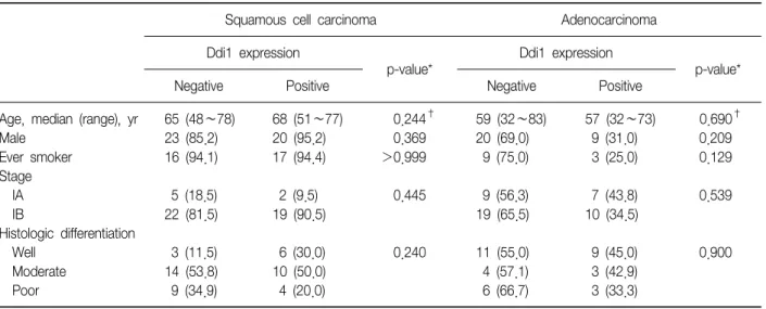
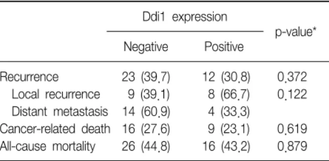
Results: Thirty-nine (40.2%) of the 97 cases were positive for Ddi1. Ddi1 expression was dominantly seen in cytoplasm rather than in the nuclei of cancer cells in all histological types, whereas adjacent nontumoral lung tissue showed negative Ddi1 staining in most cases. Ddi1 expression tended to increase in well-differentiated tumors but without statistical significance. Positive Ddi1 expression was associated with a tendency for better disease-free survival and disease-specific survival, although the difference was not significant.
Conclusion: Ddi1 expression is a property of NSCLC. Because Ddi1 could be a potential target for cancer therapy, more research is needed to evaluate its role in NSCLC.
Key Words: Lung Neoplasms; Immunohistochemistry; Ubiquitin; Proteasome Endopeptidase Complex; Anti- neoplastic Agents
Address for correspondence: Se-Kyu Kim, M.D.
Department of Internal Medicine, Yonsei University College of Medicine, 50, Yonsei-ro, Seodaemun-gu, Seoul 120-749, Korea
Phone: 82-2-2228-1954, Fax: 82-2-393-6884 E-mail: [email protected]
Received: Nov. 3, 2011 Revised: Nov. 28, 2011 Accepted: Dec. 21, 2011
Introduction
Lung cancer is one of the most commonly diagnosed cancers as well as the leading cause of cancer deaths worldwide
1. However, only about 20% of patients with non-small cell lung cancer (NSCLC) have their disease diagnosed at a stage in which surgery can be recom- mended as part of initial care. Moreover, the 5-year sur- vival rate of even the earliest stage I lung cancers after
complete surgical resection is still only approximately 70%, leading to the need for novel therapeutic targets and modalities
2.
One of the strategies for lung cancer therapy is target-
ing the ubiquitin-proteasome system (UPS), which plays
a central role in cell homeostasis, resulting in dereg-
ulation of cell processes necessary for survival
3,4. DNA
damage-inducible 1 (Ddi1) belongs to the ubiquitin-like
(UbL) and ubiquitin-associated (UBA) family of proteins
that has been implicated in the regulation of UPS
5. Ddi1
appears to release substrate from an ubiquitination com-
plex, making the substrate available for deubiquitina-
tion, and, thus, is presumed to play an important role
facilitating proteasomal degradation. Additionally, Ddi1
is the only representative in which the UbL and UBA
domains flank an aspartyl protease-like (RVP) domain.
This central Ddi1 domain has a remarkable structural similarity with retroviral proteases, suggesting that Ddi1 functions proteolytically during regulated protein turn- over in the cell. Notably, current studies show that this RVP domain is a potential therapeutic target for retro- viral aspartic protease inhibitors
6.
Much effort has been made to reposition the estab- lished retroviral aspartic protease inhibitors as anti- cancer agents for patients with NSCLC. Recent studies have shown that nelfinavir, ritonavir, and saquinavir in- hibit the growth of NSCLC and every cell type among 60 kinds of cancer cells
7. Nelfinavir was the most effec- tive of all protease inhibitors tested, resulting in apopto- sis and non-apoptotic cell death. Non-apoptotic cell death was related to induction of endoplasmic reticulum stress, which subsequently led to autophagy, a normal process of self digestion that generates energy for the cell under conditions of stress
8. Consequentially, these results suggest that nelfinavir could be repositioned as a lung cancer therapeutic, and one of the potential tar- gets for this promising drug is Ddi1.
However, the clinical role of Ddi1 in human cancer is unknown. Therefore, in this study, we investigated Ddi1 immunohistochemical expression in human lung cancer tissue and evaluated whether Ddi1 expression in- creased in lung cancer compared with that in adjacent nontumoral tissue. We also investigated Ddi1 function by assessing intracellular localization as well as tissue distribution of Ddi1 expression to confirm whether Ddi1 could be developed as a useful target for nelfinavir.
Materials and Methods 1. Study Subjects
The study group comprised 97 patients with stage I NSCLC. Patients underwent curative surgical resection at two tertiary referral hospitals (Severance and Gangnam Severance Hospital, Yonsei University College of Medicine, Seoul, Korea) from 1993∼2004. The fol- low-up duration was defined as the interval between the date of operation and the date of death or last fol- low-up. None of the patients received preoperative che-
motherapy and/or radiation therapy.
Patient charts, including pathology and operative re- ports, were reviewed, and the data were coded. One representative formalin-fixed paraffin-embedded pri- mary tumor block was obtained for each case.
This study was approved by the Institutional Review Board of Severance Hospital (4-2011-0409). The board waived informed consent from the patients. This study was conducted in accordance with the Declaration of Helsinki.
2. Immunohistochemistry (IHC)
Sections (4-μm thick) were prepared from each of the paraffin-embedded samples. After sections were de- paraffinized in xylene (three 10-minute washes) and de- hydrated in a graded ethanol series (100-, 90-, 70-, and 50%, 5-minute washes), endogenous peroxidase activity was blocked with 30% hydrogen peroxide in water for 5 min. Antigens were retrieved for Ddi1 staining by boiling in 10 mM citrate acid (pH 6.0) for 3 min using a microwave oven. Sections were then incubated with rabbit anti-Ddi1 polyclonal antibodies (GeneTex, Irvine, CA, USA) and diluted 1:100 in antibody diluents (DAKO, Glostrup, Denmark) for 1 hour at 37
oC.
Secondary biotinylated link anti-mouse IgG or anti-rab- bit IgG for the Labeled Streptavidin-Biotin2 System, horseradish peroxidase (DAKO), and a third streptavidin peroxidase conjugate (DAKO) were incubated with the sections at room temperature. The reaction was vi- sualized with the 3, 3-diaminbenzidine substrate system (DAKO). Hematoxylin was used as the counterstain.
Human placenta tissue was used as a positive control because of its easy availability and relatively stable reactivity. The negative control consisted of isotype con- trol antibody (rabbit polyclonal IgG, DAKO) substituted for the primary Ddi1 antibody. Controls were run with each batch of slides at an average of approximately 15 slides per batch.
3. IHC Interpretation
Slides were interpreted by one experienced patholo-
gist (LBJ). The intensity of Ddi1 expression as evaluated
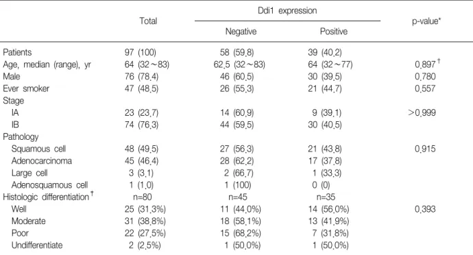
Table 1. Clinicopathological features related to DNA damage-inducible 1 (Ddi1) expression in non-small-cell lung cancer (NSCLC)
Total
Ddi1 expression
p-value*
Negative Positive
Patients 97 (100) 58 (59.8) 39 (40.2)
Age, median (range), yr 64 (32∼83) 62.5 (32∼83) 64 (32∼77) 0.897
†Male 76 (78.4) 46 (60.5) 30 (39.5) 0.780
Ever smoker 47 (48.5) 26 (55.3) 21 (44.7) 0.557
Stage
IA 23 (23.7) 14 (60.9) 9 (39.1) >0.999
IB 74 (76.3) 44 (59.5) 30 (40.5)
Pathology
Squamous cell 48 (49.5) 27 (56.3) 21 (43.8) 0.915
Adenocarcinoma 45 (46.4) 28 (62.2) 17 (37.8)
Large cell 3 (3.1) 2 (66.7) 1 (33.3)
Adenosquamous cell 1 (1.0) 1 (100) 0 (0)
Histologic differentiation
‡n=80 n=45 n=35
Well 25 (31.3%) 11 (44.0%) 14 (56.0%) 0.393
Moderate 31 (38.8%) 18 (58.1%) 13 (41.9%)
Poor 22 (27.5%) 15 (68.2%) 7 (31.8%)
Undifferentiate 2 (2.5%) 1 (50.0%) 1 (50.0%)
Data are presented as number (%) unless otherwise indicated.
*Pearson's χ
2or Fisher's exact test were used to estimate p-values.
†Mann-Whitney U-test was used to estimate the p-value.
‡