Introduction
The prevention and management of exacerbations are main objectives of chronic obstructive pulmonary disease (COPD) treatment. Each new exacerbation is harmful for the patient for diverse reasons: it increases in itself the risk of future exac- erbations
1, deteriorates the quality of life, accelerates the dete- rioration of lung function and increases the risk of hospitaliza- tion and death
2. Its prevention is, therefore, a central aspect of the management of these patients. There are various pharma- cological and non-pharmacological strategies aimed at both the control and prevention of COPD exacerbations. Although airway inflammation is one of the significant contributors to symptoms and exacerbations, current COPD guidelines do not consider the evaluation of the type of bronchitis or other complex pathophysiological processes involved in its genesis.
That leads to generalized management strategies, which are
Sputum Inflammometry to Manage
Chronic Obstructive Pulmonary Disease Exacerbations: Beyond Guidelines
Carmen Venegas, M.D.* , Nan Zhao, M.D., F.R.C.P.C.* , Terence Ho, M.D., M.Sc., F.R.C.P.C., M.Sc.
and Parameswaran Nair, M.D., Ph.D., F.R.C.P., F.R.C.P.C.
Firestone Institute for Respiratory Health, St. Joseph’s Healthcare Hamilton and the Department of Medicine, McMaster University, Hamilton, ON, Canada
Quantitative sputum cytometry facilitates in assessing the nature of bronchitis associated with exacerbations of chronic obstructive pulmonary disease (COPD). This is not assessed in most clinical trials that evaluate the effectiveness of strategies to prevent or to treat exacerbations. While up to a quarter of exacerbations may be associated with raised eosinophil numbers, the vast majority of exacerbations are associated with neutrophilic bronchitis that may indicate airway infections. While eosinophilia may be a predictor of response to corticosteroids (oral and inhaled), the limited efficacy of anti–interleukin 5 therapies would suggest that eosinophils may not directly contribute to those exacerbations.
However, they may contribute to airspace enlargement in patients with COPD through various mechanisms involving the interleukin 13 and matrix metalloprotease pathways. The absence of eosinophils may facilitate in limiting the unnecessary use of corticosteroids. The presence of neutrophiia could prompt an investigation for the specific pathogens in the airway. Additionally, sputum measurements may also provide insight into the mechanisms of susceptibility to airway infections. Iron within sputum macrophages, identified by hemosiderin staining (and by more direct quantification) may impair macrophage functions while the low levels of immunoglobulins in sputum may also contribute to airway infections. The assessment of sputum at the time of exacerbations thus would facilitate in customizing treatment and treat current exacerbations and reduce future risk of exacerbations.
Keywords: Pulmonary Disease, Chronic Obstructive; Bronchitis; Sputum Cell Count; Eosinophil; Infective Exacerbations
Address for correspondence: Parameswaran Nair, M.D., Ph.D., F.R.C.P., F.R.C.P.C.
Firestone Institute for Respiratory Health, St. Joseph’s Healthcare Hamilton, 50 Charlton Ave East, Hamilton, ON L8N 4A6, Canada Phone: 1-905-522-1155 (ext. 35044), Fax: 1-905-521-6183 E-mail: [email protected]
*Carmen Venegas and Nan Zhao contributed equally to this work.
Received: Apr. 5, 2020 Revised: Apr. 20, 2020 Accepted: May. 11, 2020 Published online: Jun. 18, 2020
cc
It is identical to the Creative Commons Attribution Non-Commercial License (http://creativecommons.org/licenses/by-nc/4.0/).
Copyright © 2020
The Korean Academy of Tuberculosis and Respiratory Diseases.
often suboptimal. Although “endotyping” is recommended for
“individualized” care of COPD exacerbations, this is not often practiced
3.
We present the following three cases to illustrate the limita- tions of current guidelines and common clinical practice in most outpatient clinics across the world.
(1) A 67-year-old male with a past smoking history of 21 years, moderate airflow obstruction (forced expiratory volume in 1 second [FEV
1] of 61% predicted), and recurrent exacerba- tions (two in the last 12 months): He is on fluticasone/salme- terol 1,000 μg/100 μg daily and tiotropium 18 mcg daily. After his first exacerbation, his FEV
1decreased to 44% predicted and subsequently worsened to 33% predicted after the sec- ond exacerbation. Current guidelines would suggest that both exacerbations “be treated with more bronchodilators,” and perhaps with a “short burst of prednisone” and a “broad-spec- trum antibiotic”
4, and perhaps adding long-term macrolide or a phosphodiesterase 4 inhibitor
4,5.
(2) A 57-year-old male, current smoker with a history of 15 pack-years: He reports productive cough, and in increase in wheeze and exertional dyspnea. His FEV
1/forced vital capac- ity (FVC) is 2.8 L/4.4 L (ratio of 63%) and improves to 2.9 L/4.2 L post bronchodilator, which is consistent with mild to moder- ate airflow obstruction (FEV
1of 78% predicted). Chest X-ray is normal. His current treatment includes salbutamol as needed, which he uses about 2 to 4 times a day. Current guidelines would suggest that he be commenced on a combination of a long-acting beta-2 agonist (with or without a long-acting anti- cholinergic inhaler)
4.
(3) An 81-year-old male, with a 34 years history of smok- ing: His previous medical history includes glaucoma, benign prostate hyperplasia, diabetes and coronary artery disease. He presents with exertional breathlessness and cough and has had two exacerbations within the last year. His pre-broncho- dilator FEV
1/FVC is 0.9 L/4.4 L, and postbronchodilator is 1.0 L/4.5 L, which are 29% and 90% predicted, respectively. Total lung capacity is 122%, residual volume is 160%, and KCO is 30% predicted. Arterial blood gases show a PCO
2of 58 mm Hg, PO
2of 64 mm Hg and pH of 7.38. Right ventricular systolic pressure is 40 mm Hg. Computed tomography of the thorax reveals heterogenous centrilobular emphysema. Current treatment is budesonide/formoterol (200 μg/6 μg) 2 puffs twice daily, terbutaline as needed, furosemide and ramipril.
Current guidelines would suggest adding a long-acting anti- cholinergic inhaler or alternatively switching to a single com- bination inhaler
4.
Current COPD Guidelines on Treatment and Prevention of Acute Exacerbations
Current recommendations are largely focused on decreas- ing exacerbations and improving symptoms by optimizing the
use of bronchodilators. It is known that both long-acting beta agonists (LABA) and long-acting anti-cholinergics (LAAC) can reduce the rate of exacerbations in patients with COPD.
Furthermore, the current literature supports that combined therapy (LABA/LAAC) is superior to monotherapy
6and to LABA/inhaled corticosteroid (ICS)
7combination, although its effect does not reach the sum of both
8. More recently, attempts have been made to demonstrate that triple inhalation therapy is even more effective than the LABA/LAAC combination;
but the results have been variable. A large study of more than 10,000 patients, which compared triple therapy against two types of dual therapy (LAAC/LABA and LABA/ICS), showed lower rates of moderate and severe exacerbations in the first group
9. A similar effect is observed when comparing single- inhaler triple combination with single-inhaler dual broncho- dilator combination, with a 15% reduction in the risk of mod- erate to severe exacerbations
10. The latter was not reflected in a subsequent trial performed in a “real-world clinical setting,”
where there were no differences between the groups treated with triple therapy and LAAC/LABA. However, when analyz- ing by subgroups, it was observed that among patients with blood eosinophilia >6% and in those with two or more previ- ous exacerbations, there was a statistically significant benefit (hazard ratio [HR] of 0.66 and 0.83, respectively)
11. Therefore, based on the current evidence, there is an inclination to add ICS to dual bronchodilator therapy in patients with moderate to very-severe stable COPD with a high risk of exacerbations, especially in those with serum eosinophils higher than 300 cells/μL
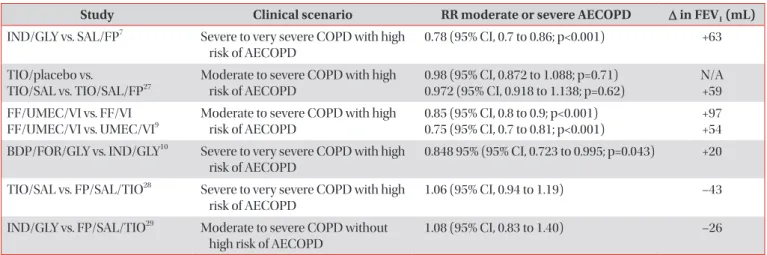
4,5. A summary of the current principal studies which compare dual and triple inhalers’ effect on FEV
1and exacer- bation rates is shown in Table 1.
Despite the availability of these pharmacologic interven-
tions, about 30% of patients with COPD have frequent ex-
acerbations
1, which entails high healthcare costs
5. As part
of the numerous efforts aimed at trying to reduce the rate of
hospitalizations in these patients, comprehensive care man-
agement programs (CCMP) have been implemented, includ-
ing strategies such as patient education, implementation of
action plans, and serial telephone evaluations by a trained
team. Unfortunately, the application of these strategies has not
been consistently shown a reduction in the risk of hospitaliza-
tion, but rather, in some cases they have increased it. This was
shown in a trial performed in 426 COPD patients who were
assigned to a CCMP versus conventional care. The study was
terminated before enrolment was completed, given higher
risks of hospitalization and mortality in the intervention group
(HR of 1.13 and 3.0, respectively)
12. Furthermore, there was
no improvement in quality of life outcomes with these inter-
ventions
13. These results contrast with other studies in which
these strategies were proven effective to reduce COPD-related
admissions, although the studied populations were signifi-
cantly different (e.g., patients with other comorbidities were
excluded)
14. Another interesting explanation for this phenom-
ena is that patients, having the resource of at-distance health monitoring, experience a false sense of safety, which delays consultation in case of an emergency
15.
Sputum, COPD, and Exacerbation Phenotype
Classically, an acute exacerbation of COPD (AECOPD) is defined as respiratory symptoms that worsen beyond the normal day-to-day variability requiring additional therapy
4,5. However, this definition of an AECOPD may be overlapping with many concurrent pathologies such as worsening left or right heart function, respiratory or metabolic acidosis, with or without bronchitis
15. The pillars of AECOPD management ac- cording to most guidelines include bronchodilation, systemic corticosteroids, and antibiotics
4, without in depth character- ization of the bronchitic component. The non-specific man- ner of AECOPD treatment with the above strategy does not adequately target the key pathology involved while subjecting the patient to additional side effects. A potential resolution to overcome this challenge is to assess the sputum characteris- tics at baseline and at each exacerbation for COPD patients and to guide treatment based on these objective measures.
Sputum cytology has already been well established, particu- larly in asthma
16,17. In brief, it involves sputum being induced from the lower respiratory tract using increasing concentra- tions of nebulized saline. Total cell count (TCC) and viability is assessed following processing, and a cytospin slide is made for differential cell count. This protocol has been validated with well-established normal limits described in literature (Table 2)
18-20. Apprehension regarding the bronchoconstric- tive properties of nebulized saline is unfounded as several studies have demonstrated the safety of sputum induction in
both stable and exacerbating COPD
21,22. If the FEV
1is too low for hypertonic saline induction, sputum can be obtained with a modified protocol using normal saline
23. Spontaneously ex- pectorated sputum is comparable to induced sputum in terms of cell differential and can be used for clinical purposes
24. When assessed during an exacerbation, the sputum cytology can allow a more discriminatory approach in therapy
25.
Using sputum analysis, it is possible to characterize the pa- tient’s airway inflammation at baseline and at each exacerba- tion, as there is good evidence that they may be discordant. A comparison of airway characteristics of COPD patients dur- ing stability and exacerbation were studied in a retrospective cross-sectional survey and showed that neutrophilic bronchi- tis is much more prominent during exacerbations compared to baseline
26. In a similar study that included a subpopulation (n=65) of COPD patients who had successive sputum analysis during convalescence and exacerbations showed that there was poor correlation between the baseline and exacerbation airway cytology. Furthermore, 85.2% of patients had subse- quent exacerbations that differed in bronchitic subtype from Table 1. Principal studies comparing dual to triple therapy effect on reducing the risk of AECOPD
Study Clinical scenario RR moderate or severe AECOPD ∆∆ in FEV
1(mL) IND/GLY vs. SAL/FP
7Severe to very severe COPD with high
risk of AECOPD
0.78 (95% CI, 0.7 to 0.86; p<0.001) +63
TIO/placebo vs.
TIO/SAL vs. TIO/SAL/FP
27Moderate to severe COPD with high risk of AECOPD
0.98 (95% CI, 0.872 to 1.088; p=0.71) 0.972 (95% CI, 0.918 to 1.138; p=0.62)
N/A +59 FF/UMEC/VI vs. FF/VI
FF/UMEC/VI vs. UMEC/VI
9Moderate to severe COPD with high risk of AECOPD
0.85 (95% CI, 0.8 to 0.9; p<0.001) 0.75 (95% CI, 0.7 to 0.81; p<0.001)
+97 +54 BDP/FOR/GLY vs. IND/GLY
10Severe to very severe COPD with high
risk of AECOPD
0.848 95% (95% CI, 0.723 to 0.995; p=0.043) +20
TIO/SAL vs. FP/SAL/TIO
28Severe to very severe COPD with high risk of AECOPD
1.06 (95% CI, 0.94 to 1.19) –43
IND/GLY vs. FP/SAL/TIO
29Moderate to severe COPD without high risk of AECOPD
1.08 (95% CI, 0.83 to 1.40) –26
AECOPD: acute exacerbations of COPD; RR: rate ratio; FEV
1: forced expiratory volume in one second; IND: indacaterol; GLY: glycopyrronium;
SAL: salmeterol; FP: fluticasone propionate; COPD: chronic obstructive pulmonary disease; CI: confidence interval; N/A: non-available; TIO:
tiotropium; FF: fluticasone furoate; UMEC: umeclidinium; VI: vilanterol; BDP: beclometasone dipropionate; FOR: formoterol fumarate.
Table 2. Normal values for total and differential cell counts in healthy adults
Mean Median 2SD* 90th percentile Total cell count
(×10
6/g)
4.1 2.4 13.8 9.7
Eosinophils (%) 0.4 0.0 2.2 1.1
Neutrophils (%) 37.5 36.7 77.7 64.4
Macrophages 58.8 60.8 100 86.1
Data source: Belda et al.
20.
*Two standard deviations.
baseline or even a previous exacerbation
30. Taken together, this suggests that exacerbations are not simply a worsening of the underlying inflammation and emphasizes the importance of sputum analysis at every exacerbation to tease out these variations that can lead to change in therapeutic strategy for each episode. The approach of combined antibiotics and cor- ticosteroids is only appropriate in 2.5%–8% of exacerbations as defined by the presence of mixed granulocytic bronchitis.
Hence, by characterizing the luminal inflammation using spu- tum examination, the most appropriate and effective thera- peutic strategy can be put in place and reduce the likelihood of adverse drug events and economic burden for the patient and the health care system, respectively
31,32.
Eosinophils in COPD: Actor or Spectator?
A proportion of COPD patients have evidence of eosino- philic bronchitis either during exacerbation or at baseline.
Sputum eosinophilia is found in 10%–40% of patients with COPD
33and has been associated with severity of the disease, exacerbation frequency and degree of emphysema on quanti- tative imaging
34,35. AECOPD with viral infections, in particular, demonstrate increased eosinophilic activity as evidenced by a significant increase in the presence of soluble eosinophil cationic protein in sputum
36. The presence of eosinophils in sputum also predicts response to corticosteroids, both sys- temic
37and inhaled
38. In randomized control trials, controlling luminal eosinophils using corticosteroids have been shown to reduce severe AECOPD
25.
Although it is more impractical to obtain, sputum eosino- phil counts cannot be replaced by blood eosinophils as a biomarker. In a study investigating the blood eosinophil count that correlates with exacerbations, a threshold of 300 cells/μL or more was found to predict an increased risk of AECOPD
39. However, when directly compared with sputum, circulatory eosinophil counts correlates poorly with luminal eosinophil counts
34,40. The role of circulatory eosinophils in airway dis- ease remains questionable as well. When analyzing the ef- fect of blocking eosinophil recruitment from circulation into target tissues, paradoxical elevation of blood eosinophil count was seen in clinical trials using anti–interleukin 13 (IL-13) monoclonal antibodies (MABs) for the treatment of asthma
41. Despite this elevation, patients in the treatment arm demon- strated improved lung function. One can argue that this lack of correlation or direct pathological role is arbitrary and, if blood eosinophil counts can predict response to therapy, then it re- mains a valid biomarker. However, this stance may be flawed when looking closer at anti–interleukin 5 (IL-5) drug trials in COPD and comparing them to those done in asthma.
The use of anti-eosinophil therapy targeting the IL-5 path- way in asthma have shown overall improvement in asthma- related quality of life, reduced corticosteroid use and improve-
ment in lung function. Importantly, it demonstrated dose related reduction in exacerbation frequency
42-45. The same cannot be said of COPD trials using the same molecules. A study that combined the results of two phase 3 clinical trials where COPD patients with an eosinophilic phenotype were given low and high dose mepolizumab (100 mg in METREX, 100 mg, and 300 mg in METRO). This study concluded that 100 mg mepolizumab is effective at reducing exacerbation fre- quency among this population. However, it appears that while the lowest dose of mepolizumab appeared to be effective at reducing exacerbation frequency (p=0.04), a three-fold higher dose paradoxically did not (p=0.14)
46which opposes the data seen in asthma
43. Moreover, the exclusion of subjects with asthma in this study was based on self-report and may have contaminated the sample. In this study, eosinophilic pheno- type was determined by blood eosinophil levels of >150 cells/
μL at time of screening or >300 cells/μL during the previous year. These thresholds may not be specific to eosinophilic pa- tients as studies on normal leukocyte differentials done in the 1970s and 1980s among healthy volunteers has established a 95% normal range for blood eosinophils of 0 to 700 cells/μL with a median of 150 cells/μL
47,48. To resolve these method- ological issues, the diagnosis of asthma need to be rigorously excluded and eosinophilia be identified at the tissue level by sputum analysis. When these amendments in patient selec- tion are applied, mepolizumab only depletes sputum and blood eosinophils in COPD without an effect on exacerbation rate
49. Similarly, benralizumab, an anti-IL-5-receptor MAB, also failed to demonstrate reduction in exacerbation regard- less if selecting for patients based on elevated circulatory
50or sputum
51eosinophilia. Thus, these negative studies have led to the speculation that eosinophils, whether circulatory or lu- minal, predict steroid responsiveness and disease severity but is otherwise not directly involved in COPD. Perhaps eosino- philia is only is a marker that rises and falls in concordance with an another, unknown process that is key in COPD patho- biology are not intrinsically contributory themselves.
More recent studies regarding the role of eosinophils in COPD have revealed evidence that challenges the specta- tor theory. In murine models, lung matrix metalloprotease 12 (MMP-12), a key mediator in emphysematous alveolar destruction produced by macrophages, was found to be el- evated
52. The levels of MMP-12 correlated with the presence of activated eosinophils and their subsequent IL-13 release.
In these experiments, the eosinophils were preferentially acti-
vated in vitro by IL-33, and not IL-5. This finding suggests that,
not only do eosinophils contribute to the pathophysiology of
COPD, the luminal eosinophils in COPD have different biol-
ogy compared to those found in asthma and have entirely dif-
ferent downstream effects. Thus, this emphasizes the impor-
tance in controlling eosinophilic inflammation in COPD not
only during exacerbations but also while stable, as ongoing
eosinophilia may continue to propagate tissue damage even if
noxious inhalants have been removed.
While these studies regarding the role of eosinophils repre- sent important breakthroughs in biology, the true impact of the research is still yet to be seen in clinical decision making and, in translation, to patient care. When following currently available guidelines, the use of ICS is not recommended as part of first line therapy
4. The current guideline recommenda- tions regarding ICS addition are based on blood eosinophil counts, as opposed to sputum
5. As mentioned above, there is an inclination to add ICS in patients with at least moderate COPD but this may be putting some at increased risk of infec- tions
53. At the same time, as blood and sputum eosinophils do not correlate well, another proportion of patients who would benefit from ICS may be overlooked when following these principles. Among those without severe disease or who are not considered at “high risk” based on spirometry and previ- ous frequency of exacerbation, and not type of exacerbation, the initiation of ICS is only recommended after a minimum of 12 months of persistent symptoms and reduced health status despite LABA and LAAC dual therapy
5. However, up to 18% of COPD patients have eosinophilic bronchitis at baseline
26and may not necessarily fall under the “high risk” profile. Given the new evidence of the role of eosinophils in COPD, rigorous ad- herence of guidelines without sputum analysis may result in both over- and under-treatment of a large number of patients.
Beyond Cells: Other Uses for Sputum
The utility of sputum is not limited to just cytology but can also be used to assess for other comorbid conditions that may contribute to a patient’s symptoms. The detection of lipids in sputum macrophages can be used as a non-invasive marker for gastroesophageal reflux. Using a reproducible method of indexing the degree of lipid staining using oil red O in mac- rophages, an index of seven or more correlated with the gold standard 24-hour ambulatory esophageal pH recording, and was highly specific and sensitive
54.
Hemosiderin-laden macrophages (HLM) can also be de- tected on induced sputum as a maker of pulmonary capillary leakage from left ventricular dysfunction. This has been previ- ously well described on bronchoalveolar lavage (BAL) fluid
55,56but acquisition of BAL fluid is an inefficient way of establishing left ventricular dysfunction. In a prospective cross-sectional pilot study, 46 dyspneic patients and nine healthy controls un- derwent echocardiography and sputum induction within 72 hours of each other. The presence of HLM proportion directly correlated with decrease cardiac function and was found to be a sensitive and specific maker for left ventricular dysfunction
57. This was further validated among patients who were present- ing with acute respiratory symptoms as well where HLM were significantly elevated in those whose dyspnea was of cardiac as opposed to pulmonary origin
58. Persistent elevations of
sputum HLM may also increase the likelihood of infectious AECOPD, which is further explored in the next section.
Apart from using sputum for immediate patient manage- ment decisions, other non-cellular components can be quan- tified to better understand the biology of airway diseases.
The fluid phase of sputum has several utilities including the measurement of soluble cellular by-products
16. Such mea- surements include downstream secretion products of various leukocytes, as a surrogate marker of activity, as well as various cytokines, chemokines, and adhesion molecules that are key in supporting local inflammation
16,59,60. More recently, autoan- tibodies have been detected in sputum, which may provide further insight on the contribution of local immunity to under- lying pulmonary conditions
61,62. While systemic immunoglob- ulin deficiency is a contributor to infectious exacerbations, as discussed below, local immunoglobulin levels also appear to impact exacerbation frequency and lung function in COPD
63. Bacterial migration across the epithelium in small airways is poorly inhibited where there is local deficiency of secretory IgA. This promotes further inflammation and remodeling, even with smoking cessation
63. Thus far, these assessments have been limited to research use but, may one day be em- ployed clinically to further fine tune management strategies and personalized medicine.
What Causes Infective Exacerbations?
The most common causes of COPD exacerbations are low- er respiratory tract infections
64, although other factors such as environmental pollution and exposure to fine particulate mat- ter may also contribute
65. Whilst viruses are the most frequent causative microbe, bacterial colonization also plays a signifi- cant role in infective exacerbations
66.
Why some individuals with COPD are more susceptible than others to develop infective exacerbations is still an unre- solved question. Though some explanations for this propen- sity are already well established, such as structural alterations and immune dysregulations secondary to smoke
63,67, there are still novel mechanism which remain poorly understood. Sev- eral biomarkers have been linked to the predisposition to re- spiratory infections in these patients, including quantification of multiple proteins, specific cells, metabolites in exhaled air, images and even genetic predictors
15,68. Recurrent respiratory tract infection may also occur because of local humoral defi- ciency. We recently measured a complete immunoglobulin profile in asthmatics with recurrent respiratory tract infection and found that there was a relative deficiency of sputum IgA compared to healthy controls
69.
It has been observed that the development of these infec-
tions could be correlated with changes in the composition
of the pulmonary microbiome through increased inflamma-
tion of the airways
70. A study that followed 87 subjects and
analyzed their pulmonary microbiota with molecular biology techniques showed that both the diversity and the propor- tion of different families of bacteria varied within the same individual throughout periods of stability versus exacerbation.
Interestingly, in acute respiratory episodes, individuals tended to carry more abundantly Proteobacteria ( Moraxella catarrha- lis and Haemophilus influenzae) than Firmicutes (Streptococ- cus pneumoniae). This relationship was reversed during pe- riods of stability. Likewise, it was observed that the treatment with oral corticosteroids was related to lower diversity and a change in the microbiome composition of the investigated subjects. These alterations were maintained even six weeks after the episodes were treated
71.
Among the genetic factors involved with an increased risk of respiratory infections, those related to iron metabolism stand out
72. As an essential nutrient, iron plays a central role in bacterial survival and multiplication. Furthermore, in states of chronic inflammation, hepcidin-mediated intracellular iron overload could potentially cause immune cell dysfunction
73,74Locally, the iron overload at the respiratory system can be measured by staining hemosiderin in sputum macrophages
57, which can undoubtedly be an interesting biomarker that could predict higher infectious risk in these patients. A retro- spective study showed a correlation between a higher per- centage of HLM (hemosiderin index) and a higher frequency of infectious exacerbations in the previous two years. Interest-
ingly, it was also shown that higher local levels of interleukin 6, the cytokine responsible for promoting hepcidin release, correlated with a higher hemosiderin index
75. Excess iron in pulmonary macrophages also appears to be a predisposing factor for infective exacerbation when studied prospectively, with those with a higher sputum hemosiderin index having a significantly shorter time to infective exacerbation (unpub- lished data).
Management of AECOPD: St. Joseph’s Strategy and Resolving Our Cases
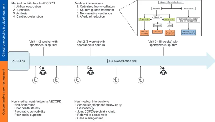
Our local approach to AECOPD could be summarized (Fig- ure 1) as follows.
First, we try to identify the cause of the symptoms, whether it is worsening of airflow limitation, bronchitis, respiratory or metabolic acidosis, left or right ventricular dysfunction, or a combination of these. A general approach to this can be easily made through simple tests available in the emergency depart- ment, including spirometry, chest X-ray, and arterial blood gases. Treatment is initiated according to guidelines, with bronchodilators, systemic steroids and antibiotics if they are indicated. Secondly, as soon as feasible to obtain, a sputum sample is collected, and treatment is adjusted accordingly. If sputum shows eosinophilic bronchitis, steroids are continued
Figure 1. Local approach to acute exacerbation of chronic obstructive pulmonary disease (AECOPD).
Clinical phenotyping & guided treatment Comprehensive care management
Medical contributors to AECOPD 1. Airflow obstruction 2. Bronchitis 3. Acidosis
4. Cardiac dysfunction
Medical interventions 1. Optimized bronchodilators 2. Sputum-guided treatment 3. Non-invasive ventilation 4. Afterload reduction
Sputum differential cell count
Bronchitis No bronchitis
Mixed
Optimize bronchodilation and self-management Bacterial Viral
Eosinophilic Neutrophilic
Steroids
(PO/inh) Antibiotics Supportive treatment
Reduce ICS if eosinophils <2%