Tuberc Respir Dis 2009;66:186-191
CopyrightⒸ2009. The Korean Academy of Tuberculosis and Respiratory Diseases. All rights reserved.
만성폐쇄성폐질환 환자에서 골다공증의 유병률과 위험인자
1
이화여자대학교 의학전문대학원 내과학교실,
2만성기도폐쇄성질환 임상연구센터
심윤수1, 이진화1,2, 류연주1, 천은미1, 장중현1
Prevalence and Risk Factors of Osteoporosis in Patients with Chronic Obstructive Pulmonary Disease
Yun Su Sim, M.D.
1, Jin Hwa Lee, M.D.
1,2, Yon Ju Ryu, M.D.
1, Eun Mi Chun, M.D.
1, Jung Hyun Chang, M.D.
11
Department of Internal Medicine, Ewha Womans University School of Medicine,
2Clinical Research Center for Chronic Obstructive Airway Diseases, Seoul, Korea
Background: Osteoporosis is a significant comorbidity in patients with chronic obstructive pulmonary disease (COPD). This study examined the prevalence and risk factors associated with osteoporosis in patients with COPD.
Methods: The bone mineral densities (BMDs) of the lumbar spine and femoral bone were measured in 53 patients with clinically stable COPD and 41 age- and gender-matched control subjects showing a normal lung function.
Osteoporosis was defined as a T-score ≤−2.5. The subjects’ clinical characteristics and laboratory data were reviewed, and multiple logistic regression analysis was used to identify the risk factors associated with osteoporosis in COPD patients.
Results: The prevalence of osteoporosis was 47% and 32% in the COPD patients and controls, respectively. In particular, using the femoral neck T-score, the prevalence of osteoporosis in COPD patients was higher than that in the controls (26% vs. 5%; p=0.006). The average T-score of the lumbar spine (p=0.025) and femoral neck of COPD patients were significantly lower than those of the controls (p=0.001). The forced expiratory volume in the 1 second (FEV1) % predicted (p=0.019; odds ratio [OR], 0.955; 95% confidence interval [CI], 0.919-0.993) and age (p=0.024; OR, 1.144; 95% CI, 1.018-1.287) were independently associated with osteoporosis in patients with COPD.
Conclusion: Using the femoral neck T-score, the prevalence of osteoporosis in patients with COPD was higher than the age-and gender-matched controls. A lower FEV1 and older age further increase the risk of osteoporosis in patients with COPD.
Key Words: Chronic obstructive pulmonary disease, Osteoporosis, Prevalence, Risk factor, Lung function
Address for correspondence: Jin Hwa Lee, M.D.
Department of Internal Medicine, School of Medicine, Ewha Womans University, 911-1, Mok-dong, Yancheon-gu, Seoul 158-710, Korea
Phone: 82-2-2650-6007, Fax: 82-2-2655-2076 E-mail: [email protected]
Received: Jan. 22, 2009 Accepted: Mar. 6, 2009
서 론
만성폐쇄성폐질환(chronic obstructive pulmonary dis- ease, COPD)이 진행되면 호흡기증상 외에 여러 합병증이
나타날 수 있다1. 특히 활동력 감소와 더불어 골다공증의 빈도가 증가하여 만성폐쇄성폐질환 환자 중에 36∼60%
에서 골다공증이 발생하는 것으로 알려져 있다2-4. 골다공증이 발생하면 척추나 대퇴골절의 위험이 증가 하고, 골절은 만성폐쇄성폐질환 환자에게 통증을 일으키 고 활동 감소로 이어져 삶의 질을 저하시키며 결국에는 사망률을 증가시키게 된다5. 만성폐쇄성폐질환 환자에서 골다공증의 위험인자로는 흡연, 낮은 체질량지수, 성선 저 하, 비타민 D 감소, 스테로이드 투약과 활동력 저하 등이 알려져 있다6.
본 연구에서는 만성폐쇄성폐질환 환자에서 골다공증의
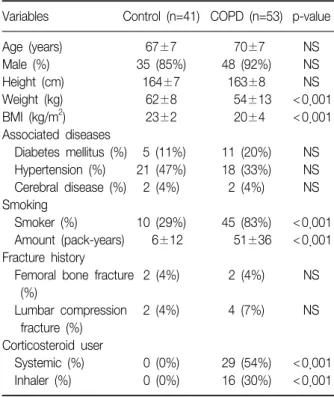
Table 1. Clinical characteristics in patients with COPD and age- and sex-matched control subjects
Variables Control (n=41) COPD (n=53) p-value
Age (years) 67±7 70±7 NS
Male (%) 35 (85%) 48 (92%) NS
Height (cm) 164±7 163±8 NS
Weight (kg) 62±8 54±13 <0.001
BMI (kg/m
2) 23±2 20±4 <0.001
Associated diseases
Diabetes mellitus (%) 5 (11%) 11 (20%) NS Hypertension (%) 21 (47%) 18 (33%) NS Cerebral disease (%) 2 (4%) 2 (4%) NS Smoking
Smoker (%) 10 (29%) 45 (83%) <0.001 Amount (pack-years) 6±12 51±36 <0.001 Fracture history
Femoral bone fracture 2 (4%) 2 (4%) NS (%)
Lumbar compression 2 (4%) 4 (7%) NS fracture (%)
Corticosteroid user
Systemic (%) 0 (0%) 29 (54%) <0.001 Inhaler (%) 0 (0%) 16 (30%) <0.001 Data are mean±standard deviation (SD) or number (frequency).
COPD: chronic obstructive pulmonary disease; BMI: body mass index; NS: not significant.
유병률을 조사하여, 연령과 성비를 일치시킨 정상 폐기능 대조군과 비교해 보고, 만성폐쇄성폐질환 환자에서 골다 공증 발생의 위험인자를 알아 보고자 하였다.
대상 및 방법
1. 대상
2004년 2월부터 2006년 8월까지 이화여자대학교 의료 원에 방문한 COPD 환자 중에 폐기능검사와 골밀도검사 를 시행한 53명을 대상으로 하였다. 본 연구에서 COPD의 정의는 NHLBI/WHO Global Initiative for Chronic Obstrutive Lung Disease (GOLD) Workshop Summary에 나온 국제지침 기준7을 적용하여 기관지확장제 흡입 후 노력성폐활량(forced vital capacity, FVC)에 대한 1초간노 력성호기량(forced expiratory volume in 1 second, FEV1) 의 비(FEV1/FVC)가 0.7 미만인 경우로 정의하였다.
같은 기간에 본원에 방문하여 골밀도와 폐활량을 측정 한 사람 중 기저 폐질환이 없고 폐기능이 정상이면서 COPD 환자군과 나이와 성별이 일치되는 41명을 대조군 으로 하였다.
환자군과 대조군 모두 단순 흉부 X-선 촬영에서 결핵반 흔으로 한쪽 폐 면적의 1/4 이상이 손상되었거나 기관지 확장증, 흉막비후, 장경이 3 cm 이상인 결절이 있는 경우 는 제외하였다.
2. 방법
임상기록을 검토하여 성별과 나이, 흡연력과 과거력, 키와 몸무게를 조사하였고, 체질량지수를 계산하였다.
폐기능은 Vmax spectra 22 (Sensormedics, Yorba Linda, USA)로, COPD 환자는 급성악화시기가 아닌, 안정 상태 에서 미국흉부학회의 폐기능검사 지침에 따라 시행하였 다8.
골밀도는 Dual energy X-ray absorptiometry (Prodigy, GE-lunar, Madison, USA)를 이용하여 둘째, 셋째, 넷째 요추와 대퇴골목에서 측정하였다. 골다공증은 세계보건 기구가 정한 기준에 따라 T-점수가 −2.5 이하인 경우로 정의하였고, T-점수가 −1.0에서 −2.5까지 감소한 경우 를 골감소증(osteopenia)으로 정의하였다9.
3. 통계분석
통계분석은 SPSS for window 12.0 (SPSS Inc, Chicago, USA) 프로그램을 이용하였다. 연속변수의 비교는 Student
t
-test를, 범주형 변수의 비교는 카이제곱검정을 이용하여 분석하였다. COPD 환자에서 골다공증과 관련된 요인 분 석은 다변량로지스틱회귀분석을 이용하여 환자군 내에서 대응비(odds ratio)와 95% 신뢰구간을 산출하였다. p값이 0.05 미만일 때 통계적으로 유의하다고 판정하였다.결 과
COPD 환자의 체중과(p<0.001) 체질량지수는 연령과 성비를 일치시킨 대조군과 비교하여 유의하게 작은 반면 (p<0.001), 흡연자 비율과(p<0.001) 흡연량은 유의하 게 높았다(p<0.001). 스테로이드를 사용한 대조군은 없 었다(p<0.001) (Table 1).
COPD 환자의 FVC (p<0.001), FEV1 (p<0.001), FEV1/FVC는 각각 대조군보다 유의하게 작았다(p<0.001) (Table 2).
말초혈액검사 결과 COPD 환자의 평균 혈색소와(p=
0.039) 혈청 칼슘과(p=0.001) 인은 대조군보다 유의하게 낮았고(p=0.048), 말초혈액백혈구 수(p=0.001), 적혈구침
Table 2. Lung function in patients with COPD and age- and sex-matched controls
Variables Control (n=41) COPD (n=53) p-value
FVC (L) 3.37±0.76 2.61±0.93 <0.001
FVC (% predicted) 94±13 76±24 <0.001 FEV
1(L) 2.65±0.57 1.32±0.74 <0.001 FEV
1(% predicted) 106±13 55±27 <0.001
FEV
1/FVC (%) 79±7 50±15 <0.001
Data are mean±SD.
COPD: chronic obstructive pulmonary disease; FEV
1: forced ex- piratory volume in 1 second; FVC: forced vital capacity.
Table 3. Laboratory data in patients with COPD and age- and sex-matched controls
Variables Control (n=41) COPD (n=53) p-value Hemoglobin (g/dL) 13.6±1.6 12.9±1.7 0.039 White blood cell 7.3±2.3 9.7±2.8 0.001 count (×10
3/m
3)
ESR (mm/hr) 14±14 37±36 <0.001
CRP (mg/dL) 1.4±3.6 4.1±5.7 0.014 Serum total calcium 9.4±0.6 9.0±0.5 0.001 (mg/dL)
Serum phosphorus 3.4±0.6 3.1±0.6 0.048 (mg/dL)
Alkaline phosphatase 208±60 200±79 NS (IU/L)
Data are mean±SD.
COPD: chronic obstructive pulmonary disease; CRP: C-reactive protein; ESR: erythrocyte sedimentation rate; NS: not signifi- cant.
Table 4. Comparison of bone density parameters and pre- valence of osteoporosis in patients with COPD and age- and sex-matched controls
Variables Control (n=41) COPD (n=53) p-value T-score
Femoral neck −0.62±1.24 −1.59±1.42 0.001 Lumbar spine (L2-4) −1.41±1.42 −2.15±1.65 0.025
Osteopenia* 16 (39%) 21 (40%) NS
Femoral neck 5 (12%) 14 (26%) NS Lumbar spine (L2-4) 16 (39%) 21 (40%) NS
Osteoporosis
†13 (32%) 25 (47%) NS
Femoral neck 2 (5%) 14 (26%) 0.006
Lumbar spine (L2-4) 13 (32%) 24 (45%) NS Data are mean±SD or number (frequency).
COPD: chronic obstructive pulmonary disease; L2-4: the sec- ond, third and forth lumbar vertebrae; NS: not significant.
*Osteopenia is defined as a T-score between −1.0 and −2.5,
†
Osteoporosis is defined as a T-score below −2.5.
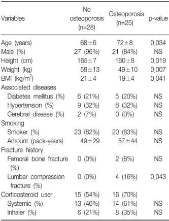
Table 5. Clinical characteristics of COPD patients accord- ing to presence of osteoporosis
No Osteoporosis
Variables osteoporosis p-value
(n=25) (n=28)
Age (years) 68±6 72±8 0.034
Male (%) 27 (96%) 21 (84%) NS
Height (cm) 165±7 160±8 0.019
Weight (kg) 58±13 49±10 0.007
BMI (kg/m
2) 21±4 19±4 0.041
Associated diseases
Diabetes mellitus (%) 6 (21%) 5 (20%) NS Hypertension (%) 9 (32%) 8 (32%) NS Cerebral disease (%) 2 (7%) 0 (0%) NS Smoking
Smoker (%) 23 (82%) 20 (83%) NS Amount (pack-years) 49±29 57±44 NS Fracture history
Femoral bone fracture 0 (0%) 2 (8%) NS (%)
Lumbar compression 0 (0%) 4 (16%) 0.043 fracture (%)
Corticosteroid user 15 (54%) 16 (70%) Systemic (%) 13 (46%) 14 (61%) NS Inhaler (%) 6 (21%) 8 (35%) NS Data are mean±SD or number (frequency).
COPD: chronic obstructive pulmonary disease; BMI: body mass index; NS: not significant.
강속도(p<0.001)와 C-반응단백질은 COPD 환자에서 대 조군보다 유의하게 높았다(p=0.014) (Table 3).
골밀도 측정 결과 COPD 환자의 대퇴골목과 요추의 평 균 T-점수는 각각 −1.59±1.42와(p=0.001) −2.15±
1.65로 대조군보다 유의하게 낮았다(p=0.025). COPD 환 자의 골감소증과 골다공증의 유병률은 각각 40%, 47%로, 대조군의 39%, 32%보다 높았으나, 대퇴골목의 T-점수를 기준으로 한 골다공증의 유병률만 통계적으로 유의한 차 이를 보였다(26% vs. 5%; p=0.006) (Table 4).
COPD 환자를 골다공증의 유무에 따라 비교하였을 때, 골다공증이 있는 환자의 평균 나이가 더 많고(p=0.034), 평균 키(p=0.019), 몸무게(p=0.007)와 체질량지수가 더 작았으며(p=0.041), 요추압박골절의 빈도가 높았다(p=
Table 6. Lung function of COPD patients according to presence of osteoporosis
No Osteoporosis
Variables osteoporosis p-value
(n=25) (n=28)
FVC (L) 2.92±1.06 2.24±0.58 0.007
FVC (% predicted) 79±26 72±22 NS
FEV
1(L) 1.61±0.85 0.98±0.37 0.002
FEV
1(% predicted) 62±29 47±21 0.043
FEV
1/FVC (%) 54±16 45±14 0.029
Data are mean±SD.
COPD: chronic obstructive pulmonary disease; FEV
1: forced ex- piratory volume in 1 second; FVC: forced vital capacity; NS: not significant.
Table 7. Risk factors of osteoporosis in patients with COPD by multiple logistic regression analyses
95% confidence
Variables Odds ratio p-value
interval
FEV
1(% predicted) 0.955 0.919∼0.993 0.019
Age (years) 1.144 1.018∼1.287 0.024
COPD: chronic obstructive pulmonary disease; FEV
1: forced ex- piratory volume in 1 second.
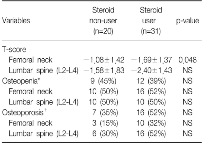
Table 8. Comparison of bone density parameters and prevalence of osteoporosis in COPD patients according to corticosteroid use
Steroid Steroid
Variables non-user user p-value
(n=20) (n=31) T-score
Femoral neck −1.08±1.42 −1.69±1.37 0.048 Lumbar spine (L2-L4) −1.58±1.83 −2.40±1.43 NS
Osteopenia* 9 (45%) 12 (39%) NS
Femoral neck 10 (50%) 16 (52%) NS
Lumbar spine (L2-L4) 10 (50%) 10 (50%) NS Osteoporosis
†7 (35%) 16 (52%) NS Femoral neck 3 (15%) 10 (32%) NS Lumbar spine (L2-L4) 6 (30%) 16 (52%) NS Data are mean±SD or number (frequency).
COPD: chronic obstructive pulmonary disease; L2-4: the sec- ond, third and forth lumbar vertebrae; NS: not significant.
*Osteopenia is defined as a T-score between −1.0 and −2.5,
†
Osteoporosis is defined as a T-score below −2.5.
0.043) (Table 5). COPD 환자 중 골다공증이 있는 환자가 골다공증이 없는 환자보다 FVC와(p=0.007) FEV1의 절대 값(p=0.002), FEV1의 정상예측치에 대한 비율(p=0.043), 그리고 FEV1/FVC 비가 유의하게 작았다(p=0.029) (Table 6). COPD 환자에서 골다공증 유무에 따라 혈청학적 지표 를 비교하였을 때 두 군 사이에 차이는 없었다.
다변량로지스틱회귀분석 결과 COPD 환자에서 골다공 증 발생과 관련된 독립적인 위험인자는 FEV1의 정상예측 치에 대한 비율과(p=0.019; odds ratio [OR], 0.955; con- fidence interval [CI], 0.919-0.993] 연령이었다(p=0.024;
OR, 1.144; CI, 1.018-1.287) (Table 7).
COPD 환자를 스테로이드 투약 유무에 따라 분류하였 을 때, 스테로이드를 투여 받은 적이 있는 환자가 그렇지 않은 환자보다 대퇴골목의 T-점수가 유의하게 낮았다(p=
0.048) (Table 8).
고 찰
본 연구에서 COPD 환자의 골다공증 유병률은 폐기능
이 정상이고 연령과 성별을 일치시킨 대조군보다 높았고, COPD 환자 중 47%가 골다공증을, 40%가 골감소증을 가 지고 있어 골밀도 감소의 유병률이 87%에 이르렀다. 이는 J
ϕ
rgensen 등10이 COPD 환자를 대상으로 하여 발표한 68%에 비하여 높은 유병률로, 본 연구대상의 평균 연령이 70세로 이전 연구보다 7세 정도 연령이 높기 때문으로 생 각된다.COPD 환자의 흡연력이 대조군보다 높았는데, 기존의 연구에서 흡연은 골밀도를 감소시키는 원인으로 잘 알려 져 있으며6, 20갑년 이상 흡연한 COPD 환자에서 요추 골 밀도가 12% 이상 낮다고 한다11,12. 흡연은 뼈조직을 산성 화시켜 골용해를 촉진시키고, 성선호르몬을 저하시킬 수 있다11,12.
본 연구에서 COPD 환자는 대조군보다 체질량지수가 낮았다. 뼈가 재형성되는 과정에서 골량의 변화는 부하된 무게의 변화에 따른다고 한다. 기계적인 힘이 작용하는 정도에 따라서 골량이 증가하거나 감소하고 작용방향에 따라서 골의 요소가 치환되거나 유지되어 뼈가 만들어지 기 때문에, 체질량지수가 낮을수록 골량에 부하된 무게가 작아져서 골밀도가 감소한다13. 또한, 성선호르몬의 활성 화는 지방조직에서 이루어지는데, 상대적으로 지방이 적 은 COPD 환자에서 성선호르몬이 감소하게 되어 골밀도 가 떨어지는 것으로 알려져 있다14.
본 연구에서 COPD 환자의 54%가 최근에 전신 스테로 이드를 사용하였으며 30%가 흡입스테로이드를 사용하고 있었다. 스테로이드는 골모세포의 기능을 떨어뜨려 골형 성을 방해하고 파골세포의 분화를 촉진시켜 골융해를 가 져오며, 소변에서 칼슘의 배설을 증가시키고, 장에서 칼슘 의 흡수를 방해하는 것으로 알려져 있다15-17. 본 연구에서 COPD 환자의 칼슘과 인의 혈청 농도가 대조군보다 낮았 는데, COPD 환자에서 나타나는 영양결핍에 의한 것인지, 스테로이드 투약에 의한 것인지 명확하게 구별하기는 어 려우나, 두 가지 모두 작용하였을 가능성이 높다.
혈액 검사에서 COPD 환자의 말초혈액백혈구 수, 적혈 구침강속도와 C-반응단백질 등 염증지표가 대조군보다 높았다. 만성염증은 TNF-α 등 사이토카인 농도를 높여 뼈의 대사율을 증가시켜 골밀도를 저하시키는 것으로 알 려져 있어18, COPD에서 나타나는 높은 염증지표 수치는 만성염증과 관련된 골밀도 감소를 간접적으로 설명해 준 다고 할 수 있다.
골다공증의 유병률은 대퇴골의 T-점수를 기준으로 하 면 COPD 환자에서 대조군보다 더 높았지만, 요추골을 기 준으로 했을 때는 유의한 차이를 보이지 않았다. 체중비 부하운동보다 체중부하운동이 요추골에 비해 대퇴골의 골밀도를 증가시키는 것으로 알려져 있다19,20. 호흡곤란으 로 활동력이 감소한 COPD 환자의 경우 걷거나 이동에 의한 체중부하가 대조군에 비해 현저히 줄어들 수 밖에 없다. 따라서, 연령이나 전반적인 전신상태에 의한 골밀 도의 감소는 대퇴골이나 요추골에서 비슷하게 진행할 것 으로 예상되나21, COPD 환자에서 활동 감소로 체중부하 운동이 감소하면 상대적으로 대퇴골의 골밀도 감소가 더 두드러지게 될 것이다. 이러한 이유로 COPD 환자군과 대 조군 간에 평균 골밀도의 차이와 골다공증 유병률의 차이 가 대퇴골에서 더욱 민감하게 나타난 것으로 생각된다.
COPD 환자를 골다공증 유무에 따라 분류하여 비교하 였을 때, 골다공증이 나타난 환자에서 골다공증이 없는 환자군보다 나이가 많고, 체중과 체질량지수가 작았다.
저체중과 낮은 체질량지수를 보인 COPD 환자에서는 앞 서 언급한 부하된 무게와 체지방의 차이로 골형성이 잘 되지 않을 가능성이 높기 때문에 골다공증의 유병률이 높 을 것으로 생각된다.
골다공증을 가진 COPD 환자군에서 전신적 스테로이드 및 흡입 스테로이드의 사용이 그렇지 않은 COPD 환자군 에서 보다 많았지만 통계적으로 유의한 차이를 보이지는 않았다. 이는 대상 수가 적고 후향적 연구라는 제한점으
로 사용한 스테로이드의 정확한 누적량과 기간에 대한 정 보가 부족하기 때문일 수 있어서, 추후 전향적 연구를 통 해 정확한 누적사용량에 대한 평가가 필요할 것으로 생각 된다.
골다공증을 가진 COPD 환자와 그렇지 않은 COPD 환 자에서 FVC와 FEV1의 절대값이 유의한 차이를 보인 것은 두 군 간에 키 차이 때문일 수 있으나, FEV1의 정상예측치 에 대한 비율과 FEV1/FVC가 차이를 보인 것은 골다공증 환자에서 기류폐쇄가 더 심하다는 것을 반영한다. 더 심 한 기류폐쇄는 활동력 감소로 이어지기 때문에 골다공증 의 위험을 현저히 높일 수 있다. 본 연구에서도 COPD 환 자에서 골다공증의 여러 위험요인 중 FEV1의 정상예측치 에 대한 비율이 가장 중요한 위험인자로, FEV1이 낮을수 록 골다공증의 위험이 현저하게 증가하였다.
COPD 환자에서 골절이 일어나게 되면 골절에 의한 출 혈, 사망률의 증가 외에도 이후 활동력 감소로 이어져 골 밀도가 더욱 감소하고 폐기능은 저하되어 추가적인 사망 률 증가로 연결된다. 따라서, 골절을 미연에 방지하는 것 은 COPD 환자에게 매우 중요하므로, 기류폐쇄가 심하고 나이가 많은 COPD 환자라면, 골밀도를 반드시 측정하여 골다공증의 예방에 힘써야겠다.
요 약
연구배경: 골다공증은 만성폐쇄성폐질환 환자의 중요 한 동반질환 중 하나이다. 저자들은 COPD 환자에서 골다 공증의 유병률과 위험인자를 알아 보고자 하였다.
방 법: 안정 상태인 COPD 환자 51명과 이들과 나이와 성별을 일치시킨 대조군 41명에서 요추골과 대퇴골의 골 밀도를 측정하였다. 임상기록과 검사결과를 검토하여, COPD 환자에서 골다공의 위험인자를 분석하였다.
결 과: COPD 환자에서, T점수가 −2.5 이하인, 골다공 증의 유병률은 47%였고, 대조군에서 32%였다. 특히 대퇴 골목의 T점수를 기준으로 한 골다공증의 유병률은 COPD 환자에서 대조군에 비해 유의하게 높았다(26%
vs.
5%;p=0.006). COPD 환자의 요추와(p=0.025) 대퇴골목의 평 균 T 점수는 대조군보다 유의하게 낮았다(p=0.001). COPD 환자에서 FEV1의 정상예측치에 대한 비율과(p=0.019; odds ratio [OR], 0.955; 95% confidence interval [CI], 0.919- 0.993) 연령이 골다공증 발생과 관련된 독립적인 위험인자 였다(p=0.024; OR, 1.144; 95% CI, 1.018-1.287).
결 론: COPD 환자에서 골다공증의 유병률은 연령과
성별을 일치시킨 대조군보다 높다. 특히 대퇴골의 T점수 가 요추보다 COPD 환자와 대조군의 골밀도의 차이를 더 분명하게 보여 주었다. COPD 환자에서 FEV1이 낮을수록, 나이가 많을수록 골다공증 발생 위험이 크게 증가한다.
참 고 문 헌
1. Chatila WM, Thomashow BM, Minai OA, Criner GJ, Make BJ. Comorbidities in chronic obstructive pulmo- nary disease. Proc Am Thorac Soc 2008;5:549-55.
2. Incalzi RA, Caradonna P, Ranieri P, Basso S, Fuso L, Pagano F, et al. Correlates of osteoporosis in chronic obstructive pulmonary disease. Respir Med 2000;94:
1079-84.
3. Iqbal F, Michaelson J, Thaler L, Rubin J, Roman J, Nanes MS. Declining bone mass in men with chronic pulmonary disease: contribution of glucocorticoid treatment, body mass index, and gonadal function.
Chest 1999;116:1616-24.
4. Shane E, Silverberg SJ, Donovan D, Papadopoulos A, Staron RB, Addesso V, et al. Osteoporosis in lung transplantation candidates with end-stage pulmonary disease. Am J Med 1996;101:262-9.
5. Block JE, Stubbs H. Hip fracture-associated mortality reconsidered. Calcif Tissue Int 1997;61:84.
6. Biskobing DM. COPD and osteoporosis. Chest 2002;
121:609-20.
7. Pauwels RA, Buist AS, Calverley PM, Jenkins CR, Hurd SS. Global strategy for the diagnosis, management, and prevention of chronic obstructive pulmonary disease.
NHLBI/WHO Global Initiative for Chronic Obstructive Lung Disease (GOLD) Workshop summary. Am J Respir Crit Care Med 2001;163:1256-76.
8. American Thoracic Society. Standardization of Spirome- try, 1994 Update. Am J Respir Crit Care Med 1995;152:
1107-36.
9. Kanis JA, Melton LJ 3rd, Christiansen C, Johnston CC, Khaltaev N. The diagnosis of osteoporosis. J Bone Miner Res 1994;9:1137-41.
10. Jϕrgensen NR, Schwarz P, Holme I, Henriksen BM, Petersen LJ, Backer V. The prevalence of osteoporosis in patients with chronic obstructive pulmonary disease:
a cross sectional study. Respir Med 2007;101:177-85.
11. Slemenda CW, Hui SL, Longcope C, Johnston CC Jr.
Cigarette smoking, obesity, and bone mass. J Bone Miner Res 1989;4:737-41.
12. Daniell HW. Osteoporosis and smoking. JAMA 1972;
221:509.
13. Bassett CA, Becker RO. Generation of electric potentials by bone in response to mechanical stress. Science 1962;137:1063-4.
14. Cauley JA, Salamone LM, Lucas FL. Postmenopausal en- dogenous and exogenous hormones, degree of obe- sity, thiazide diuretics, and risk of osteoporosis. In:
Marcus R, Feldman D, Kelsey J, editors. Osteoporosis.
1st ed. New York: Academic Press; 1996. p. 551-76.
15. Adachi JD. Corticosteroid-induced osteoporosis. Am J Med Sci 1997;313:41-9.
16. Lane NE, Lukert B. The science and therapy of gluco- corticoid-induced bone loss. Endocrinol Metab Clin North Am 1998;27:465-83.
17. Canalis E. Mechanisms of glucocorticoid action in bone: implications to glucocorticoid-induced osteo- porosis. J Clin Endocrinol Metab 1996;81:3441-7.
18. Sevenoaks MJ, Stockley RA. Chronic Obstructive Pulmonary Disease, inflammation and co-morbidity--a common inflammatory phenotype? Respir Res 2006;7:
70.
19. Dalsky GP, Stocke KS, Ehsani AA, Slatopolsky E, Lee WC, Birge SJ Jr. Weight-bearing exercise training and lumbar bone mineral content in postmenopausal women. Ann Intern Med 1988;108:824-8.
20. White MK, Martin RB, Yeater RA, Butcher RL, Radin EL.
The effects of exercise on the bones of postmenopausal women. Int Orthop 1984;7:209-14.
21. Choi JS, An KC, Lee CS, Choi JM, Kim JY, Shin DR.
DEXA T-score concordance and discordance between hip and lumbar spine. J Korean Soc Spine Surg 2003;
10:75-81.