Three-month Treatment Response and Exacerbation in Chronic
Obstructive Pulmonary Disease
The aim of this study was to investigate relationships between acute exacerbation and Forced Expiratory Volume 1 second (FEV1) improvement after treatment with combined
long-acting beta-agonist (LABA) and inhaled corticosteroid (ICS) in patients with chronic obstructive pulmonary disease (COPD). A total of 137 COPD patients were classified as responders or nonresponders according to FEV1 improvement after 3 months of LABA/ICS
treatment in fourteen referral hospitals in Korea. Exacerbation occurrence in these two subgroups was compared over a period of 1 yr. Eighty of the 137 COPD patients (58.4%) were classified as responders and 57 (41.6%) as nonresponders. Acute exacerbations occurred in 25 patients (31.3%) in the responder group and in 26 patients (45.6%) in the nonresponder group (P = 0.086). FEV1 improvement after LABA/ICS treatment was a
significant prognostic factor for fewer acute exacerbations in a multivariate Cox proportional hazard model adjusted for age, sex, FEV1, smoking history, 6 min walk
distance, body mass index, exacerbation history in the previous year, and dyspnea scale. Three-month treatment response to LABA/ICS might be a prognostic factor for the occurrence of acute exacerbation in COPD patients.
Keywords: Pulmonary Disease, Chronic Obstructive; Forced Expiratory Volume; Disease Progression
Jung Su Lee,1 Chin Kook Rhee,2
Kwang Ha Yoo,3 Ji-Hyun Lee,4
Ho Il Yoon,5 Tae-Hyung Kim,6
Woo Jin Kim,7 JinHwa Lee,8
Seong Yong Lim,9 Tai Sun Park,1
Jae Seung Lee,1 Sei Won Lee,1
Sang-Do Lee,1 and Yeon-Mok Oh1 1Department of Pulmonary and Critical Care
Medicine, and Clinical Research Center for Chronic Obstructive Airway Diseases, Asan Medical Center, University of Ulsan College of Medicine, Seoul;
2Department of Internal Medicine, Seoul St. Mary’s
Hospital, The Catholic University of Korea, Seoul;
3Department of Internal Medicine, Konkuk
University School of Medicine, Seoul; 4Department
of Internal Medicine, CHA Bundang Medical Center, CHA University, Seongnam; 5Department of Internal
Medicine, Seoul National University Bundang Hospital, Seoul National University College of Medicine, Seongnam; 6Division of Pulmonology,
Department of Internal Medicine, Hanyang University Guri Hospital, Hanyang University College of Medicine, Guri; 7Department of Internal Medicine
and Environmental Health Center, Kangwon National University Hospital, School of Medicine, Kangwon National University, Chuncheon;
8Department of Internal Medicine, Ewha Womans
University Mokdong Hospital, College of Medicine, Ewha Womans University, Seoul; 9Division of
Pulmonary and Critical Care Medicine, Department of Medicine, Kangbuk Samsung Hospital, Sungkyunkwan University School of Medicine, Seoul, Korea
Received: 29 May 2014 Accepted: 29 August 2014 Address for Correspondence: Yeon-Mok Oh, MD
Department of Pulmonary and Critical Care Medicine, and Clinical Research Center for Chronic Obstructive Airway Diseases, Asan Medical Center, University of Ulsan College of Medicine, 88 Olympic-ro 43-gil, Songpa-gu, Seoul 138-736, Korea
Tel: +82.2-3010-3136, Fax: +82.2-3010-6968 E-mail: [email protected]
Funding: This study was supported by a grant from the Korean
Health 21 R&D Project, Ministry of Health and Welfare, Republic of Korea (HI10C2020 and A102065).
http://dx.doi.org/10.3346/jkms.2015.30.1.54 • J Korean Med Sci 2015; 30: 54-59
INTRODUCTION
Chronic obstructive pulmonary disease (COPD) is believed to be complex and hetero-geneous, not a simple and homogeneous disorder. The varied pathological features of COPD lead to recognition of patient subgroups that have different characteristics and may have distinct responses to treatment (1). Thus, clinical responses to bronchodila-tors and inhaled corticosteroids (ICS) may vary among individuals (2). Therapeutic re-sponse is an important component of improved health-related quality of life and influ-ences the clinical outcomes of the disease (3).
Many recent studies support the effectiveness of bronchodilator and anti-inflamma-tion therapy to improve lung funcanti-inflamma-tion in specific subgroups of COPD patients (1, 4). Several studies have found a significant relationship between the decline in lung func-tion and poor clinical outcomes in COPD patients (5-9), but few have shown that im-proved lung function translates into imim-proved clinical outcomes, including decreased exacerbation (10, 11).
The ability to predict whether COPD patients with improved lung function in respon-se to bronchodilator and anti-inflammatory treatment are likely to have better clinical outcomes, including a reduction in exacerbations, is important. Treatment response to a short-acting bronchodilator like salbutamol has been used to diagnose bronchodila-tor reversibility (BDR). However, there is evidence that BDR does not distinguish among clinically relevant outcomes (12, 13).
A standardized therapeutic response indicator that can be used to predict the clini-cal benefit of long-term bronchodilator treatment is needed. This study examined whe-ther the response of COPD patients to a 3 months treatment with a bronchodilator and anti-inflammatory agent could predict exacerbation. A cohort of 137 patients was
eval-uated over a 1 yr period. After 3 months of treatment, the pa-tients were divided into two subgroups based on improvement of Forced Expiratory Volume 1 second (FEV1), and the occur-rence of exacerbations in the two subgroups was compared.
MATERIALS AND METHODS
Subjects
This study was a post hoc analysis of 133 male and four female COPD patients who were selected from the Korean Obstructive Lung Disease (KOLD) cohort. All had stable COPD and were prospectively recruited from the pulmonary clinics of 14 hospi-tals in Korea between June 2005 and December 2012. The in-clusion criteria for the KOLD cohort have been described else-where (14, 15). COPD was diagnosed based on the presence of airflow limitation that was not fully reversible (post-bronchodi-lator FEV1/forced vital capacity (FVC) < 0.70) and more than 10-pack-years of smoking history.
Three-month treatment response and classification of subgroups
For all patients, the use of respiratory medicine was restricted for 2 weeks before enrollment. Baseline clinical data included demographic information, smoking history, past exacerbation history, a 6 min walk distance (6MWD) test, body mass index (BMI), pulmonary function tests and chest radiography. The modified Medical Research Council (mMRC) dyspnea scale was administered to assess the degree of dyspnea.
FEV1 was measured by spirometry after 12 weeks of treatment and expressed as a percentage compared to the predicted value for healthy Korean subjects (16). The COPD patients were clas-sified as responders or nonresponders based on FEV1 improve-ment after 3 months of combined treatimprove-ment with a long-acting beta-agonist and inhaled corticosteroid (LABA/ICS) (salmeter-ol/fluticasone, 50 μg/500 μg or formoterol/budesonide, 9 μg/320 μg). Responders were defined as having a minimal clinically important difference (MCID) (3, 17) of 120 mL or greater im-provement in FEV1 after treatment, and nonresponders were defined as having an FEV1 improvement of < 120 mL.
Obligatory study visits were scheduled every 3 months for 1 yr. Trained research coordinators recorded medical history, smok-ing history and status, information on previous treatments, and compliance with inhaler use. The investigators were not includ-ed in the interview process. Patients were askinclud-ed to bring their inhalation device to each study visit so that medication use could be measured. Ninety percent of subjects answered that they used more than 80% of the recommended medication doses. We ed-ucated patients about the method of using inhaler devices at the enrollment. Also we had checked that patients properly used the inhaler at the visit of 3 months.
Pulmonary function tests
Spirometry was performed as recommended by the American Thoracic Society using a Vmax 22 instrument (Sensor-Medics, Yorba Linda, CA, USA) or a PFDx machine (MedGraphics, St. Paul, MN, USA) (18). To assess short-acting BDR, FEV1, FVC, and FEV1/FVC were evaluated before and 15 min after inhala-tion of salbutamol (400 μg) using a metered-dose inhaler (MDI) fitted with a spacer. The predicted values of FEV1, FVC, and FEV1/ FVC were calculated from equations formulated using data from a population of healthy nonsmoking Koreans (16, 19).
Exacerbation identification
Patients were interviewed at the clinic every 3 months for 1 yr of follow-up. Exacerbations were identified according to previous-ly accepted criteria of moderate or severe exacerbation in which the patients visited a clinic or an emergency department, or were hospitalized (17, 20, 21). Visit or admission to hospitals that are involved in research was traced by medical record. If patients visit other clinics other than the participating institutes, we checked exacerbation occurrence using pre-structured in-terview sheet including the following sentence. ‘In the past three months, did you visit other clinic or emergency room due to in-creased sputum amount or purulent sputum or deterioration of dyspnea?’ Also, we use pre-structured interview sheet for check-ing other clinic name, reason for visit, visit date and frequency. Statistical analysis
Categorical data were analyzed using chi-square or Fisher’s ex-act tests. The primary outcome was time to occurrence of the first exacerbation within the year of follow-up. Hazard ratios (HRs) and confidence intervals (CIs) were calculated by Cox proportional hazards regression. Univariate regression analyses were done to explore potential risk factors. Multivariate regres-sion analyses were also done to examine risk factors adjusted for potential confounding covariates. All statistical analyses were performed using the SPSS statistical package (version 18.0, SPSS Inc, Chicago, IL, USA); P < 0.05 was considered statistically sig-nificant.
Ethics statement
The institutional review board of the Asan Medical Center (IRB No. 2012-0226) and the other 13 hospitals approved the study. Informed consent was obtained from all of participating patients.
RESULTS
Baseline characteristics of patients
Table 1 shows the baseline characteristics of the 137 COPD pa-tients. Patients were classified into two subgroups according to the MCID of FEV1 change before and after the 3-month treat-ment. Eighty patients (58.4%) were classified as responders and
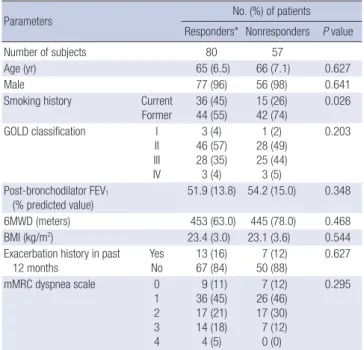
Table 1. Baseline characteristics of the study subjects
Parameters No. (%) of patients
Responders* Nonresponders P value
Number of subjects 80 57
Age (yr) 65 (6.5) 66 (7.1) 0.627
Male 77 (96) 56 (98) 0.641
Smoking history Current Former 36 (45) 44 (55) 15 (26) 42 (74) 0.026 GOLD classification I II III IV 3 (4) 46 (57) 28 (35) 3 (4) 1 (2) 28 (49) 25 (44) 3 (5) 0.203 Post-bronchodilator FEV1 (% predicted value) 51.9 (13.8) 54.2 (15.0) 0.348 6MWD (meters) 453 (63.0) 445 (78.0) 0.468 BMI (kg/m2) 23.4 (3.0) 23.1 (3.6) 0.544
Exacerbation history in past 12 months Yes No 13 (16) 67 (84) 7 (12) 50 (88) 0.627 mMRC dyspnea scale 0 1 2 3 4 9 (11) 36 (45) 17 (21) 14 (18) 4 (5) 7 (12) 26 (46) 17 (30) 7 (12) 0 (0) 0.295
Data are presented as numbers of subjects (No) with percentage (%) or means with standard deviation in parentheses. *Subjects who showed the improvement of FEV1
more than the minimal clinical important difference. 6MWD, 6 min walk distance; BMI, body mass index; GOLD, the Global Initiative for Chronic Obstructive Lung Dis-ease; mMRC, modified Medical Research Council.
Exacerba tion occurrence (%) 60 50 40 30 20 10 0 3MTR < 120 (n = 57) 51% 120 ≤ 3MTR < 300 (n = 39) 29% 3MTR ≥ 300 (mL) (n = 41) 20% Exacerba tion occurrence (%) 50 40 30 20 10 0 3MTR < 4 (n = 60) 47% 4 ≤ 3MTR < 8 (n = 26) 35% 3MTR ≥ 8 (%) (n = 51) 28% A B
Fig. 1. Exacerbation rates according to 3-month treatment response (3MTR). (A) The 3MTR was evaluated with the change of FEV1 in mL after 3-month treatment (P = 0.035).
A cut-off value of 120 mL was defined by the minimal clinically important difference (MCID)(3, 17).The other cut-off of 300 mL was defined as it was near the upper tertile val-ue. Exacerbation occurrence represents % of COPD patients who experienced exacerbation during the one year of follow-up. (B) The 3MTR was evaluated with the change of FEV1 in % of the predicted reference value after 3-month treatment (P = 0.039). According to MCID (3, 17), the responder group was defined when FEV1 improvement was 4%
or more of the predicted value after 3-month treatment. We further classified the responder group arbitrarily into two categories based on the change in FEV1, with a cut-off of
8% predicted value.
57 (41.6%) as nonresponders.
There were no significant differences in age, sex proportion, BMI, post-bronchodilator FEV1, 6MWD, exacerbation in previ-ous 12 months, patient distribution of GOLD stage, or dyspnea scale scores between the two subgroups. But, current smokers are more in responder group than in nonresponder group. Exacerbation occurrence
Acute exacerbations occurred in 25 patients (31.3%) in the re-sponder group and 26 patients (45.6%) in the nonrere-sponder group. Although it was not significant difference in
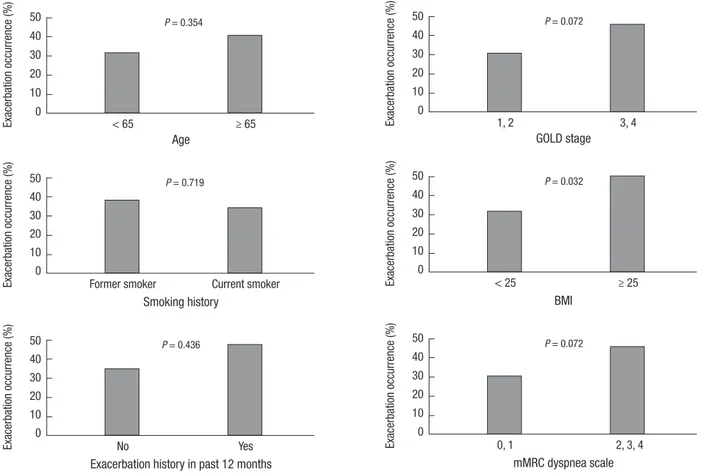
exacerba-tion occurrence between the groups, responder group tended to show the low incidence of exacerbation occurrence (P = 0.086). We further classified the responder group into two categories based on the change in FEV1, with a cut-off of 300 mL. The oth-er cut-off of 300 mL was defined as it was near the uppoth-er toth-ertile value. The exacerbation occurrence decreased significantly with an incremental change of the FEV1 after treatment (P = 0.035, Fig. 1A). We reanalyzed relationship exacerbation occurrence with 3-month treatment response using the change of FEV1 in % of the predicted reference value. According to MCID, the re-sponder group was defined when FEV1 improvement was 4% or more of the predicted value after 3-month treatment. The re-sult was compared to the earlier rere-sult using absolute volume change of FEV1 (Fig. 1B). In the univariate analyses, BMI was the only other factor that was significantly related with exacer-bation occurrence (Fig. 2).
Relationship of exacerbation occurrence with 3-month treatment response and other factors
The multivariate Cox proportional hazard model adjusted for age, sex, FEV1, smoking history, 6MWD, BMI, 12-month exac-erbation history, and dyspnea scale revealed that the 3-month treatment response was the only significant risk factor for exac-erbation (Table 2). In this model, BDR was not independently related to occurrence of exacerbation.
DISCUSSION
We have shown that the 3-month treatment response to LABA/ ICS was associated with decreased occurrence of acute exacer-bations in COPD patients. Several previous studies have shown that the decline in lung function or deterioration of COPD-as-sociated symptoms was related to a worse clinical outcome. However, this study found that lung function improvement fol-lowing pharmacological intervention was related to a better
clinical outcome. The results support a 3-month treatment re-sponse as a prognostic factor for exacerbation. The occurrence of exacerbation was significantly lower among responders than among nonresponders during the 1-yr follow-up period. Recent evidence shows that COPD phenotypes cannot be classified as chronic bronchitis or emphysema, but should be seen as a complex and heterogeneous disorder. The different pathological processes of COPD make it difficult to choose med-ications and predict therapeutic response. An important area in
COPD research is the development of more powerful, multi-variate methods for predicting clinical outcomes, and respon-siveness of individual patients to particular therapies, using clinical, laboratory and radiographic characteristics. Kitaguchi et al. (22) classified COPD subgroups based on bronchial wall thickening on high-resolution CT (HRCT). Similarly, Lee et al. (1) classified COPD patients based on the emphysema index on HRCT and pre-bronchodilator FEV1. They demonstrated that airway obstruction-dominant or mixed-subtype COPD
pa-Table 2. Prediction of exacerbation with 3-month treatment responsiveness Variables
Univariate analysis Multivariate analysis Hazard ratio of acute
exacerbation 95% CI P value Hazard ratio of acute exacerbation 95% CI P value
3MTR 0.63 0.36-1.09 0.10 0.53 0.30-0.95 0.03
Age (yr) 1.03 0.99-1.07 0.21 1.02 0.98-1.08 0.22
Male sex 0.67 0.16-2.74 0.57 0.72 0.16-3.15 0.66
Post-bronchodilator FEV1 (% predicted value) 0.99 0.97-1.01 0.15 0.98 0.96-1.01 0.14
Current vs. former smokers 0.85 0.48-1.51 0.58 1.08 0.59-1.97 0.82
6MWD (meters) 1.00 0.99-1.00 0.33 1.00 1.00-1.01 0.54
BMI (kg/m2) 0.98 0.90-1.07 0.68 1.02 0.93-1.12 0.68
Past exacerbation history 1.40 0.68-2.87 0.37 1.45 0.69-3.06 0.33
mMRC dyspnea scale 1.28 0.98-1.67 0.07 1.29 0.96-1.74 0.10
Cox proportional hazard analysis was performed. Data are presented as numbers of subjects (No) with percentage (%) or means with standard deviation in parentheses. 3MTR, 3-month treatment response; 6MWD, 6 min walk distance; BMI, body mass index; mMRC, modified Medical Research Council.
Fig. 2. Exacerbation occurrence according to baseline characteristics.
Exacerba tion occurrence (%) Age < 65 ≥ 65 50 40 30 20 10 0 P = 0.354 Exacerba tion occurrence (%) GOLD stage 1, 2 3, 4 50 40 30 20 10 0 P = 0.072 Exacerba tion occurrence (%) Smoking history
Former smoker Current smoker 50 40 30 20 10 0 P = 0.719 Exacerba tion occurrence (%) BMI < 25 ≥ 25 50 40 30 20 10 0 P = 0.032 Exacerba tion occurrence (%)
Exacerbation history in past 12 months
No Yes 50 40 30 20 10 0 P = 0.436 Exacerba tion occurrence (%) mMRC dyspnea scale 0, 1 2, 3, 4 50 40 30 20 10 0 P = 0.072
tients showed significant reversibility of airflow limitation in re-sponse to short-acting bronchodilator or combined LABA/ICS treatment compared with emphysema-dominant patients. Thus, recent work has attempted to divide COPD patients into sub-groups and to analyze their clinical differences.
The current COPD treatment guidelines are based on mark-ers of disease severity such as FEV1, symptom severity, quality of life, and the frequency of exacerbation. The guidelines do not accurately predict treatment response nor do they allow for spe-cific therapy of various COPD characteristics or personalized medicine. Therefore, it is possible to apply therapeutic strate-gies that are similarly effective for all patients. According to re-cent reports, COPD patients were classified into their categories based on their response to the specific bronchodilators; how-ever they could not find the relation between the categories and clinical outcome (4, 12, 13, 15). For example, Albert et al. (12) evaluated BDR as a potential COPD phenotype and indicator of therapeutic response, but found that BDR was not related to any particular clinical outcome.
This study showed an improvement of FEV1 in COPD patients after 3 months of LABA/ICS treatment, which was associated with a decrease of acute exacerbation. In addition to previous studies that also reported a treatment response, our result dem-onstrated that the therapeutic response was related to an im-proved outcome, i.e., decrease of acute exacerbation.
The BODE index, which has been related to the survival rate of COPD patients, is a scoring system composed of BMI, FEV1, dyspnea, and exercise capacity, and is correctable with the ap-propriate treatment or the effort of the patients (23). The poten-tial of the 3-month treatment response as a prognostic factor, together with the improvement in the BODE index, requires further evaluation. Because it is difficult to predict the appro-priate therapeutic approach for individual COPD patients be-fore beginning treatment, comparing the drug responses of in-dividual patients is the only way to find the effective bronchodi-lator. We supposed the possible relation between the treatment response and the clinical outcome of the COPD patients. Some methodological limitations of this study should be not-ed. First, the use of the MCID criterion and its relation to clini-cal outcome is subject to expert debate, and these results may assist in resolving some of the questions about the relevance of the MCID. Second, our study design was based on a post hoc analysis of the prospective cohort. In order to compensate the weakness of post hoc analysis, we analyzed multivariate model including known risk factors and probable risk factors related to exacerbation occurrence. Furthermore, a prospective study with larger sample size may be needed to confirm the relation-ship between the 3-month treatment response and the clinical prognosis. Third, in our study, a past history of exacerbations known as an important risk factor for exacerbation occurrence was not statistically significant. Our study excluded severe
symp-tomatic patients who could not tolerate a washout period of 2 weeks before enrollment. Consequently, only 2% of GOLD stage IV patients were included in the study. This difference in sub-jects’ inclusion may affect the characteristics of the study popu-lation which were different to that of the other studies. Although it was not statistically significant, it had a weak trend that the known risk factors be positive predictors to future exacerbation in our result. We believe that our findings would be more sig-nificant if such patients had been included in our study. Fourth, responder group included more current smokers than nonre-sponder group. We analyzed multivariate model including smok-ing history. It was not a significant risk factor for acute exacer-bation. In recent studies, it is controversial whether current smo-king is a risk factor for exacerbation or not (7, 24, 25). Fifth, the enrolled COPD patients were consistently confirmed the air-flow limitation on the registration and the 3-month follow up. However, eleven patients who have possibility of asthma COPD overlap syndrome (ACOS) depend on the BDR results were in-cluded (26). Nevertheless, 3MTR was confirmed as the progno-sis factor of acute exacerbation significantly on multivariate Cox proportional hazard model which excluded the potential ACOS patients (P = 0.047). Finally, history of acute exacerbation was relied on patient memory, only when patients visited other clin-ics other than the participating institutes. Considering that the patients were elderly, this could have been underestimated. However, in our study, there were no significant differences in the baseline characteristics of the two subgroups, including age. Although patients were old, we supposed that memory of visiting hospital was hard to forget. We prevented the loss of in-formation including exacerbation occurrence by using pre-struc-tured interview sheet.
In conclusion, acute exacerbation occurs significantly less often among the 3-month treatment responders than among nonresponders. Further studies are needed to confirm the rela-tionship between treatment response and additional clinical outcomes such as hospitalization or mortality.
DISCLOSURE
No conflicts of interest exist for each author.
AUTHOR CONTRIBUTION
Conceived and designed the experiments: Lee Jung Su, Rhee CK, Yoo KH, Lee Ji-Hyun, Yoon HI, Kim TH, Kim WJ, Lee Jin-Hwa, Lim SY, Park TS, Lee Jae Seung, Lee SW, Lee SD, Oh YM. Performed the experiments: Lee Jung Su, Oh YM. Analyzed the data: Lee Jung Su, Oh YM. Contributed reagents/materials/ analysis tools: Lee Jung Su, Oh YM. Wrote the first draft of the manuscript: Lee Jung Su, Oh YM. Wrote the paper: Lee Jung Su, Oh YM. ICMJE criteria for authorship read and met: Lee Jung
Su, Rhee CK, Yoo KH, Lee Ji-Hyun, Yoon HI, Kim TH, Kim WJ, Lee JinHwa, Lim SY, Park TS, Lee Jae Seung, Lee SW, Lee SD, Oh YM. Agree with manuscript results and conclusions: Lee Jung Su, Rhee CK, Yoo KH, Lee Ji-Hyun, Yoon HI, Kim TH, Kim WJ, Lee JinHwa, Lim SY, Park TS, Lee Jae Seung, Lee SW, Lee SD, Oh YM. Enrolled patients: Rhee CK, Yoo KH, Lee Ji-Hyun, Yoon HI, Kim TH, Kim WJ, Lee JinHwa, Lim SY, Park TS, Lee Jae Seung, Lee SW, Lee SD, Oh YM.
ORCID
Jung Su Lee http://orcid.org/0000-0001-5644-0150 Yeon-Mok Oh http://orcid.org/0000-0003-0116-4683
REFERENCES
1. Lee JH, Lee YK, Kim EK, Kim TH, Huh JW, Kim WJ, Lee JH, Lee SM, Lee S, Lim SY, et al. Responses to inhaled long-acting beta-agonist and
corti-costeroid according to COPD subtype. Respir Med 2010; 104: 542-9.
2. Donohue JF, Jones PW. Changing patterns in long-acting
bronchodila-tor trials in chronic obstructive pulmonary disease. Int J Chron Obstruct Pulmon Dis 2011; 6: 35-45.
3. Miles MC, Donohue JF, Ohar JA. Optimum bronchodilator combinations
in chronic obstructive pulmonary disease: what is the current evidence? Drugs 2012; 72: 301-8.
4. Lee JS, Huh JW, Chae EJ, Seo JB, Ra SW, Lee JH, Kim EK, Lee YK, Kim TH, Kim WJ, et al. Different therapeutic responses in chronic obstructive
pulmonary disease subgroups. Int J Tuberc Lung Dis 2011; 15: 1104-10.
5. Alvarez-Gutiérrez FJ, Miravitlles M, Calle M, Gobartt E, López F, Martin A; Grupo de Estudio EIME. Impact of chronic obstructive pulmonary
disease on activities of daily living: results of the EIME multicenter study. Arch Bronconeumol 2007; 43: 64-72.
6. Mannino DM, Davis KJ. Lung function decline and outcomes in an
el-derly population. Thorax 2006; 61: 472-7.
7. Niewoehner DE, Lokhnygina Y, Rice K, Kuschner WG, Sharafkhaneh A, Sarosi GA, Krumpe P, Pieper K, Kesten S. Risk indexes for exacerbations
and hospitalizations due to COPD. Chest 2007; 131: 20-8.
8. Niewoehner DE, Collins D, Erbland ML. Relation of FEV(1) to clinical
outcomes during exacerbations of chronic obstructive pulmonary dis-ease. Department of Veterans Affairs Cooperative Study Group. Am J Respir Crit Care Med 2000; 161: 1201-5.
9. Donaldson GC, Seemungal TA, Bhowmik A, Wedzicha JA. Relationship
between exacerbation frequency and lung function decline in chronic obstructive pulmonary disease. Thorax 2002; 57: 847-52.
10. Jones PW, Donohue JF, Nedelman J, Pascoe S, Pinault G, Lassen C.
Cor-relating changes in lung function with patient outcomes in chronic ob-structive pulmonary disease: a pooled analysis. Respir Res 2011; 12: 161.
11. Westwood M, Bourbeau J, Jones PW, Cerulli A, Capkun-Niggli G, Wor-thy G. Relationship between FEV1 change and patient-reported outcomes
in randomised trials of inhaled bronchodilators for stable COPD: a sys-tematic review. Respir Res 2011; 12: 40.
12. Albert P, Agusti A, Edwards L, Tal-Singer R, Yates J, Bakke P, Celli BR,
Coxson HO, Crim C, Lomas DA, et al. Bronchodilator responsiveness as
a phenotypic characteristic of established chronic obstructive pulmonary disease. Thorax 2012; 67: 701-8.
13. Hanania NA, Celli BR, Donohue JF, Martin UJ. Bronchodilator
revers-ibility in COPD. Chest 2011; 140: 1055-63.
14. Kim WJ, Oh YM, Sung J, Kim TH, Huh JW, Jung H, Lee JH, Kim EK, Lee JH, Lee SM, et al. Lung function response to 12-week treatment with
com-bined inhalation of long-acting beta2 agonist and glucocorticoid accord-ing to ADRB2 polymorphism in patients with chronic obstructive pul-monary disease. Lung 2008; 186: 381-6.
15. Lee YK, Oh YM, Lee JH, Kim EK, Lee JH, Kim N, Seo JB, Lee SD; KOLD Study Group. Quantitative assessment of emphysema, air trapping, and
airway thickening on computed tomography. Lung 2008; 186: 157-65.
16. Choi JK, Paek D, Lee JO. Normal predictive values of spirometry in
Kore-an population. Tuberc Respir Dis 2005; 58: 230-42.
17. Cazzola M, MacNee W, Martinez FJ, Rabe KF, Franciosi LG, Barnes PJ, Brusasco V, Burge PS, Calverley PM, Celli BR, et al.; American Thoracic Society; European Respiratory Society Task Force on outcomes of COPD.
Outcomes for COPD pharmacological trials: from lung function to bio-markers. Eur Respir J 2008; 31: 416-69.
18. Miller MR, Hankinson J, Brusasco V, Burgos F, Casaburi R, Coates A, Crapo R, Enright P, van der Grinten CP, Gustafsson P, et al.; ATS/ERS Task Force. Standardisation of spirometry. Eur Respir J 2005; 26: 319-38. 19. Park J, Choi I, Park K. Normal predicted standards of single breath
car-bon monoxide diffusing capacity of lung in healthy nonsmoking adults. Korean J Intern Med 1985; 28: 176-83.
20. Vestbo J, Hurd SS, Agusti AG, Jones PW, Vogelmeier C, Anzueto A, Barnes PJ, Fabbri LM, Martinez FJ, Nishimura M, et al. Global strategy for the
diagnosis, management, and prevention of chronic obstructive pulmo-nary disease: GOLD executive summary. Am J Respir Crit Care Med 2013; 187: 347-65.
21. Wedzicha JA, Donaldson GC. Exacerbations of chronic obstructive
pul-monary disease. Respir Care 2003; 48: 1204-13; discussion 13-5.
22. Kitaguchi Y, Fujimoto K, Kubo K, Honda T. Characteristics of COPD
phe-notypes classified according to the findings of HRCT. Respir Med 2006; 100: 1742-52.
23. Celli BR, Cote CG, Marin JM, Casanova C, Montes de Oca M, Mendez RA, Pinto Plata V, Cabral HJ. The body-mass index, airflow obstruction,
dyspnea, and exercise capacity index in chronic obstructive pulmonary disease. N Engl J Med 2004; 350: 1005-12.
24. Kessler R, Faller M, Fourgaut G, Mennecier B, Weitzenblum E.
Predic-tive factors of hospitalization for acute exacerbation in a series of 64 pa-tients with chronic obstructive pulmonary disease. Am J Respir Crit Care Med 1999; 159: 158-64.
25. Hurst JR, Vestbo J, Anzueto A, Locantore N, Mullerova H, Tal-Singer R, Miller B, Lomas DA, Agusti A, Macnee W, et al.; Evaluation of COPD Longitudinally to Identify Predictive Surrogate Endpoints (ECLIPSE) Investigators. Susceptibility to exacerbation in chronic obstructive
pul-monary disease. N Engl J Med 2010; 363: 1128-38.
26. Soler-Cataluña JJ, B. C, Izquierdo JL, López-Campos JL, JM. M, Agüero R, Baloira A, Carrizo S, Esteban C, Galdiz JB, et al. Consensus document
on the overlap phenotype COPD-asthma in COPD. Arch Bronconeumol 2012; 48: 331-7.