Introduction
Chronic obstructive pulmonary disease (COPD) is char- acterized by irreversible airflow obstruction and persistent airway inflammation caused by the inhalation of noxious particles, commonly from cigarette smoking
1. COPD is a het- erogeneous condition, as the pathophysiological and clinical
manifestations vary considerably between individuals
2. For example, the presence and severity of emphysema shows marked variability between individuals. This heterogeneity causes different responses to pharmacological interventions.
Inhaled corticosteroids (ICS) are anti-inflammatory drugs that are commonly used to treat COPD patients
3. Randomized controlled trials (RCTs) have shown that ICS combined with a long-acting β
2-agonist (LABA) reduce exacerbation rates and improve both lung function and quality of life compared to LABA monotherapy
4. Furthermore, triple therapy consist- ing of ICS plus LABA plus long-acting muscarinic antagonist (LAMA) in a single inhaler also demonstrates these clinical benefits compared to LAMA/LABA combination treatment or LAMA monotherapy
5-8. These RCTs were conducted in pa- tients with a history of exacerbations; these individuals have an increased future risk of exacerbations, as the past exacerba- tion history is the best predictor of future risk
9.
It is well recognized in clinical practice that the beneficial effects of ICS vary between patients
3. Furthermore, long term ICS treatment has the potential for adverse effects, including osteoporosis, pneumonia, and cataracts
2. While RCTs show an overall benefit for ICS on a population basis (in patients with a history of exacerbations)
4, clinical practice requires an
Blood Eosinophil Counts in Chronic
Obstructive Pulmonary Disease: A Biomarker of Inhaled Corticosteroid Effects
Dave Singh, M.D.
Division of Infection, Immunity & Respiratory Medicine, University of Manchester, Manchester University NHS Hospital Trust, Manchester, UK
Blood eosinophil counts have emerged as a chronic obstructive pulmonary disease (COPD) biomarker that predict the effects of inhaled corticosteroids (ICS) in clinical practice. Post-hoc and prospective analysis of randomized control trials have shown that higher blood eosinophil counts at the start of the study predict a greater response to ICS. COPD patients with frequent exacerbations (2 or more moderate exacerbations/yr) or a history of hospitalization have a greater response to ICS. Ex-smokers also appear to have a greater ICS response. Blood eosinophil counts can be combined with clinical information such as exacerbation history and smoking status to enable a precision medicine approach to the use of ICS. Higher blood eosinophil counts are associated with increased eosinophilic lung inflammation, and other biological features that may contribute to the increased ICS response observed. Emerging data indicates that lower blood eosinophil counts are associated with an increased risk of bacterial infection, suggesting complex relationships between eosinophils, ICS response, and the airway microbiome.
Keywords: Biomarker; Chronic Obstructive Pulmonary Disease; Eosinophils
Address for correspondence: Dave Singh, M.D.
Division of Infection, Immunity & Respiratory Medicine, University of Manchester, Manchester University NHS Hospital Trust, Southmoor Road, Manchester M23 9QZ, UK
Phone: 44-0-161-946-4073, Fax: 44-0-161-946-1459 E-mail: [email protected]
Received: Mar. 20, 2020 Revised: Apr. 1, 2020 Accepted: Apr. 10, 2020 Published online: Apr. 29, 2020
cc It is identical to the Creative Commons Attribution Non-Commercial License (http://creativecommons.org/licenses/by-nc/4.0/).
Copyright © 2020
The Korean Academy of Tuberculosis and Respiratory Diseases.
individualized approach to the use of these drugs, in order to identify individuals more likely to gain benefit while also limit- ing the potential for harm
2,3.
Precision medicine combines individual clinical and bio- logical information to enable a more personalized approach to pharmacological treatment, with the aim of identifying patients most likely to benefit while also minimizing the risk of causing harm
2. Blood eosinophil counts have emerged as a COPD biomarker that can be combined with clinical informa- tion to enable a precision medicine approach to the use of ICS in clinical practice. This review will focus on the evidence sup- porting blood eosinophils as a biomarker to guide ICS use in COPD patients, and discuss practical issues regarding imple- mentation in clinical practice.
Eosinophilic Inflammation in COPD
A number of cytokines and chemokines, including inter- leukin 5 (IL-5), control the maturation, trafficking, and activ- ity of eosinophils
10. Eosinophils secrete various proteins that promote inflammation and tissue remodeling. Studies have reported that COPD patients have increased numbers of eo- sinophils in sputum samples, broncho-alveolar lavage, and bronchial biopsies compared to healthy controls
11,12; close inspection of the data shows that a subset of COPD patients have increased eosinophil numbers, while the remainder have levels similar to controls. Blood eosinophil numbers in COPD patients are also higher than age-matched healthy con- trols, even when patients with a history of asthma or atopy are excluded
13.
Kolsum et al.
14reported that COPD patients with higher blood and lung eosinophil counts showed numerous other pathological differences, including increased levels of bio- markers of type 2 (T2) inflammation and greater reticular basement membrane thickening. These features are also seen in patients with asthma
15,16, but Kolsum et al.
14carefully excluded individuals with a history of asthma or atopy, so it would be incorrect to apply an asthma label to these COPD patients with eosinophilic inflammation. Furthermore, a study comparing COPD patients with a confirmed childhood
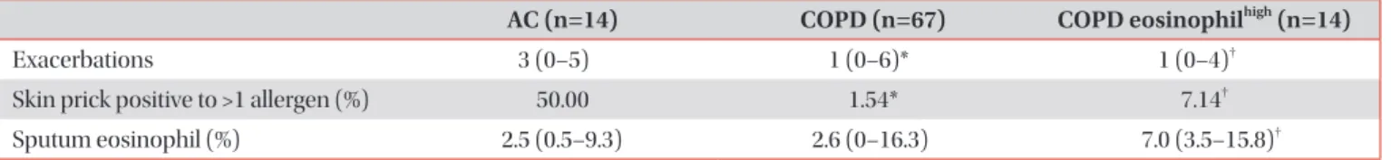
history of asthma versus COPD patients with increased eo- sinophils and no history of asthma showed that the former group had more evidence of allergy and more exacerbations while displaying less eosinophilic inflammation (data shown in Table 1)
17. These data highlight that the terms “asthma” and
“eosinophilic” should not be used interchangeably in COPD patients.
Relationship between Blood and Lung Eosinophil Numbers
Many studies have reported statistically significant asso- ciations between blood and sputum eosinophil counts, with weak to moderate correlation coefficients (0.17–0.54)
18-22. Factors that may negatively impact this relationship are poor quality sputum slides, probably more common in multicentre studies, and using only one significant figure for blood eosino- phil counts. Studies of blood and lung eosinophil counts have shown diverse results, with both positive associations and no relationship
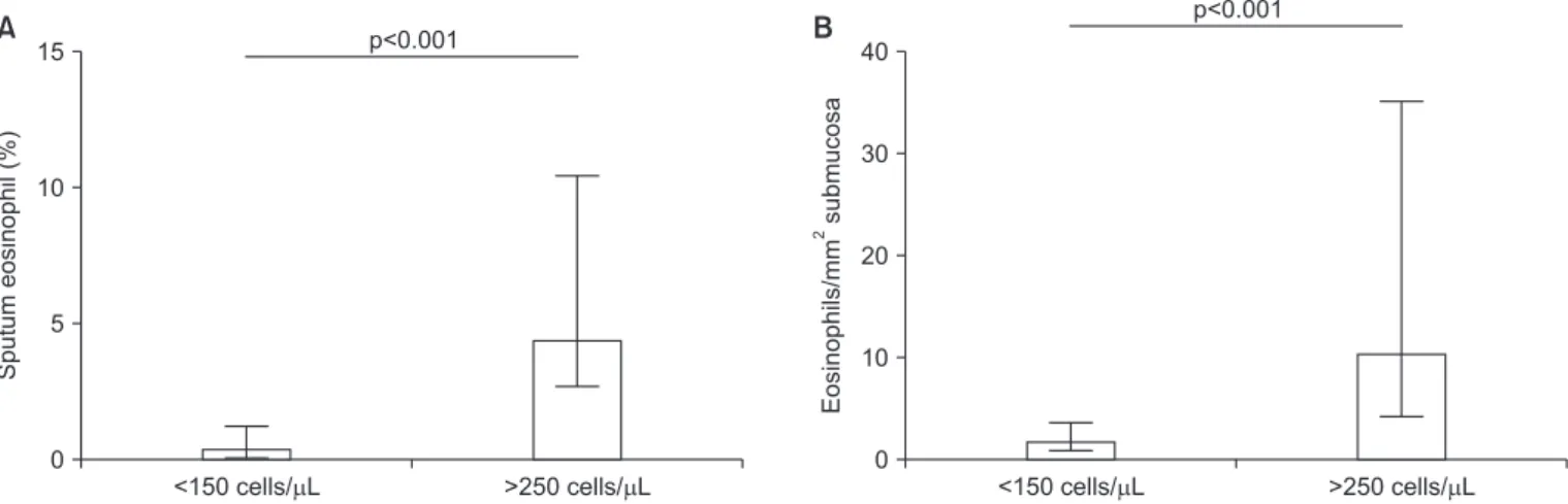
14,23,24. Again, technical factors affecting lung eosinophil measurements may reduce the ability to observe an association. Nevertheless, Kolsum et al.
14showed clearly that COPD patients with blood eosinophil counts <150 cells/
μL had lower bronchial mucosa, broncho-alveolar lavage, and sputum eosinophil numbers compared to COPD patients with blood eosinophil counts >250 cells/μL (Figure 1). Over- all, most of these studies have shown associations between blood and pulmonary eosinophil counts, indicating that blood eosinophils are a biomarker that reflects the degree of eosino- philic lung inflammation.
Modeling the Relationship between Blood Eosinophils and ICS Effects
RCTs using induced sputum eosinophil counts demonstrat- ed that COPD patients with more eosinophils had a greater forced expiratory volume in 1 second (FEV
1) improvement af- ter corticosteroid treatment
25,26. Sputum sampling for eosino- phil counts is not widely available or practical in clinical prac-
Table 1. Clinical characteristics of COPD patients with AC compared to COPD patients
AC (n=14) COPD (n=67) COPD eosinophil
high(n=14)
Exacerbations 3 (0–5) 1 (0–6)* 1 (0–4)
†Skin prick positive to >1 allergen (%) 50.00 1.54* 7.14
†Sputum eosinophil (%) 2.5 (0.5–9.3) 2.6 (0–16.3) 7.0 (3.5–15.8)
†Adapted from Kolsum et al. Respir Res 2017;18:73, according to the Creative Commons license BioMed Central
17. Values are presented as median (range) for exacerbations and sputum eosinophils.
Chronic obstructive pulmonary disease (COPD) patients (out of n=67) with blood eosinophil counts >300 cells/μL are denoted COPD eosin- ophil
high.
*p<0.05 for COPD vs. childhood asthma (AC).
†p<0.05 for COPD eosinophil
highvs. AC.
tice. Post-hoc analyses of RCTs including only COPD patients with a history of exacerbations were performed to evaluate the ability of blood eosinophil counts to predict ICS effects
27-29. The effect of ICS/LABA treatment compared to LABA monotherapy on exacerbation prevention was greater in pa- tients with higher blood eosinophil counts at the start of the study
27-29. Data modeling (the INCONTROL study; n=4,528) showed that the effect of ICS treatment was observed at above approximately 100 eosinophils/μL, with increasingly larger benefits at higher eosinophil counts
27; this continuous rela- tionship is described by Figure 2.
An important consideration for biomarkers in clinical prac- tice is whether the results split the population in a binomial manner; this is the case for diagnostic biomarkers where a binomial categorization of “disease” or “no disease” is required.
In contrast, pharmacological treatment responses form a continuous spectrum (i.e., ranging from no response to small response to large response). A biomarker for predicting drug responses should therefore predict different magnitudes of response. Using blood eosinophil counts to predict ICS
“responders” and “non-responders” is a simplistic approach that does not mirror the range of clinical responses observed.
Instead, the INCONTROL data modeling results show that blood eosinophil counts can be used to predict different mag- nitudes of response, reflecting the population distribution of drug responses
27.
Pre-specified analysis of triple therapy studies conducted in COPD patients with a history of exacerbations have also reported greater ICS effects on exacerbation prevention in patients with higher blood eosinophil counts. In the IMPACT study (n=10,333), data modeling showed that the benefit of triple therapy compared to LABA/LAMA on exacerbation prevent was observed at above approximately 100 eosino- phils/μL
30. Again, the magnitude of benefit increased at higher blood eosinophil counts, with approximately 50% exacer-
bation rate reduction observed at >300 eosinophils/μL, as shown in Figure 2. Interestingly, ICS benefits were lower in current smokers, with ICS benefits in this subgroup observed at a higher threshold, approximately >200 eosinophils/μL. A similar negative influence of current smoking on ICS effects was reported in the INCONTROL data modeling comparing ICS/LABA versus LABA
27. The reduced effects of ICS in cur- rent smokers has not been consistently reported in COPD clinical trials and may be related to insufficient statistical power in previous subgroup analysis.
The TRIBUTE study compared triple therapy versus LABA/
LAMA
7, while the TRINITY study compared triple therapy versus LAMA
6. These studies were conducted in patients with a history of exacerbations, and in both studies it was
ICS+LABA
100
Exacerbationrate(yr)
200 300
25%*
50%*
*Approximate treatment difference LABA
Blood eosinophil count (cell/ L)
Figure 2. Representative illustration of the results of studies investi- gating the relationship between inhaled corticosteroid (ICS) effects (on exacerbations) and blood eosinophil counts. Effect sizes are es- timates. Comparison shown is ICS/long-acting beta agonist (LABA) versus LABA; similar findings exist for ICS/LABA/long-acting mus- carinic antagonist (LAMA) versus LABA/LAMA.
<150 cells/ L 15
10
5
>250 cells/ L
Sputumeosinophil(%)
0
<150 cells/ L 40
30
20
10
>250 cells/ L
Eosinophils/mmsubmucosa2
0 p<0.001
p<0.001
A B
Figure 1. Chronic obstructive pulmonary disease patients with blood eosinophil counts <150 cells/μL and >250 cells/μL. Median is shown,
while error bars are interquartile range. (A) Sputum eosinophil counts (%). (B) Bronchial mucosa eosinophil counts (per mm
2submucosa)
14.
demonstrated that a single eosinophil threshold distinguished between patients with higher and lower ICS responses e.g., in TRINITY, eosinophils ≥2% or ≥200 cells/μL split the popula- tion into groups with approximately >30% and ≤10% exac- erbation rate reductions above and below these thresholds respectively. As already discussed, these single thresholds are not the optimum way to analyze the data. The KRONOS study evaluated triple therapy in a COPD population that in- cluded patients with and without a history of exacerbations
31. Data modeling again showed the “continuous” relationship between blood eosinophil counts and ICS response, with no benefit observed at lower eosinophil counts and increasingly larger benefits at higher eosinophil counts.
Blood Eosinophils, Exacerbation History, and ICS Response
Two large studies have compared ICS/LABA versus LABA/
LAMA in patients with a history of exacerbations, with differ- ent outcomes on the prevention of moderate to severe exac- erbations in the overall population; in the IMPACT study, ICS/
LABA had a greater effect than LABA/LAMA (10% mean dif- ference)
8, while the FLAME study reported that LAMA/LABA had a greater effect than ICS/LABA (17% mean difference)
32. IMPACT data modeling showed a greater effect of ICS/LABA (compared to LAMA/LABA) at higher blood eosinophil counts
30, while in the FLAME study the treatments appeared similar at higher blood eosinophil counts
33. These studies enrolled populations with different levels of exacerbation risk;
IMPACT included more patients with ≥2 moderate exacerba- tions or ≥1 severe exacerbation (hospitalization) in the previ- ous year. This key difference appeared to increase the ICS effect (in patients with higher exacerbation risk) in IMPACT
34. The predictive ability of blood eosinophils when comparing double combination inhalers therefore changes according to the exacerbation risk i.e., at higher eosinophil counts, any benefit of ICS/LABA over LAMA/LABA is more likely to be observed in patients at higher exacerbation risk. Other differ- ences in the study designs of IMPACT and FLAME have been discussed and debated, such as differences in the run-in pe- riods; previous treatment was continued during the run-in in IMPACT, while LAMA monotherapy was used in FLAME
3,34. This led to ICS withdrawal in some patients at randomiza- tion in IMPACT but before run-in for all patients in FLAME.
Regardless, the results of IMPACT and FLAME indicate that exacerbation risk and blood eosinophil counts interact to de- termine ICS response.
ICS withdrawal studies have demonstrated that exacerba- tions rates are greater in patients with higher blood eosinophil counts
35-37. Furthermore, higher eosinophil counts plus a his- tory of ≥2 exacerbations appears to identify individuals who are at the greatest risk of exacerbation after ICS withdrawal
37.
The concept of ICS withdrawal in clinical practice has gained popularity in recent years, due to concerns about ICS side ef- fects and a confidence that many patients can be treated suc- cessfully with LABA/LAMA combination inhalers without the need for additional ICS treatment. However, these ICS with- drawal studies indicate that this strategy has increased risk in patients with higher blood eosinophil counts (>300 cells/μL)
3. Furthermore, these ICS withdrawal studies were performed mainly in patients with 0 or 1 exacerbation in the previous year
35,38, but in the subgroup with ≥2 exacerbations there ap- peared to be the greatest risk
37. Again, these data highlight that blood eosinophil counts and exacerbation risk both influence ICS response.
Real word data analysis, using UK primary care information, reported that the effectiveness of triple therapy compared to LABA/LAMA was greater in patients with more exacerbations or higher blood eosinophils
39. These real world data compli- ment the RCT data already reviewed.
Blood Eosinophil Count Stability
The intra-class correlation coefficient (ICC) for repeated blood eosinophil counts in COPD patients has been re- ported to be 0.64–0.89 in studies with follow up ranging from 3 months to 5 years
40-44. ICC values >0.75 are interpreted as showing excellent correlation
41. On one hand, it has been noted that these ICC values are similar to other biomarkers used in clinical practice such as cholesterol or glycated hemo- globin
3, supporting the case for using blood eosinophil counts in clinical practice. On the other hand, concerns have been expressed that blood eosinophil counts may show excessive variability, limiting their clinical usefulness
45.
It is worth dissecting published data in detail in order to understand eosinophil stability properly. A recent publica- tion investigating stability (n=225) using the categories <100, 100 to <300 and ≥300 eosinophils/μL showed that 69.3% of COPD patients remained in the same category after 1 year
41. Importantly, movement from one category to an adjacent category was more likely in patients with eosinophil measure- ments close to the threshold value, suggesting that movement between categories was more related to natural measurement variation rather than altered disease pathophysiology. GOLD 2020 recommends the use of <100 cells/μL and >300 cells/
μL thresholds, but cautions that these are “estimates, rather
than precise cut-off values” for predicting ICS effects
46. Previ-
ous criticisms of blood eosinophil count stability have focused
on the re-categorization of patients due to movement across
thresholds
45, but a more practical approach (as advocated by
GOLD) is needed when the numerical changes are small; for
example, moving from just below to just above the 300 eosino-
phils/μL threshold does not change the clinical interpretation
(namely, that there is increased likelihood of ICS benefit).
This one year stability study
41and a different study with fol- low up from 2–5 years (n=59)
40both showed that the repeat- ability coefficients were lower (i.e., less numerical variation) in patients with lower blood eosinophil counts. Small changes in these patients may cause movement across a threshold value (e.g., 100 eosinophils/μL). However, as already discussed, it is important to understand that the clinical prediction of ICS re- sponse is unlikely to be altered by a small numerical change.
Individuals with higher blood eosinophil counts (>300 cells/
μL) have more numerical variation, but again this may not re- sult in any change in clinical interpretation, e.g., a change from 500 to 250 cells/μL still suggests that the patient is more likely to derive benefit from ICS treatment.
Studies of blood eosinophil stability have used various thresholds, with some studies using percentage eosinophil counts rather than absolute numbers
21. Percentage counts are clearly influenced by the presence of other immune cells, and the field has now moved towards using absolute numbers to more accurately define the degree of eosinophilic inflamma- tion.
Blood Eosinophils and Clinical Outcomes
Numerous cohort studies have investigated associations between blood eosinophil counts and clinical features or outcomes
18,21,45,47-49. In particular, there has been interest in whether blood eosinophil counts are associated with either exacerbation rates or mortality. The results of these studies have been inconsistent, and it is reasonable to conclude that blood eosinophil counts should not be routinely used in clini- cal practice as a prognostic biomarker for events such as exac- erbations and mortality.
The RCTs already reviewed that were conducted in COPD patients with a history of exacerbations showed an associa- tion between higher blood eosinophil counts and increased exacerbation rates in the treatment arms without ICS (Figure 2)
27-30. These data suggest that blood eosinophil are a prog- nostic biomarker in patients with both (1) a history of exac- erbations and (2) receiving no ICS treatment. These RCTs showed no relationship between blood eosinophil counts and exacerbation rates in patients treated with ICS, as these drugs modify exacerbation risk in an eosinophil dependent manner.
These observations provide the explanation for the lack of as- sociation between blood eosinophil counts and exacerbations in observational cohort studies, which include many patients with (1) no prior exacerbation history and/or (2) taking ICS treatment. Despite these limiting factors, some large cohort studies have still found that patients with a history of ≥2 ex- acerbations plus higher blood eosinophil counts have more exacerbations at during follow-up
47.
Blood Eosinophils, Type 2 Inflammation, and Microbiome
The mechanistic explanation for the association between higher blood eosinophil counts and increased ICS response in COPD patients has not been definitively elucidated. Never- theless, there are pieces of evidence that provide insights. The bronchoscopy study by Kolsum et al.
14showed that higher blood eosinophil counts are associated with increased eo- sinophilic airway inflammation, greater reticular basement membrane thickening, and increased levels of the T2 cyto- kines IL-5 and eotaxin-2; these pathophysiological features are also found in patients with asthma
50,51. However, this study excluded individuals with a history of asthma or atopy. It has also been shown that T2 gene expression in COPD bronchial biopsies is associated with lung and blood eosinophil counts
52. Overall, eosinophilic airway inflammation seems to be associ- ated with a wider profile of T2 inflammation; T2 inflammation is known to be corticosteroid sensitive in asthma
53, and prob- ably the same situation exists in COPD patients.
The asthma-COPD overlap (ACO) describes a group of individuals with clinical features found in both conditions
54,55. While “eosinophilic COPD” shares some pathological features with asthma, there is no added value in labeling these patients as ACO, as ACO is a broad label encompassing different clini- cal phenotypes. Referring to these patients as “eosinophilic COPD” or “COPD with higher blood eosinophil counts” is a more precise description of this COPD subset than using ACO which also includes (multiple) other subtypes.
It has been observed that higher sputum eosinophil counts are associated with lower levels of colonizing bacteria in the airways of COPD patients
56,57. Furthermore, a longitudinal observational study reported that blood eosinophil counts
<100 cells/μL were associated with increased probability of chronic bacterial airway infection and pneumonia
58. These findings suggest that susceptibility to bacterial airway infec- tion is increased in COPD patients with lower eosinophil counts, but the mechanisms to explain these findings have not been defined
59. Eosinophils have no direct anti-bacterial activ- ity against common pathogens that infect COPD patients
60. Therefore, it is likely that there are other differences in anti- microbial host defence associated with eosinophil numbers.
Interestingly, low sputum eosinophil counts in COPD patients have been associated with reduced bacterial diversity and increased Proteobacteria including haemophilus
61,62. Overall, these studies show both increased bacterial load and altered microbiome profiles in patients with lower eosinophil counts.
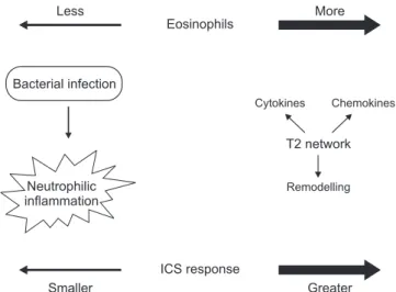
The presence of bacterial infection promotes neutrophilic
airway inflammation
56,62,63. ICS have limited effects on neutro-
philic airway inflammation in humans
64-66. It therefore appears
that COPD patients with low eosinophil counts are more
likely to skew towards bacterial infection and neutrophilic in-
flammation that responds poorly to ICS treatment. In contrast,
high eosinophil counts, associated with increased T2 gene ex- pression and a lower burden of bacterial infection, is a profile of airway inflammation that is more ICS sensitive; this model to explain the relationships between eosinophils, bacteria and ICS response are shown in Figure 3.
It is relevant to consider whether ICS are targeting eosino- phils themselves, and/or other aspects of inflammation as- sociated with higher eosinophil counts. An RCT using sputum eosinophils to predict ICS effects showed improvements in FEV
1in COPD patients with higher eosinophil counts, but no reduction in eosinophil counts
25. In contrast, ICS/LABA re- duced sputum eosinophil counts compared to placebo, in ad- dition to suppression of lymphocytes and mast cells numbers in the bronchial mucosa
65. There was no reduction in sputum neutrophil numbers. Another bronchoscopy study in COPD patients also showed an effect of ICS treatment on submu- cosal lymphocytes, but no effect on neutrophils and eosino- phils
66. These studies have provided consistent evidence that ICS reduce airway lymphocyte numbers and have no effect on neutrophil counts, but the results for eosinophils have been mixed. This mixed evidence can be interpreted as showing that ICS can reduce airway eosinophil numbers, but that this finding is not consistent in all patients. This may be due to low baseline eosinophil numbers in some patients. Alternatively, the therapeutic benefit of ICS in eosinophilic COPD patients may be due to pharmacological effects on inflammation com- ponents (T2 inflammation) beyond the eosinophil cell.
RCTs investigating the effects of monoclonal antibodies targeting IL-5, conducted in COPD patients with a history of exacerbations, have reported both positive and negative results for the effect on exacerbation rate reduction
67,68. A pre- specified combined analyses of the GALATHEA and TER- RANOVA studies of benralizumab (which targets IL-5 recep- tor alpha) demonstrated that the greatest treatment benefit
was observed in the subgroup receiving triple therapy with blood eosinophil counts ≥220 cells/μL and ≥3 exacerbations in the prior year
69. These findings highlight that significant eo- sinophilic inflammation, which may respond to monoclonal antibody targeting, can persist despite ICS treatment.
Studies focused on COPD exacerbations have found that blood and sputum eosinophil counts are increased in a subset of patients during exacerbations
70. Interestingly, higher blood eosinophil counts in the stable state are associated with an increased probability of exacerbations with increased sputum eosinophil numbers
71. This association between eosinophils in the stable and exacerbation states suggests that ICS treat- ment probably suppresses exacerbation subtypes involving increased eosinophilic inflammation.
GOLD 2019 and Blood Eosinophils
The evidence already reviewed formed the basis of the GOLD 2019 recommendations to use blood eosinophil counts as a biomarker to help direct ICS treatment in COPD patients with a history of exacerbations
3. These recommenda- tions combine clinical information (exacerbation risk) with biological data (blood eosinophils) as a precision medicine approach in order to optimize the potential for benefit over risk.
The FLAME and IMPACT studies reported different results for the comparison of the effects of ICS/LABA versus LABA/
LAMA combinations on exacerbation rates
8,32. A major reason for these divergent results was the different exacerbations risks of the study populations
34. Accordingly, GOLD states that the benefits of ICS are likely to be greater in high exacerbation risk patients with a history of ≥2 moderate exacerbations and/
or ≥1 severe exacerbation in the previous year compared to patients with 1 moderate exacerbation
3. Other clinical factors relevant to the use of ICS include a history of asthma, which favors ICS use, and risk factors for ICS side effects, such as repeated pneumonia, which argue against ICS use
72. All of this clinical information should be collected and then used alongside blood eosinophil counts in order to make optimal decisions for each individual.
GOLD uses thresholds of >300 eosinophils/μL and <100 eosinophils/μL to identify individuals with a higher and lower probability, respectively, of experiencing treatment benefit with ICS
46. The GOLD 2020 revision states that these are “esti- mates, rather than precise cut-off values”
46. The purpose of this statement is highlight that small numerical changes in blood eosinophil counts should not lead to a change in ICS treat- ment, even if the change leads to movement across a thresh- old value.
Some clinicians have focused on the >300 eosinophils/μL threshold stated in GOLD, preferring to use ICS only in these individuals. However, this approach does not account for the
Less More
Eosinophils
Bacterial infection
Neutrophilic inflammation
Smaller Greater
ICS response
Cytokines Chemokines
Remodelling T2 network