Predictors of Pulmonary Function Response to Treatment with
Salmeterol/fluticasone in Patients with Chronic Obstructive
Pulmonary Disease
Chronic obstructive pulmonary disease (COPD) is a heterogeneous disease and responses to therapies are highly variable. The aim of this study was to identify the predictors of pulmonary function response to 3 months of treatment with salmeterol/fluticasone in patients with COPD. A total of 127 patients with stable COPD from the Korean Obstructive Lung Disease (KOLD) Cohort, which were prospectively recruited from June 2005 to September 2009, were analyzed retrospectively. The prediction models for the FEV1, FVC and IC/TLC changes after
3 months of treatment with salmeterol/fluticasone were constructed by using multiple, stepwise, linear regression analysis. The prediction model for the FEV1
change after 3 months of treatment included wheezing history, pre-bronchodilator FEV1, post-bronchodilator FEV1 change and emphysema extent on CT (R = 0.578).
The prediction models for the FVC change after 3 months of treatment included pre-bronchodilator FVC, post-pre-bronchodilator FVC change (R = 0.533), and those of IC/ TLC change after 3 months of treatment did pre-bronchodilator IC/TLC and post-bronchodilator FEV1 change (R = 0.401). Wheezing history, pre-bronchodilator
pulmonary function, bronchodilator responsiveness, and emphysema extent may be used for predicting the pulmonary function response to 3 months of treatment with salmeterol/fluticasone in patients with COPD.
Key Words: Pulmonary Disease, Chronic Obstructive; Emphysema; Corticosteroids; Adrenergic beta-Agonists; Respiratory Function Tests
Jae Seung Lee1, Jin Won Huh1, Eun Jin Chae2,
Joon Beom Seo2, Seung Won Ra3, Ji-Hyun Lee4,
Eun-Kyung Kim4, Young Kyung Lee5,
Tae-Hyung Kim6, Woo Jin Kim7, Jin Hwa Lee8,
Sang-Min Lee9, Sangyeub Lee10,
Seong Yong Lim11, Tae Rim Shin12, Ho Il Yoon13,
Seung Soo Sheen14, Yeon-Mok Oh1,
and Sang-Do Lee1
1Department of Pulmonary and Critical Care Medicine, Asthma Center, and Clinical Research Center for Chronic Obstructive Airway Diseases, 2Department of Radiology, Asan Medical Center, University of Ulsan College of Medicine, Seoul; 3Department of Pulmonary and Critical Care Medicine, Ulsan University Hospital, University of Ulsan College of Medicine, Ulsan; 4Division of Pulmonary and Critical Care Medicine, Department of Internal Medicine, Bundang CHA Hospital, College of Medicine, CHA University, Seongnam; 5Department of Radiology, East-West Neo Medical Center, Kyunghee University, Seoul; 6Division of Pulmonology, Department of Internal Medicine, Hanyang University Guri Hospital, Hanyang University College of Medicine, Guri; 7Department of Internal Medicine, College of Medicine, Kangwon National University, Chuncheon; 8Department of Internal Medicine, Ewha Womens University Mokdong Hospital, College of Medicine, Ewha Womans University, Seoul; 9Division of Pulmonary and Critical Care Medicine, Department of Internal Medicine, Seoul National University College of Medicine, Clinical Research Institute, Seoul; 10Division of Respiratory and Critical Care Medicine, Department of Internal Medicine, College of Medicine, Korea University Anam Hospital, Seoul; 11Division of Pulmonary and Critical Care Medicine, Department of Medicine, Kangbuk Samsung Hospital, Sungkyunkwan University School of Medicine, Seoul; 12Department of Internal Medicine, Kangnam Sacred Heart Hospital, Hallym University College of Medicine, Seoul; 13Department of Internal Medicine, Seoul National University Bundang Hospital, Seoul National University College of Medicine, Seongnam; 14Department of Pulmonary and Critical Care Medicine, Ajou University School of Medicine, Suwon, Korea Received: 18 October 2010
Accepted: 24 December 2010 Address for Correspondence: Sang-Do Lee, MD
Department of Pulmonary and Critical Care Medicine, and Clinical Research Center for Chronic Obstructive Airway Diseases, Asan Medical Center, University of Ulsan College of Medicine, 86 Asanbyeongwon-gil, Sonpa-gu, Seoul 138-736, Korea
Tel: +82.2-3010-3140, Fax: +82.2-3010-6968 E-mail: [email protected]
This study was supported by a grant of the Korea Healthcare technology R&D Project, Ministry for Health, Welfare and Family Affairs, Republic of Korea (A040153) and by the Asan Institute for Life Science (07-306).
DOI: 10.3346/jkms.2011.26.3.379 • J Korean Med Sci 2011; 26: 379-385
INTRODUCTION
Chronic obstructive pulmonary disease (COPD) is usually pro-gressive and is associated with an abnormal inflammatory re-sponse of the lung to noxious particles or gases (1). Previous studies have shown that regular treatment of COPD with an in-haled corticosteroid (ICS) improve pulmonary function and re-duces the frequency of exacerbations, even though it does not modify the long-term decline of forced expiratory volume in 1 sec (FEV1) (2, 3). Combination therapy with an ICS and a long acting β2-agonist (LABA) is better in improving lung function and health status, and reducing daily symptoms and exacerba-tions than single therapy (4, 5).
COPD is a heterogeneous disease in terms of clinical, physi-ological, and pathological presentation. The chronic airflow lim-itation associated with COPD is caused by a variable contribu-tion of small airway disease (obstructive bronchiolitis) and pa-renchymal destruction (emphysema). Thus, the therapeutic re-sponse to combination treatment with an ICS and a LABA is also variable. Previous COPD guidelines recommended the use of short-term (two weeks) oral corticosteroid therapy to identify COPD patients who might benefit from long-term treatment with an ICS (6). However, it was recently shown that the response to a short-term oral corticosteroid is a poor predictor of the long-term response to an ICS (7). Currently, there are no known good predictors of the responsiveness of COPD patients to treatment with an ICS and a LABA.
The aim of this study was to identify predictors of pulmonary function response to 3 months of treatment with salmeterol/ fluticasone in patients with COPD.
MATERIALS AND METHODS
Patients
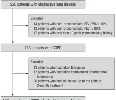
A total of 127 stable COPD patients who had been treated with salmeterol/fluticasone for 3 months were analyzed retrospec-tively. All patients were selected from the Korean Obstructive Lung Disease (KOLD) Cohort, which had 226 stable patients with obstructive lung disease (OLD) were prospectively recruit-ed from the pulmonary clinics of 11 hospitals in Korea from June 2005 to September 2009 (Fig. 1). The inclusion criteria for the KOLD cohort have been described elsewhere (8). COPD was diagnosed based on smoking history (more than 10 pack-years) and the presence of airflow limitation that was not fully revers-ible (post-bronchodilator FEV1/forced vital capacity [FVC] < 70% and post-bronchodilator FEV1 < 80% predicted). We included COPD patients who had wheezing history or who showed par-tial reversibility of airflow limitation after inhalation of salbuta-mol.
Study protocol
Baseline clinical data were obtained after cessation of the fol-lowing respiratory medications: an ICS for 2 weeks, an inhaled LABA for 2 days, an inhaled short-acting β2-agonist or inhaled short-acting anti-cholinergic for 12 hr. The baseline clinical data included demographic data, smoking history, chronic bronchitis history, wheezing history, pulmonary function tests, chest radi-ography and volumetric computed tomradi-ography (CT). Chronic bronchitis was defined as cough and sputum production on most days for a minimum of 3 months per year for at least 2 yr (9). Wheezing history was obtained through the following ques-tion: “Have you had wheezing or whistling in your chest at any time in the last year?” (10) Atopic status was assessed by a skin prick test to 11 common allergens, with a 10% histamine and saline control. Patients were considered to be atopic if they re-acted with a wheal of larger than the histamine control for more than one of the allergens. After obtaining baseline data, patients were treated with a salmeterol/fluticasone propionate 50/500 µg dry powder inhaler twice per day for 3 months, and then spi-rometry and lung volume measurement were performed again after the morning medication. During the 3-month treatment period, only salbutamol was allowed as needed. Adherence to the treatment medication monitored and recorded by research coordinators.
Pulmonary function tests
Spirometry was performed using a Vmax 22 instrument (Sensor-Medics; Yorba Linda, CA, USA) or a PFDX machine (MedGraph-ics, St. Paul, MN, USA). To assess the post-bronchodilator change, spirometry was performed pre-bronchodilator and 15 min after
Excluded
14 patients with post-bronchodilator FEV1/FVC ≥ 70% 12 patients with post-bronchodilator FEV1 ≥ 80% 17 patients with less than 10 pack-years smoking history
127 patients with COPD who had taken combination of salmeterol/fluticasone were analyzed
Excluded
13 patients who had taken tiotropium
17 patients who had taken combination of formoterol/ budesonide
26 patients who had lost follow-up at the point of 3-month treatment
226 patients with obstructive lung disease
183 patients with COPD
inhalation of salbutamol (400 µg) through a metered-dose in-haler (MDI) with a spacer. Lung volumes were measured by body plethysmography (V6200; SensorMedics, or PFDX). Diffusing capacity for carbon monoxide (DLco) was measured by the sin-gle-breath method using a Vmax229D (Sensor-Medics) or a Mas-terlab Body (Jaeger AB, Würtsburg, Germany). The predicted values of FEV1, forced vital capacity (FVC), FEV1/FVC and DLco were calculated from Korean equations formulated using data from a healthy non-smoking population (11, 12). The predicted values of lung volumes were calculated from ECSC equations (13). All pulmonary function tests were performed as recom-mended by the American Thoracic Society/European Respira-tory Society (ATS/ERS).
Computed tomography
Volumetric CT scans were performed using 16-slice multi-de-tector CT (MDCT) scanners, according to previous described scanning protocols (14). Image data were stored in the Digital Imaging and Communications in Medicine (DICOM) format. Using in-house software, images of the whole lung were extract-ed automatically and the attenuation coefficient of each pixel was measured and calculated. The cutoff level between normal lung density and a low-attenuation area (LAA) was defined as -950 HU (15). From the CT data, the volume fraction of the lung below -950 HU (V950) was calculated automatically. The airway dimensions, wall area (WA), lumen area (LA) and wall area per-cent (WA%, defined as WA/[WA + LA] × 100), were measured near the origin of two segmental bronchi (right apical and left apico-posterior) selected by a consensus reading of two radiol-ogists. The software automatically detects the airway lumen and the inner and outer boundaries of the airway wall by use of a full-width-half-maximum (FWHM) method (16). The mean value of each segmental bronchus was used for analysis.
Statistical analysis
To investigate factors associated with the initial maximal posi-tive pulmonary function response to 3 months of treatment with salmeterol/fluticasone, changes in pre-bronchodilator FEV1, FVC and inspiratory capacity (IC)/total lung capacity (TLC) be-fore and after 3 months were used as the dependent variable in univariate and multivariate analyses. The FEV1 and FVC chang-es were exprchang-essed as % of predicted normal valuchang-es (% predict-ed) to avoid the potential problem that baseline FEV1 and FVC appear to influence on FEV1 and FVC changes if expressed as % of baseline (17). Selected independent variables were sex, age, body mass index (BMI), chronic bronchitis history, wheezing history, smoking status (current or ex-smokers, smoking pack-years), pulmonary function parameters and CT parameters. For data had a normal distribution, we used Student’s t-test or a one-way analysis of variance (ANOVA) for categorical variables. For data that had a non-normal distribution, we used
Mann-Whit-ney U test or the Kruskall-Wallis test for categorical variables. Relationships between two continuous variables were examined by Pearson’s correlation analysis. The prediction models for pul-monary function response to 3 months of treatment with sal-meterol/fluticasone were constructed from multiple, stepwise, linear regression models. All statistical analyses were performed with the SPSS statistical package (version 12.0, SPSS Inc, Chica-go, IL, USA), and P values less than 0.05 were considered signif-icant.
Ethics statement
This study was approved by the Asan Medical Center institu-tional review board (Approval No. 2005-0345) and institution review boards of other10 hospitals. Written informed consent was obtained from all patients.
RESULTS
Patient characteristics
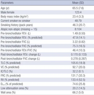
Table 1 lists the baseline characteristics of the 127 COPD pati-ents. Of the enrolled patients, 123 (97%) were male. Mean age was 65.5 (SD: 7.6) yr, and mean smoking history was 46.3 (20.7) pack years. A total of 74 patients were classified as GOLD II (mod-erate COPD), 46 as GOLD III (severe COPD), and 7 as GOLD IV (very severe COPD). Ninety-one percent of subjects indicated that they had taken over 80% of the recommended medication dose. Table 1. Baseline characteristics of patients (N = 127)
Parameters Mean (SD)
Age (yr) 65.5 (7.6)
Male:female 123:4
Body mass index (kg/m2) 23.4 (3.3)
Current smoker:ex-smoker 48:79
Smoking history (pack-years) 46.3 (20.7) Atopic:non-atopic (missing = 15) 8:104 Pre-bronchodilator FEV1 (L) 1.49 (0.50) Pre-bronchodilator FEV1 (% predicted) 47.9 (14.1) Pre-bronchodilator FVC (L) 3.22 (0.82) Pre-bronchodilator FVC (% predicted) 75.3 (16.5) Pre-bronchodilator FEV1/FVC (%) 46.4 (10.3) Post-bronchodilator FEV1 change (L) 0.170 (0.133) Post-bronchodilator FVC change (L) 0.278 (0.287) TLC (% predicted) 105.6 (18.9) VC (% predicted) 92.7 (20.0) IC/TLC (%) 32.2 (0.1) FRC (% predicted) 131.7 (33.3) RV (% predicted) 133.4 (52.5) DLco (% of predicted) 74.6 (25.8) Low attenuation area (%) 20.2 (14.5)
Wall area (%) 66.2 (5.0)
FEV1, forced expiratory volume in 1 sec; FVC, forced vital capacity; TLC, total lung capacity; VC, vital capacity; IC, inspiratory capacity; FRC, functional residual capacity; RV, residual volume; DLco, diffusing capacity for carbon monoxide; Low attenuation area, volume fraction of the lung below -950 HU at full inspiration computed tomo-graphy; Wall area, wall area/(wall area + lumen area) on computed tomography.
Univariate analysis of baseline clinical variables and pulmonary function response to 3 months of treatment with salmeterol/fluticasone
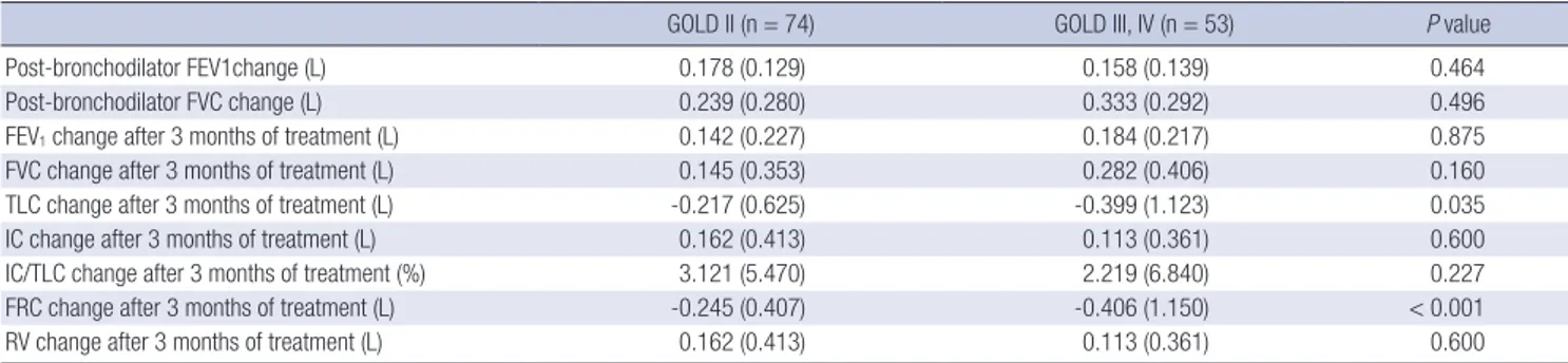
The FEV1, FVC and IC/TLC changes after 3 months of treatment were normally distributed without evidence of a separate re-sponder group. The mean FEV1, FVC and IC/TLC changes after 3 months of the treatment were 0.160 (0.223) L, 5.1 (7.3)% pre-dicted and 0.202 (0.380) L, 4.8 (9.0)% prepre-dicted, and 2.7 (6.1)%, respectively. There were no significant differences in the mean postbronchodilator FEV1 and FVC changes according to base-line GOLD severity stage. Patients with GOLD stage III or IV showed significantly larger TLC and functional residual capaci-ty (FRC) reduction after the 3 months of treatment with salme-terol/fluticasone than patients with GOLD stage II. However, there were no significant differences in the mean FEV1, FVC and IC/TLC changes after 3 months of the treatment (Table 2). Among categorical variables, COPD patients with wheezing history had significantly greater FEV1 and FVC changes after 3
months of treatment than those without wheezing history (7.8% vs 2.7% predicted for FEV1, P < 0.001; 7.3% vs 2.3% predicted for FVC, P = 0.002). Simple correlation analysis indicated that pre-bronchodilator FEV1 and FVC had a weak negative correlation with the FEV1 and FVC changes after 3 months of the treatment but that post-bronchodilator FEV1 and FVC had no significant correlation with FEV1 and FVC changes after 3 months of the treatment. The post-bronchodilator FEV1 and FVC changes had a positive correlation with the FEV1 and FVC changes after 3 months of the treatment. LAA had a weak negative correlation with the FEV1 change after 3 months of the treatment, but mean WA was not correlated with the FEV1, FVC and IC/TLC changes after 3 months of the treatment (Table 3).
Prediction models for pulmonary function response to 3 months of treatment with salmeterol/fluticasone
Multiple, stepwise, linear regression analysis showed that four variables, wheezing history, pre-bronchodilator FEV1, post-bron-Table 2. Pulmonary function response to short-acting bronchodilator and 3 months of treatment with salmeterol/fluticasone according to baseline GOLD severity stage
GOLD II (n = 74) GOLD III, IV (n = 53) P value
Post-bronchodilator FEV1change (L) 0.178 (0.129) 0.158 (0.139) 0.464
Post-bronchodilator FVC change (L) 0.239 (0.280) 0.333 (0.292) 0.496
FEV1 change after 3 months of treatment (L) 0.142 (0.227) 0.184 (0.217) 0.875
FVC change after 3 months of treatment (L) 0.145 (0.353) 0.282 (0.406) 0.160
TLC change after 3 months of treatment (L) -0.217 (0.625) -0.399 (1.123) 0.035
IC change after 3 months of treatment (L) 0.162 (0.413) 0.113 (0.361) 0.600
IC/TLC change after 3 months of treatment (%) 3.121 (5.470) 2.219 (6.840) 0.227 FRC change after 3 months of treatment (L) -0.245 (0.407) -0.406 (1.150) < 0.001
RV change after 3 months of treatment (L) 0.162 (0.413) 0.113 (0.361) 0.600
Data are expressed as means with standard deviations. FEV1, forced expiratory volume in 1 sec; FVC, forced vital capacity; TLC, total lung capacity; IC, inspiratory capacity; FRC, functional residual capacity; RV, residual volume.
Table 3. Correlation between baseline clinical variables and pulmonary function response to 3 months of treatment with salmeterol/fluticasone FEV1 change after 3-month
treatment (% predicted) FVC change after 3-month treatment (% predicted) IC/TLC change after 3-month treatment (%)
R P value R P value R P value
Age (yr) -0.028 0.755 -0.068 0.447 -0.068 0.760
Smoking pack-years 0.020 0.820 0.054 0.545 -0.133 0.137
Body mass index (kg/m2) 0.146 0.101 0.011 0.902 0.026 0.776
Pre-bronchodilator FEV1 (% predicted) -0.267 0.002 -0.340 < 0.001 -0.031 0.729
Post-bronchodilator FEV1 (% predicted) -0.141 0.114 -0.265 0.003 0.044 0.628
Post-bronchodilator FEV1 change (% predicted) 0.432 < 0.001 0.257 0.004 0.254 0.004 Pre-bronchodilator FVC (% predicted) -0.326 < 0.001 -0.358 < 0.001 -0.027 0.767
Post-bronchodilator FVC (% predicted) -0.191 0.032 -0.173 0.052 0.022 0.811
Post-bronchodilator FVC change (% predicted) 0.338 < 0.001 0.464 < 0.001 0.109 0.226
TLC (% predicted) 0.080 0.372 0.216 0.015 0.206 0.021
IC/TLC (%) -0.192 0.030 -0.244 0.006 -0.328 < 0.001
FRC (% predicted) 0.153 0.087 0.286 0.001 0.288 0.001
RV (% predicted) 0.205 0.021 0.325 < 0.001 0.232 0.009
DLco (% of predicted) 0.072 0.432 -0.025 0.785 -0.027 0.772
Low attenuation area (%) -0.222 0.015 -0.094 0.307 -0.022 0.811
Wall area (%) 0.039 0.675 0.053 0.565 0.061 0.513
R, correlation coefficient; FEV1, forced expiratory volume in 1 sec; FVC, forced vital capacity; TLC, total lung capacity; VC, vital capacity; IC, inspiratory capacity; FRC, functional residual capacity; RV, residual volume; DLco, diffusing capacity for carbon monoxide; Low attenuation area, volume fraction of the lung below -950 HU at full inspiration computed tomography; Wall area, wall area/(wall area + lumen area) × 100 on computed tomography.
chodilator FEV1 change, and LAA were independently associat-ed with the FEV1 change after 3 months of the treatment (R = 0.578). Multiple, stepwise, linear regression analysis also showed that two variables, pre-bronchodilator FVC and post-broncho-dilator FVC change were independently associated with FVC change after the 3-month treatment (R = 0.533), and pre-bron-chodilator IC/TLC and post-bronpre-bron-chodilator FEV1 change were independently associated with IC/TLC change after 3 months of the treatment (R = 0.401, Table 4).
DISCUSSION
Our results show that wheezing history, baseline pulmonary function, bronchodilator responsiveness, and emphysema ex-tent on CT could be used for predicting the pulmonary function response to 3 months of treatment with salmeterol/fluticasone in patients with COPD. This allowed us to make a significant pre-diction model for FEV1, FVC and IC/TLC changes after 3 months of treatment.
The response of pulmonary function to an ICS and a LABA in COPD patients has been well established in previous studies (4, 5). Current guidelines recommend combination of an ICS and a LABA for symptomatic COPD patients with post-bronchodilator FEV1 < 50% predicted and repeated exacerbation (1). However, clinical benefits from combination pharmacotherapy are not re-stricted to patients of severe stage of disease (18, 19). It has been difficult to predict which patients will show better responses to an ICS and a LABA. In this study, COPD patients with wheezing history, lower baseline lung function, larger bronchodilator re-sponsiveness and lower LAA on CT responded better to the treat-ment with salmeterol and fluticasone. This combination would be expected to be more effective in improving the small airway component (chronic obstructive bronchiolitis) of COPD. Wheezing is the most common symptom reported by asth-matic patients, but is also commonly reported by COPD pati-ents (20). Marini et al. (21) reported that wheezing patipati-ents with chronic airflow obstruction differed from non-wheezing pa-tients with chronic airflow obstruction by their bronchodilator
response. Our results showed the COPD patients with a wheez-ing history had a significantly higher mean FEV1 and FVC chang-es after bronchodilator and 3 months of treatment than those without such a history. Our study also showed a positive corre-lation between bronchodilator responsiveness (post-broncho-dilator FEV1 and FVC changes) and the FEV1 and FVC changes after 3 months of treatment. Previous studies showed that corti-costeroid reversibility in COPD patients was related to features of asthma (22). Recently, Kitaguchi et al. (23) also reported that wheezing and bronchodilator responsiveness were significant determinant for the reversibility in response to an ICS.
This study also showed that COPD patients with lower pre-bronchodilator FEV1 and FVC have better responses to our com-bination treatment. On the contrary, there was no significant effect of post-bronchodilator FEV1 on the response to combina-tion treatment. A post-hoc analysis in the TORCH study showed similar results in that the severity of COPD which was assessed by post-bronchodilator FEV1 was not related to the FEV1 improve-ments after a combination treatment (19). This result might be explained as follows. The lack of a relation between COPDse-verity and treatment responsiveness may be caused by the fact that pre-bronchodilator FEV1 and post-bronchodilator FEV1 change relate to treatment responsiveness. The post-broncho-dilator FEV1 and FVC (which is unrelated to responsiveness) are the sum of the value of pre-bronchodilator FEV1 and FVC (which is related to responsiveness) and the post-bronchodila-tor FEV1 and FVC changes (which is related to responsiveness). CT provided an objective method for measuring the extent and severity of emphysema (15, 24). Several recent studies indi-cated that it is possible to separate COPD patients into emphy-sema-dominant and airway-dominant phenotypes by use of high resolution CT (HRCT) (23, 25, 26). Kitaguchi et al. (23) re-ported that lower total LAA score and the grade of bronchial wall thickening were significant determinants for bronchodila-tor responsiveness and for the responsiveness to the treatment with an ICS. However, other two studies found no significant dif-ferences in bronchodilator responsiveness among groups clas-sified according to severity of emphysema (25, 26). Previously, Table 4. Multiple linear regression models for pulmonary function response to 3 months of treatment with salmeterol/fluticasone
Variables β 95% CI R
FEV1 change (% predicted) Constant Wheezing history
Pre-bronchodilator FEV1 (% predicted) Post-bronchodilator FEV1 change (% predicted) Low attenuation area (%)
8.98 3.54 -0.13 0.55 -0.09 2.70-15.22 1.18-5.90 -0.22--0.04 0.26-0.84 -0.18--0.01 0.578
FVC change (% predicted) Constant
Pre-bronchodilator FVC (% predicted) Post-bronchodilator FVC change (% predicted)
12.27 -0.15 0.55 5.34-19.20 -0.23--0.06 0.34-0.76 0.533 IC/TLC (%) Constant Pre-bronchodilator IC/TLC (%)
Post-bronchodilator FEV1 change (% predicted)
8.42 -0.23 0.34 4.04-12.80 -0.36--0.11 0.10-0.58 0.401
FEV1, forced expiratory volume in 1 sec; FVC, forced vital capacity; IC/TLC, inspiratory capacity/total lung capacity; Low attenuation area, volume fraction of the lung below -950 HU at full inspiration computed tomography; β, unconditioned coefficient; CI, confidence interval.
our study group has shown that emphysema dominant COPD patients (LAA ≥ 20% and pre-bronchodilator FEV1 ≥ 45% pre-dicted) responded poorly to the 3 months of combination treat-ment (27). In this study, LAA had a week negative correlation with the FEV1 change after 3 months of the treatment. This re-sult might be explained as follows. For COPD patients, the ma-jor determinants of FEV1 are small airway disease and emphy-sema. If a COPD patient has a more significant emphysema com-ponent than that of small airway disease, he or she would be ex-pected to have a poorer response to pharmacologic treatments that predominantly target small airway disease. In this study, mean WA was not correlated with the FEV1 and FVC change af-ter 3 months of the treatment. A previous report showed that large airway dimension serve as a useful surrogate for small air-way remodeling (28). Nonetheless, it is possible that the lack of a significant relationship between WA and treatment response may be due to the relatively imprecise and indirect estimate of small airway wall area. There are two factors that could have po-tentially confounded our volumetric CT result. First, we used three different MDCT scanners, and different scanners may have graded the emphysema index differently. A method that cor-rects for differences between different CT scanners has not yet been established. Second, we used only two large airways to evaluate airway dimension.
In this study, 19.7% of included patients had overlapping diag-noses of COPD and asthma (overlap syndrome). Those patients with overlap syndrome could affect our study results. However, subgroup analysis after excluding patients with overlap syndrome showed the similar results. Our study has two limitations. First, we did not consider airway inflammatory markers. Previous studies showed an increased number of eosinophils in bron-choalveolar lavage fluid in a subset of patients with COPD who responded to short-term administration of oral corticosteroids (29) and sputum eosinophil counts were significantly correlat-ed with reversibility in response to ICS treatment (23). If we had included airway inflammatory markers, we might have been able to develop more powerful prediction models. Second, 97% of our patients were male. Gender may have a substantial influ-ence on treatment response in COPD patients (30), so we can-not generalize our results to females.
In conclusion, wheezing history, baseline pulmonary func-tion, bronchodilator responsiveness, and emphysema extent in COPD patients may be used for predicting the pulmonary func-tion response to 3 months of treatment with salmeterol/flutica-sone.
Conflict of Interest Statement
J. S. Lee, S.W. Ra, E. J. Chae, J-H Lee, E-K Kim, Y. K. Lee, T-H Kim, J. W. Huh, W. J. Kim, J. H. Lee, S-M Lee, S. Y. Lee, S. Y. Lim, T. R. Shin, H. I. Yoon and S. S. Sheen have no conflicts of interest to disclose. J. B. Seo has been an investigator in a
government-spon-sored study (2006-2008 Korea Science and Engineering Foun-dation).
Y-M. Oh has been an investigator in university-sponsored studies (Asan Institute for Life Science, University of Ulsan Col-lege of Medicine) and an industry-sponsored study (MSD Ko-rea, and AstraZeneca Korea), and has participated as a speaker at scientific meetings organized and financed by various phar-maceutical companies (Handok, GlaxoSmithKline, AstraZene-ca Korea, MSD Korea, and Boehringer Ingelheim) and a maga-zine company (Korea Doctors’ Weekly). S-D. Lee serves as a con-sultant to GlaxoSmithKline, and has participated as a speaker at scientific meetings organized and financed by various phar-maceutical companies (GlaxoSmithKline, AstraZeneca Korea, and Boehringer Ingelheim).
REFERENCES
1. Rabe KF, Hurd S, Anzueto A, Barnes PJ, Buist SA, Calverley P, Fukuchi Y, Jenkins C, Rodriguez-Roisin R, van Weel C, Zielinski J; Global Initiative for Chronic Obstructive Lung Disease. Global strategy for the diagnosis,
management, and prevention of chronic obstructive pulmonary disease: GOLD executive summary. Am J Respir Crit Care Med 2007; 176: 532-55.
2. Burge PS, Calverley PM, Jones PW, Spencer S, Anderson JA, Maslen TK.
Randomised, double blind, placebo controlled study of fluticasone pro-pionate in patients with moderate to severe chronic obstructive pulmo-nary disease: the ISOLDE trial. BMJ 2000; 320: 1297-303.
3. Lung Health Study Research Group. Effect of inhaled triamcinolone on
the decline in pulmonary function in chronic obstructive pulmonary dis-ease. N Engl J Med 2000; 343: 1902-9.
4. Mahler DA, Wire P, Horstman D, Chang CN, Yates J, Fischer T, Shah T.
Effectiveness of fluticasone propionate and salmeterol combination de-livered via the Diskus device in the treatment of chronic obstructive pul-monary disease. Am J Respir Crit Care Med 2002; 166: 1084-91.
5. Calverley PM, Anderson JA, Celli B, Ferguson GT, Jenkins C, Jones PW, Yates JC, Vestbo J; TORCH investigators. Salmeterol and fluticasone
pro-pionate and survival in chronic obstructive pulmonary disease. N Engl J Med 2007; 356: 775-89.
6. The COPD guidelines group of the standards of care committee of the BTS. BTS guidelines for the management of chronic obstructive
pulmo-nary disease. Thorax 1997; 52 Suppl 5: S1-28.
7. Burge PS, Calverley PM, Jones PW, Spencer S, Anderson JA.
Predniso-lone response in patients with chronic obstructive pulmonary disease: results from the ISOLDE study. Thorax 2003; 58: 654-8.
8. Kim WJ, Oh YM, Sung J, Kim TH, Huh JW, Jung H, Lee JH, Kim EK, Lee JH, Lee SM, Lee S, Lim SY, Shin TR, Yoon HI, Kwon SY, Lee SD. Lung
function response to 12-week treatment with combined inhalation of long-acting beta2 agonist and glucocorticoid according to ADRB2 poly-morphism in patients with chronic obstructive pulmonary disease. Lung 2008; 186: 381-6.
9. Terminology, definitions, and classification of chronic pulmonary
em-physema and related conditions: a report of the conclusions of a Ciba guest symposium. Thorax 1959; 14: 286-99.
10. Asher MI, Keil U, Anderson HR, Beasley R, Crane J, Martinez F, Mitchell EA, Pearce N, Sibbald B, Stewart AW, Strachan D, Weiland SK, Williams
HC. International study of asthma and allergies in childhood (ISAAC):
rationale and methods. Eur Respir J 1995; 8: 483-91.
11. Park JO, Choi IS, Park KO. Normal predicted standards of single breath
carbon monoxide diffusing capacity of lung in healthy nonsmoking adults. Korean J Intern Med 1985; 28: 176-83.
12. Choi JK, Paek D, Lee JO. Normal predicted values of spirometry in
Kore-an population. Tuberc Respir Dis 2005; 58: 230-42.
13. Quanjer P, Dalhuijsen A, Van Zoramen B. Standardized lung function
testing. Report of the working party for the European Community for Coal and Steel. Bull Eur Physiopathol Respir 1983; 19 Suppl 5: 1-95.
14. Lee YK, Oh YM, Lee JH, Kim EK, Lee JH, Kim N, Seo JB, Lee SD; KOLD Study Group. Quantitative assessment of emphysema, air trapping, and
airway thickening on computed tomography. Lung 2008; 186: 157-65.
15. Gevenois PA, de Maertelaer V, De Vuyst P, Zanen J, Yernault JC.
Com-parison of computed density and macroscopic morphometry in pulmo-nary emphysema. Am J Respir Crit Care Med 1995; 152: 653-7.
16. Wood SA, Zerhouni EA, Hoford JD, Hoffman EA, Mitzner W.
Measure-ment of three-dimensional lung tree structures by using computed tomog-raphy. J Appl Physiol 1995; 79: 1687-97.
17. Calverley PM, Burge PS, Spencer S, Anderson JA, Jones PW.
Bronchodi-lator reversibility testing in chronic obstructive pulmonary disease. Tho-rax 2003; 58: 659-64.
18. Calverley P, Pauwels RA, Jones PW, Anderson JA, Vestbos J. The severity
of airways obstruction as a determinant of treatment response in COPD. Int J Chron Obstruct Pulmon Dis 2006; 1: 209-18.
19. Jenkins CR, Jones PW, Calverley PM, Celli B, Anderson JA, Ferguson GT, Yates JC, Willits LR, Vestbo J. Efficacy of salmeterol/fluticasone
propio-nate by GOLD stage of chronic obstructive pulmonary disease: analysis from the randomised, placebo-controlled TORCH study. Respir Res 2009; 10: 59.
20. Meslier N, Charbonneau G, Racineux J. Wheezes. Eur Respir J 1995; 8:
1942-8.
21. Marini JJ, Pierson DJ, Hudson LD, Lakshminarayan S. The significance
of wheezing in chronic airflow obstruction. Am Rev Respir Dis 1979; 120:
1069-72.
22. Chanez P, Vignola AM, O’Shaugnessy T, Enander I, Li D, Jeffery PK, Bousquet J. Corticosteroid reversibility in COPD is related to features of
asthma. Am J Respir Crit Care Med 1997; 155: 1529-34.
23. Kitaguchi Y, Fujimoto K, Kubo K, Honda T. Characteristics of COPD
phe-notypes classified according to the findings of HRCT. Respir Med 2006; 100: 1742-52.
24. Nakano Y, Muro S, Sakai H, Hirai T, Chin K, Tsukino M, Nishimura K, Itoh H, Paré PD, Hogg JC, Mishima M. Computed tomographic
measure-ments of airway dimensions and emphysema in smokers. Correlation with lung function. Am J Respir Crit Care Med 2000; 162: 1102-8.
25. Boschetto P, Miniati M, Miotto D, Braccioni F, De Rosa E, Bononi I, Papi A, Saetta M, Fabbri LM, Mapp CE. Predominant emphysema phenotype
in chronic obstructive pulmonary. Eur Respir J 2003; 21: 450-4.
26. Makita H, Nasuhara Y, Nagai K, Ito Y, Hasegawa M, Betsuyaku T, On-odera Y, Hizawa N, Nishimura M; Hokkaido COPD Cohort Study Group.
Characterisation of phenotypes based on severity of emphysema in chron-ic obstructive pulmonary disease. Thorax 2007; 62: 932-7.
27. Lee JH, Lee YK, Kim EK, Kim TH, Huh JW, Kim WJ, Lee JH, Lee SM, Lee S, Lim SY, Shin TR, Yoon HI, Sheen SS, Kim N, Seo JB, Oh YM, Lee SD.
Responses to inhaled long-acting beta-agonist and corticosteroid accord-ing to COPD subtype. Respir Med 2010; 104: 542-9.
28. Nakano Y, Wong JC, de Jong PA, Buzatu L, Nagao T, Coxson HO, Elliott WM, Hogg JC, Paré PD. The prediction of small airway dimensions using
computed tomography. Am J Respir Crit Care Med 2005; 171: 142-6.
29. Lapperre TS, Snoeck-Stroband JB, Gosman MM, Stolk J, Sont JK, Jansen DF, Kerstjens HA, Postma DS, Sterk PJ; Groningen and Leiden Univer-sities Corticosteroids in Obstructive Lung Disease Study Group.
Disso-ciation of lung function and airway inflammation in chronic obstructive pulmonary disease. Am J Respir Crit Care Med 2004; 170: 499-504.
30. Han MK, Postma D, Mannino DM, Giardino ND, Buist S, Curtis JL, Mar-tinez FJ. Gender and chronic obstructive pulmonary disease: why it
mat-ters. Am J Respir Crit Care Med 2007; 176: 1179-84.
AUTHOR SUMMARY
Predictors of Pulmonary Function Response to Treatment with Salmeterol/
fluticasone in Patients with Chronic Obstructive Pulmonary Disease
Jae Seung Lee, Jin Won Huh, Eun Jin Chae, Joon Beom Seo, Seung Won Ra, Ji-Hyun Lee, Eun-Kyung Kim, Young Kyung Lee, Tae-Hyung Kim, Woo Jin Kim, Jin Hwa Lee, Sang-Min Lee, Sangyeub Lee, Seong Yong Lim, Tae Rim Shin, Ho Il Yoon, Seung Soo Sheen, Yeon-Mok Oh, and Sang-Do Lee
Chronic obstructive pulmonary disease (COPD) is a heterogeneous disease and their responses to combination therapy (inhaled corticosteroid [ICS] + long acting β2-agonist [LABA]) are variable. Currently, reliable predictors for the responsiveness of COPD
patients to the combination therapy are lacking. Here we investigated the parameters of pulmonary function test and radiological findings in the COPD patients treated for 3 months with salmeterol and fluticasone. The results show that wheezing history, pre-bronchodilator pulmonary function, pre-bronchodilator responsiveness, and emphysema extent may be used for predicting the pulmonary function response to 3 months of treatment with the combination therapy.