PCR 기법을 이용한 2009년 우리나라 서해안과 남해안 바지락, Ruditapes philippinarum의 Perkinsus olseni 감염에 관한 보고
이남실⋅황지연†1)⋅최동림⋅박명애 국립수산과학원 병리연구과
Survey of Perkinsus olseni infection in Manila clam, Ruditapes philippinarum in 2009 on the west and south coast of Korea using PCR technique
Nam-Sil Lee, Jee-Youn Hwang†, Dong Lim Choi and Myoung Ae Park
Division of Pathology, National Fisheries Research and Development Institute, Busan 619-705, Korea
Prevalence of a protozoan parasite Perkinsus olseni in Manila clam Ruditapes philippinarum was surveyed from July to December 2009 on the west and south coast of Korea. P. olseni infection was diagnosed using two primer sets, P.olseni NTS Forward/P.olseni NTS Reverse set and PolsITS-140F/PolsITS-600R set in polymerase chain reaction(PCR).
The results using PolsITS-140F and PolsITS-600R primer set was retained up to 60% at all stations from July to December, except for Padori. Especially, Goheung showed 100% prevalence from October to December. The results about comparison of the 4 station's DNA sequences which were analyzed from PCR products(457bp) using PolsITS-140F and PolsITS-600R primer set, there were only 2base differences at Sunjedo.
Key words : Perkinsus olseni, Manila clam, Ruditapes philippinarum, PCR
패류의 다양한 질병은 상업적 이유로 많이 연구되 어지고 있으며, 그 중에서도 퍼킨수스 감염증은 아시 아 수역의 해산 이매패류에서 흥미로운 연구대상이 되고 있다(Choi, 2005; Choi and Park, 2010). 퍼킨수스 감염증(Perkinsosis)은 굴, 바지락, 전복을 포함한 다 양한 패류에서 Perkinsus sp.의 감염으로 나타나는 유행성질병이다. Mackin et al.(1950)에 의해 Perkinsus marinus가 첫 보고된 이래, P. olseni(Lester and Davis, 1981)를 비롯하여 P. atlanticus(Azevedo, 1989), P.
†Corresponding Author : Jee-Youn Hwang, Tel : 051-720-2494, Fax : 051-720-2498, E-mail : [email protected]
qugwadi(Blackbourn et al., 1998), P. andrewsi(Coss et al., 2001), P. chesapeaki (McLaughlin et al., 2000), P.
mediterraneus(Casas et al., 2004)와 같은 종이 보고되었 고, 최근에는 P. honshuensis와 P. beihaenisis 2종의 신종이 아시아 지역에서 발견되어 보고된 바 있다 (Dungan and Reece, 2006; Moss et al., 2008).
P. olseni는 P. marinus 와 함께 국제수역사무국 (OIE)에서 수산동물질병 관리대상(Aquatic animal health code)으로 지정하고 있는 항목으로 바지락 및 그 외의 이매패류와 복족류; Tapes decussatus, Tapes philippinarum, Anadara trapezia, Austrovenus stutchburyi, Tridacna maxima, Tridacna crocea, Pitar
rostrata, Crassostrea gigas, C. ariakensis, C. sikamea, Pinctada margaritifera, P. martensii, Haliotis rubra, H.
laevigata, H. scalaris, H. cyclobates 등을 대상으로 하는 넓은 숙주역을 가지고 있으며, 우리나라를 비롯 하여 호주, 일본, 포르투갈, 우루과이 등의 태평양 지역에서의 발병이 보고되고 있다(Park and Choi, 2001; Goggin et al., 1995; Hamaguchi et al., 1998;
Azevedo, 1989; Cremonte et al., 2005). 특히 P.
atlanticus의 경우 P. olseni와 유전적으로 매우 유사한 것으로 보고되고 있어(Murrell et al., 2002), 최근에는 두 종을 동일종으로 나타내고 있다(Park et al., 2006).
Perkinsosis를 진단하는 데는 다양한 방법이 사용되고 있는데, Ray’sFluid Thioglycollate Medium(RFTM) 배양 분석법, 조직학적 방법, PCR 분석법, Perkinsus-specific antibody에 대한 Immuno Probe 사용법 등의 다양한 방법이 이용되고 있다. 이 들 방법 중 감염여부 검사 에 대한 신속한 방법으로 PCR 분석법이 매월 실시하 는 모니터링에 편리하게 사용할 수 있는 방법이다. PCR 검사에 있어서는 Perkinsus genus specific 과 P.
olseni species specific 한 방법을 구분하여 검사에 정 밀성을 높이고 있다(OIE, 2009).
이에 본 논문에서는 지금까지 Perkinsus 감염에
대한 모니터링에 사용하던 primer set (Robledo et al.
2000)을 사용한 분석결과와 OIE(2009)에서 새롭게 지정하고 있는 Perkinsus olseni specific primer set (Moss, 2007)을 사용한 PCR 검사결과의 차이를 확인 하고 비교함과 동시에 2009년 7월부터 12월까지의 Perkinsus olseni 검출율과 유전적 분석에 대한 결과를 함께 보고하고자 한다.
재료 및 방법
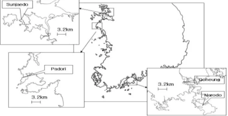
시료채취 와 deoxyribonucleic acid(DNA) 추출 2009년 6월부터 12월까지 매 월 남해안의 고흥, 나로도와 서해안의 선재도, 파도리의 네 지점(Fig.1) 에서 시료를 채취하였다. 단, 나로도에서는 8월과 10 월에, 파도리에서는 10월에 시료채집이 이루어지지 않았다. 매 달 채취된 바지락을 30개체씩 개체별로 nucleic acid 추출을 위해 아가미와 외투막 부분을 microtube(약 0.1g/microtube)에 취하였다. DNA는 High Pure PCR Template preparation Kit(Roche, Germany)를 사용하였으며 동사의 제시 방법에 따라 추출하였다. 추출한 DNA는 냉동보관(-20℃)하였다 가 분석에 사용하였다.
Fig. 1. Location of the 4 sampling site.
Polymerase chain reaction(PCR)분석
분리된 DNA를 대상으로 매달 개체별로 증폭산물 690bp로 확인되는 P. olseni NTS Forward (5’-ATG CTA TGG TTG GTT GCG GAC C-3’)와 P. olseni NTS Reverse (5’-GTA GCA AGC CGT AGA ACA GC-3’)(Robledo et al.2000) set와, 증폭산물 457bp로 확인되는 PolsITS-140F primer(5’-GAC CGC CTT AAC GGG CCG TGT T-3’)와 PolsITS-600R primer(5’-GGR CTT GCG AGC ATC CAA AG-3’) (Moss,2007) set의 2종류를 사용하여 PCR을 실시, 분 석하였다. PCR에는 Premix(Bioneer, Korea)를 사용하 였으며 전자의 경우는 94℃, 5분의 변성(denature)과 정을 거친 후, 94℃에서 1분, 60℃에서 1분, 72℃에서 1분을 30cycles 반복 하고, 72℃에서 7분의 최종 연장 단계(elongation)를 거친 후 4℃에서 냉각시켰다. 후 자의 경우는 95℃에서 4분 변성을 거친 후, 95℃에서 1분, 62℃에서 1분, 62℃에서 3분으로 40 cycles 을 실시하였으며, 65℃에서 10분간 최종 연장 단계를 거쳐 4℃에서 냉각을 실시하였다. PCR 산물의 확인 은 1% agarose gel(EtBr 함유)에 흘린 후 자외선(UV) 등으로 조사하여 에티듐브로마이드(EtBr)의 발광 형 태로 확인하였다.
염기서열 분석
PolsITS-140F와 PolsITS-600R의 primer set을 이용 하여 실시한 PCR을 통해 얻어진 네 지점의 457bp 크기의 bands를 각각 gel 상에서 잘라내어 Gel, PCR, Cleanup SV kit(GeneAll, Korea)를 사용하여 DNA
purification을 실시한 후 얻어진 DNA를 cloning kit(T-blunt PCR Cloning Kit, SolGent, Korea)를 이용하 여 대장균 클론개체를 얻었다. 얻어진 대장균을 LB 배지에 배양하여 Plasmid를 얻어내고(AccuPrep Plasmid Mini Extraction kit, Bioneer, Korea), 이 plasmid 를 전문업체((주)솔젠트)에 의뢰하여 염기서열 분석 데이터를 얻었다. 얻어진 데이터는 분석 소프트웨어 (Genetix)를 이용하여 염기서열 비교를 실시하였다.
결 과
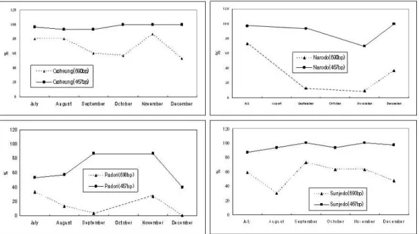
2009년도 7월부터 12월의 우리나라 서해와 남해 의 네 지점의 바지락 채굴지에서 채취한 바지락에 대한 P.olseni의 검출율은, 690bp 결과를 통한 결과가 고흥지역에서 가장 높았으나 457bp의 결과는 고흥과 선재도에서 유사하게 86.4∼100%의 검출율로 확인 되었으며, 나로도가 690bp의 결과에서 11월에 10%로 검출된 것이 457bp로 확인했을 때는 70%로 나타나 두 가지의 프라이머 sets 사용으로 검출율에 차이가 나타남을 확인하였다. 전체적인 경향으로는 파도리 에서 검출율이 가장 낮았다. 특히 457bp의 결과에서 는 파도리의 7, 8, 12월을 제외한 네 지점의 검사기간 전체 결과에서 60%를 넘는 검출율을 나타내어 지속 적인 감염상태를 확인할 수 있었다(Table 1, Fig.2).
네 지점에서 의 PolITS-140F와 PolITS-600R을 사용한 PCR 증폭산물의 염기서열을 비교한 결과, 선재도의 결과에서 두 군데의 염기차이를 나타내는 것 이외에 는 모두 같은 것으로 확인되었다(Fig.3).
Narodo Goheung Sunjedo Padori 690bp 457bp 690bp 457bp 690bp 457bp 690bp 457bp July 73(22/30) 96.7(29.30) 80(24/30) 96.7(29/30) 59(13/22) 86.4(19/22) 33(10/30) 53.3(16/30) August - - 80(24/30) 93.3(28/30) 30(9/30) 93.3(28/30) 13(4/30) 56.7(17/30) September 13(4/30) 93.3(28/30) 60(18/30) 93.3(28/30) 73(22/30) 100(30/30) 3(1/30) 86.7(26/30)
October - - 57(17/30) 100(30/30) 63(19/30) 93.3(28/30) - -
November 10(3/30) 70(21/30) 87(26/30) 100(30/30) 63(19/30) 100(30/30) 27(8/30) 86.7(26/30) December 37(11/30) 100(30/30) 53(16/30) 100(30/30) 47(14/30) 96.7(29/30) 0(0/30) 40(12/30) data : % (positive reaction sample number/total sample number)
- : No samples.
Bolic type : Peak value
Table 1. Results of prevalence from July to December 2009 at the 4 sampling sites.
Fig. 2. Monthly detection rates of P.olseni using PCR method with different 2 primer sets from July to December 2009.
Fig. 3. Results of DNA seqences about 457bp of P. olseni ITS region using PolsITS-140F/PolsITS-600R primer set at 4 stations.
Fig. 4. A comparioson of gel loading images between 690bp(A) and 457bp(B) PCR products of Padori samples in July. (M; marker, number(1∼30); sample numbers, +; positive control, -; no sample negative control)
고 찰
일반적으로 P. olseni의 발병은 15℃이상으로 수온 이 올라가는 봄부터 발생하여 수온이 10℃이하로 내 려가는 겨울은 발병률이 감소하며(Villalba et al., 2005), 염도와의 관련성도 아직 명확하지 않으나 실 험적으로 25psu(practical salitnity units)까지가 상한으 로 나타나며 15psu 아래에서도 저항성을 나타내는 것으로 보고하고 있다(La Peyre M et al., 2006).
Burreson & Calvo(1996)도 여름철 높은 수온과 염도 가 Perkinsus의 증식, 숙주의 방어능과 생리활성에 영향을 주어 높은 유병율과 감염도를 가져오는 것으 로 보고하였다. 본 조사에서는 퍼킨수스증의 발병정 도와 감염도와는 상관없이 PCR 검사를 통한 검출율 을 조사하였으며, 그 결과는 7월부터 소폭 증가하기 시작하여 12월까지 계속적으로 높게 유지되는 것으 로 확인되었다. 이러한 결과는 Park et al.(2006)이나 Uddin et al.(2010)에서 보고하고 있는 여름보다 가을 에 감염도가 높이 나타난다는 내용과 일치하며, 여기 에는 여름 장마 후 염분농도의 하강과 산란 후 스트레 스의 영향이 있었을 것으로 설명하고 있다. 또한, 서 해의 두 지점과 남해의 두 지점을 각각 비교했을 때, 파도리 보다는 선재도가, 나로도보다는 고흥에서 검 출율이 높았으며, 네 지점 중에서는 파도리의 검출율 이 가장 낮았다(Table 1, Fig. 2). 이러한 결과는 내만 안쪽보다는 외해를 접하는 쪽이 낮은 검출율을 나타 내어, 내만과 외만의 환경해수의 차이가 감염율에도 영향을 줄 것으로 추정된다. 퍼킨수스증은 병적 증상 을 나타내지 않고 숙주 내에 장기간 잠복되어 있다가 환경에 따라 발병하는 경우가 많은 것으로 설명하고 있다(OIE, 2009). 본 결과에서도 나타나는 것처럼 항 시 내재하는 Perkinsus가 환경조건에 따라 발병상황 이 달라질 수 있으므로 계속적으로 환경조건과 검출 율의 모니터링이 진행되어야 할 것이다.
조사에 사용된 690bp의 산물을 생산하는 primer
set의 경우 P. olseni의 NTS(nontranscribed spacer) region을 대상으로 디자인 된 것으로, NTS region은 ITS(internal transcribed spacer) region 과 비교하여 Perkinsus sp. 내에서의 종특이성을 나타내는 경향이 높은 것으로 보고하고 있다(Marsh et al., 1995, Robledo et al., 1998; Park et al., 2005). 그러나 새로이 국제수역사무국(OIE)에서는 NTS region 내의 다양 성에 관한 정보가 부족하여 결과의 정확성이 떨어질 우려가 있어 개정한 P. olseni에 대한 종특이적인 Primer set은 NTS region 보다 유전적 정보가 잘 알려 진 ITS region을 대상으로 만들어졌고 감도가 높은 것으로 설명하고 있다(OIE, 2009). 본 연구 결과에서 도 높은 감도를 확인 할 수 있었으며 EtBr로 감작시킨 gel 상의 사진도 690bp의 분석결과보다 457bp의 분석 결과에서 밴드가 명확하게 나타났다(Fig.4). 두 결과 의 검출감도에서는 명확한 차이를 나타내었지만 검출 경향은 네 지점에서 유사한 것을 알 수 있었다(Fig. 2).
실제 Perkinsus 모니터링에서 중요한 것은 검출여 부보다 실제 감염정도, 즉 Perkinsus cell/개체, 또는 Perkinsus cells/g tissue 등을 분석하거나 그렇지 못할 경우 RFTM 결과를 Mackin의 Scale 에 따라 semi- quantitative 하게 정량을 하기도 한다. 그러나 매달 모니터링을 실시하면서 개체별로 감염Cell 수를 세 거나, 배양과정을 거치는 것은 시간적인 소모가 많아 현실적으로 활용하기는 어려운 실정이다. 본 모니터 링에서는 정량적인 분석부분을 도입하지는 못하였지 만 감염여부의 확인은 바지락 조직에서 직접 추출한 DNA를 대상으로 PolsITS-140F와 PolsITS-600R로 실 시한 PCR 결과로 확인 가능하였다. 그러나 기생체 감염의 모니터링에서 정량적인 부분을 무시할 수 없 으므로, 이후 분석방법에서 realtime-PCR 법과 같은 방법을 병행할 수 있는 primer set을 제작하여 분석한 다면 신속한 정량분석의 결과를 얻는데 도움이 될
것으로 사료된다.
PolsITS-140F와 PolsITS-600R로 실시한 PCR 결과 로 생성된 457bp의 네 지점에서의 염기서열을 비교 한 결과 선재도에서 두 군데의 염기차이가 나타난 것 이외에는 동일하게 나타나 이 부분의 유전적 차이 는 확인되지 않았다. Choi(2005)에 따르면 형태학적 으로나 유전적으로 우리나라 바지락에서 나타나는 Perkinsus 종은 Norén et al.(1999)의 분류에 따른 P.
olseni로 설명하고 있다. 국내에서 검출되는 P. olseni 사이의 유전적 다양성에 대한 설명은 아직 미비하다. PCR의 감도가 높은 부위와 유전적 변형이 많은 부위 는 다를 수 있으므로 유전적 차이에 대한 분석은 이 후 따로 행해져야 할 것으로 사료된다.
요 약
본 내용은 2009년 하반기, 우리나라 남해안(고흥, 나로도)과 서해안(선재도, 파도리)의 네 지점에서 시 료채취 한 바지락에서의 Perkinsus olseni 감염에 대한 모니터링 결과로, 이전에 국제수역사무국(OIE)의 manual에서 지정하던 P. olseni에 대하여 특이적인 primer set으로 사용한 PCR 분석 결과와 2009년에 새로이 지정하는 P. olseni에 대하여 특이적인 primer set을 사용한 분석 결과를 비교하고, 결과를 통한 P.
olseni의 검출율을 조사하였다. 특히 2009년에 새로 이 지정된 PolsITS-140F와 PolsITS-600R의 primer set 을 사용한 결과는 신속한 검출여부 분석에 적합한 것으로 생각되었다. P. olseni의 검출경향은 파도리에 서 가장 낮았으며, 고흥에서 가장 높았다. 파도리의 7월, 8월 그리고 12월의 결과를 제외하고는 전 지점에 서 7월에서 12월까지 지속적으로 높은 검출율을 나 타내었다. PolsITS-140F와 PolsITS-600R의 primer set 을 이용한 PCR 산물의 염기서열을 분석한 결과, 선재
도의 결과에서 두 군데의 염기차이를 나타내는 것 이외에는 모두 같은 것으로 확인되었다.
감사의 글
본 연구는 국립수산과학원 연구지원 RP-2010-AQ-052 에 의하여 운영되었습니다.
참고문헌
Azevedo, C.: Fine structure of Perkinsus atlanticus n. sp.
(apicomplexa, Perkinsea) parasite of the clam Ruditapes decussatus from Portugal. J. Parasitol., 75:627-635, 1989.
Blackbourn, J., Bower, S.M. and Meyer, G.R.: Perkinsus qugwadi sp. nov.(incertace sedis), a pathogenic protozoan parasite of Japanese scallops, Patinopecten yessoensis, cultured in British Columbia, Canada. Can. J. Zool., 7:942-953, 1998.
Burreson, E.M. and Calvo, L.M.R.: Epizootiology of Perkinsus marunus disease of oysters in Chesapeake Bay, with emphasis on data since 1985.
J. Shellfish Res., 15:17-34, 1996.
Casas, S.M., Grau, A., Kimberly, S.R., Apalupakul, K., Azevedo, C. and Villalba A.: Perkinsus mediterraneus n. sp., a protistan parasite of the European flat oyster Ostrea edulis from the Balearic Islands, Mediterranean Sea, Dis. Aquat. Org., 58:231-234, 2004.
Choi, K.S. and Park, K.I.: Review on the Protozoan Parasite Perkinsus olsei(Lester and Davis 1981) Infection in Asian Water. Coastal Environmental and
Ecosystem Issues of the East China Sea, Eds., A. Ishimatsu and H.-J. Lie, pp.269-281. 2010.
Choi, K.S., Park K.I., Cho M. and Soudant P.: Diagnosis, Pathology, and Taxonomy of Perkinsus sp. Isolated from the Manila clam Ruditapes philippinarum in Korea. J. Aquaculture, 18:207-214, 2005.
Coss, C.A., Robledo, J.A.F., Ruita, G.M. and Vasta, G.R.:
Description of Perkinsus andrewsi n. sp. isolated from the Balthic clam(Macoma balthica) by characterization of the ribosomal RNA locus, and development of a species-specific PCR-based diagnostic assay. J. Eukaryot. Microbiol., 48:52-61, 2001.
Cremonte, F., Balseiro, P. and Figueras, A.: Occurrence of Perkinsus olseni(Protozoa:Apicomplexa) and other parasites in the venerid commercial clam Pitar rostrata from Uruguay, southwestern Atlantic coast. Dis. Aquat. Org., 64:85-90, 2005.
Dungan, C.F. and Reece K.S.: In vitro propagatio of two Perkinsus spp. Parasites from Japanese Manila clams Venerupis philippinarum and Description of Pekinsus honshuensis n. sp. J. Eukaryot.
Microbiol. 53: 316-326, 2006
Goggin, D.L. and Lester, R.J.G.: Perkinsus, a protistan parasite of abalone in Australia: a review. Aust. J. Mar, Freshwater Res., 46:639-646, 1995.
Hamaguchi, M., Suzuki, N., Usuk,i H. and Ishioka, H.:
Perkinsus protozoan infection in shor-necked clam tapes(=Ruditapesphillippinarum) in Japan. Fish Pathol., 33:73-480, 1998.
La Peyre, M., Casas, S. and La Peyre, J.: Salinity effects on viability, metabolic activity and proliferation of three Perkinsus species. Dis. Aquat. Org.,
71:59-74, 2006.
Lester, R.J.G. and Davis, G.H.G.: A new Perkinsus species(Apicomplexa, Perkinsea) from the abalone, Halitis ruber. J. Invert. Pathol. 37:181-187, 1981.
Mackin, J.G., Owen, H.M. and Collier, A.: Preliminary note on the occurrence of a new protistan parasite, Dermocystidium marinum n. sp. in Crassostrea nirginica(Gmelin). Science, 111:328-329, 1950.
Marsh, A.G., Gauthier, J.D. and Vasta, G.R.: A seniquantitative PCR assay for assessing Perkinsus marinus infections in the eastern oyster, Crassostrea virginica. J. Parasitol., 81:577-583, 1995.
McLayghlin, S.M., Tall, B.D., Shaheen, A., Elsayed, E.E.
and Faisal, M:,Zoosporulation of a new Perkinsus species isolated from the gills of the softshell clam Mya arenaria. Parasite, 7:115-122, 2000.
Moss, J.A.: Characterization of exotic pathogens associated with the suminoe oyster, Crassostrea ariakensis.
Ph.D. Sissertaion. Virginia Institute of Marine Science. College of William and Mary, Gloucester Point, Virginia USA. p.230, 2007.
Moss, J.A., Xiao, J., Dungan, C.F. and Reece, K.S.:
Description of Perkinsus beihaiensis n. sp., a new Perkinsus sp. parasite in oysters of southern China.
J. Eukayot. Microbiol. 55:117-130, 2008.
Murell, A., Kleenan, S.N., Barker, S.C. and Lester, R.J.G.:
Synonymy of Perkinsus olseni Lester & Davis, 1981 and Perinsus atlanticus Azevedo, 1989 and an update on the phylogenetic position of the genus Perkinsus. Bull. Eur. Assoc. Fish Pathol., 22:258-265, 2002.
Norén, F., Moestrup Ø. and Rehnstam-Holm A.-S.:
Parivilucifera infectans Noren et Moestrup gen.
et sp. nov.(Perkinsozoa phylum nov.): a parasitic flagellate capable of killing toxic microalgae. Eur.
J. Protistol., 35, 233-254,1999.
OIE: Manual of Diagnostic Tests for Aquatic Animals, World organization for animal health, Paris, 2009.
Park, K.-I., Ngo, T.T.T., Choi, S.-D., Cho, M. and Choi, K.-S.: Occurrence of Perkinsus olseni in the Venus clam Protothaca jedoensis in Korean waters. J.
Inv. Pathol. 93:81-87, 2006.
Park, K.-I. and Choi, K.-S.: Spatial distribution of the protozoan parasite Pekinsus sp. found in Manila clams, Ruditapes phillippinarum, in Korea.
Aquaculture, 203:9-22, 2001.
Park, K.I., Park, J.K., Lee, J. and Choi, K.S.: Use of molecular markers for species identification of Korean Perkinsus sp. isolated from Manilaclam Ruditapes philippinarum. Dis. Aquat. Org., 66:255-263, 2005.
Robledo J.A.F., Coss C.A. and Vasta G.R.: Characterization
of the ribosomal RNA locus of Perkinsus atlanticusand development of a polymerase chain reaction-based diagnostic assay. J. Parastol., 86:972-978, 2000.
Uddin, M.J., Yang, H.S., Choi, K.S., Kim, H.J., Hong, J.S.
and Cho, M.: Seasonal Changed in Perkinsus olseni Infection and Gametogenesis in Manil Clam, Ruditapes philippinarum, From Seonjaedo Island in Incheon, off the West Coast of Korea. J. World Aquaculture society, 41:93-101, 2010.
Villalba, A., Casas, S.M., Lopez, C. and Carballal, M.J.:
Study of pekinsosis in the carpet shell clam Tapes decussatus in Galicia(NW Spain) Ⅱ. Temporal pattern of disease dynamics and association with clam mortality. Dis. Aquat. Organ., 65:257-267, 2005.
Manuscript Recevied : July 5, 2010 Revised : August 19, 2010 Accepted : August 20, 2010