The Detection and Diagnosis Methods of Infectious Viroids caused Plant Diseases
Se Hee Lee, Yang-Hoon Kim and Ji-Young Ahn*
Department of Microbiology, Chungbuk National University, 1 Chungdae-ro, Seowon-gu, Cheongju-si, Chungcheongbuk-do 28644, Korea Received May 11, 2016 /Revised May 27, 2016 /Accepted May 27, 2016
Viroids are about 250-400 base pair of short single strand RNA fragments have been associated with economically important plant diseases. Due to the lack of protein expression capacity associated with replication, it is very difficult to diagnosis viroid diseases in serological methods. For detecting viroid at plants, molecular-based techniques such as agarose gel electrophoresis, polyacrylamide gel electro- phoresis (PAGE), DNA-hybridization, blotting analysis and conventional RT-PCR are reliable.
Real-time RT-PCR methods that grafted on RT-PCR methods with improved confirmation methods have been also utilized. However, they are still labor-intensive, time-consuming, and require personnel with expertise. Loop-mediated Isothermal Amplification (LAMP) method is a nucleic acid amplifica- tion method under the isothermal condition. The LAMP methodology has been reported to be simple, rapid, sensitive and field applicable in detecting a variety of pathogens. The results of LAMP method can be colorized by adding a visible material such as SYBR green I, Evagreen, Calcein, Berberine and Hydroxy naphthol blue (HNB) with simple equipment or naked eyes. The combination of LAMP method and nucleic pathogens, viroids, can be used to realize simple diagnosis platform for the genet- ic point-of care testing system. The aim at this review is to summary viroid-caused diseases and the simple visible approach for diagnosing viroids using Loop-mediated Isothermal Amplification (LAMP) method.
Key words :
Diagnosis, Loop-mediated isothermal amplification (LAMP), plant disease, RNA infectious agents, viroid
*Corresponding author
*Tel : +82-43-261-2301, Fax : +82-43-264-9600
*E-mail : [email protected]
This is an Open-Access article distributed under the terms of the Creative Commons Attribution Non-Commercial License (http://creativecommons.org/licenses/by-nc/3.0) which permits unrestricted non-commercial use, distribution, and reproduction in any medium, provided the original work is properly cited.
Journal of Life Science 2016 Vol. 26. No. 5. 620~631 DOI : http://dx.doi.org/10.5352/JLS.2016.26.5.620
서 론
과수 바이로이드 감염 질병은 미국, 중국, 일본, 한국 등 전 세계적으로 발생하여 과실 및 화훼에 큰 피해를 끼치고 있다 [12, 51]. 바이로이드에 대한 특별한 매개체는 잘 알려져 있지 않고 대다수의 바이로이드가 접목이나 삼목과도 같은 영양 번식을 할 때 사용되는 도구에 의해 전염되는 것으로 알려져 있다. 또한 식물이 바이로이드에 감염되어도 감염 초기에는 별다른 증상을 보이지 않고 수확기에 접어들어 병징이 발현되 는 경우가 많기 때문에 조기 진단이 어려워 수확되는 과실의 상품 가치가 하락 되는 등, 농가에 치명적인 피해가 유발된다.
바이로이드 유발 질병을 억제하기 위하여 현재 수행되고 있는 최선의 방책은 바이로이드가 전염되지 않은 기주의 확보 및 병원체 저항성 주를 생성하여 농가에 보급하는 것으로 그로 인하여 조기 진단의 중요성이 강조되고 있다. 또한 증폭 및
감염 과정에서 매우 미세한 염기 변화로도 수많은 변종, 즉 변이체의 생산을 쉽게 초래할 수 있기에 특정한 예방 및 조기 방법의 개발 필요성이 대두되고 있다. 최근에 들어서는 감자, 자두, 사과, 호프, 국화 등의 경제적으로 부가가치가 높은 농작 물 및 화훼 상품에 대한 조기 진단을 위하여 식물 감염성 질병 을 진단하기 위한 여러 기술들이 개발되고 있으며, 바이로이 드를 검출하기 위한 방법 또한 개발 및 개선되어 활용되고 있다. 그러나 아직 바이로이드 질병의 심각성이 널리 인지되 지 않고 바이로이드 질병과 바이러스 질병의 구분이 확실하게 인식되어 있지 않는 등의 바이로이드 질병에 대한 연구가 미 비한 실정이다. 이러한 배경으로, 이 총설에서는 개괄적인 바 이로이드와 그와 관련된 질병 및 병징에 대하여 살펴본 후, 기존부터 최신까지 사용되고 있는 검출·진단 방법과 관련된 연구동향을 정리하고자 한다.
바이로이드의 개관
바이로이드는 1971년, 최초로 감자에서 발견되었으며[52], 바이러스와 유사한 질병을 발생시키기에 viroid라고 명명되었 다. 이러한 RNA 병원체는 외피 단백질이 없고 250~400 여개 의 염기로만 구성되어 현재까지 보고된 어떤 생물 병원체보다 도 크기가 작다. 전체 서열 중 G+C% 함량이 매우 높으며 80%
- Review -
Fig. 1. Model of five viroid domains (T1, P, C, V, T2) de- termined from sequence homologies among viroids [30].
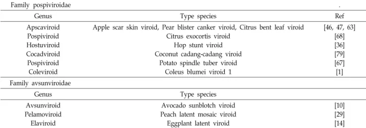
Table 1. Classification of viroids
Family pospiviroidae .
Genus Type species Ref
Apscaviroid Pospiviroid Hostuviroid Cocadviroid Pospiviroid Coleviroid
Apple scar skin viroid, Pear blister canker viroid, Citrus bent leaf viroid Citrus exocortis viroid
Hop stunt viroid Coconut cadang-cadang viroid
Potato spindle tuber viroid Coleus blumei viroid 1
[46, 47, 63]
[68]
[36]
[79]
[67]
[1]
Family avsunviroidae
Genus Type species
Avsunviroid Pelamoviroid Elaviroid
Avocado sunblotch viroid Peach latent mosaic viroid
Eggplant latent viroid
[10]
[29]
[14]
이상이 염기 결합으로 구성되어 막대 모양의 2차적인 구조를 형성하는데[52], 어떠한 단백질도 암호화하고 있지 않아 스스 로 복제할 수 없으며, 복제 기작이 일어날 때는 숙주의 효소 시스템을 이용한다[70]. 바이로이드는 오로지 식물체에만 감 염되어 RNA-mediated transcriptional gene silencing (TGS) 와 post-transcriptional gene silencing (PTGS) 등의 RNA si- lencing를 유도한다[60]. 바이로이드는 전체 유사성 (homology) 확인에서 90% 이하일 경우 신종 바이로이드로 분류하며 90% 이상일 경우에는 변종 혹은 변이체(varients)로 구분한다[9]. 바이로이드의 구조는 총 5가지 도메인(domain) 으로 구분되며 연구 초기에 Fig. 1에서 나타낸 것처럼, 왼쪽 말단인 Left hand terminal domain (T1), 병징 유발에 관여하 는 Pathogenic domain (P), 중앙의 보존 부위를 Central con- served domain (C), 염기 변화가 심한 Variable domain (V), 오른쪽 말단인 Right hand terminal domain (T2)로 나누었다 (Fig. 1) [30].
바이로이드가 주로 감염되는 숙주로는 사과, 배, 포도, 토마 토, 감자, 시트러스, 국화, 코코넛 팜, 아보카도, 홉 등이 있다 [58]. 주로 야기되는 바이로이드는 Pospiviroidae과의 Apple scar skin viroid (ASSVd) [46], Pear blister canker viroid (PBCVd) [24], Citrus bent leaf viroid (CVd-Ib) [63], Citrus
exocortis viroid (CEVd) [68], Hop stunt viroid (HSVd) [36], Coconut cadang-cadang viroid (CCCVd) [79], Potato spindle tuber viroid (PSDVd) [67], Coleus blumei viroid 1 (CbVd) [1], Avsunviroidae과의 Avocado sunblotch Viroid (ASVd) [10], Peach latent mosaic viroid (PLMVd) [29], Eggplant la- tent viroid (ELVd) [14] 등이 있다(Table 1).
Apple scar skin viroid (ASSVd)
사과에 주로 감염되는 이 병원체는 1935년, 중국 만주에서 최초로 발견되었다[46]. 1987년, 일본에서 처음으로 Apple scar skin viroid (ASSVd)라고 명명되었고[23], 국내에서는 1998년 경북 의성 지역에서 일본에서 도입한 미끼라이프라는 품종에서 처음 발견되었다. ASSVd에 감염된 사과는 크기가 정상보다 50~70% 정도로 줄어들거나 정상 과실의 표면에 붉 은색이 아닌 얼룩덜룩 비착색 부위가 남는다. 또한 이러한 부 위는 과실이 성숙될수록 더욱 분명하게 드러나며 크기가 1~2 cm까지 점차 확대되어 수확기에는 과피 전체의 50% 이상을 덮게 되고 그로 인해 사과의 상품성을 떨어트린다(Fig. 2) [31].
또한 ASSVd 감염은 접목에 의해 성목에서 1~2년 내에 나무 전체로 전염이 되며, 결실과에서 병징을 발견할 수 있다. 대부 분의 병징이 과실에서 발견되지만 예외로 특정한 환경에서 재배될 때, 잎이 말리거나 잎에 상위생장성 병징이 나타나는 경우가 있다[20, 28]. 또한 감염 가지에 즙액이 묻은 전성 가위 를 사용할 경우에는 60~70%의 전염율을 내며, 최근에는 사과 뿐만 아니라 살구와 복숭아, 체리에서도 검출되었다[44].
Pear blister canker viroid (PBCVd)
유럽 배와 마르멜로 생식질에 감염되어 각 배들에 blister와
splits와 껍질 궤양을 유발하는 Pear blister canker viroid
(PBCVd)는 처음 R. Flores 등에 의해서 명명되었다[17]. 이들
은 Pospiviroidae과, Apscaviroid속에 속하며 잎과 과실에는
A
B
a b c d
e f g h
Fig. 2. Newly known symptom of Apple scar skin viroid (ASSVd) on apples [31].
A B
Fig. 3. (A) Symptoms of PBC disease on the pear indicator A 20 graft inoculated with the P2098T isolate (two-year-old stock) [11] and (B) Symptoms induced by PBCVd (isolate P2098T) 3-4 months after in- oculation on the pear indicator Fieud 37, grown un- der greenhouse conditions. Similar symptoms are ob- served in the pear indicator Fieud 110 [J. C.
Desvignes, unpublished data].
병리학적인 변화가 보이지 않으나 오로지 배와 나무껍질에서 만 병징이 나타난다(Fig. 3). 얕은 껍질에 상처가 생기거나, 농 포는 기본으로 발생하며 산발적인 동고병과 비늘로 뒤덮인 듯한 껍질이 되는 경우도 있으며, 이에 감염되었을 시, 5-8년 후에는 식물 자체가 사멸한다[11]. 배(Pyrus communis)와 마르 멜로(Cydonia oblonga)가 주로 감염되는 자연 숙주로, 마르멜로 의 줄기는 유럽에서는 배 뿌리에 종종 접목되어 영양 번식을 통한 바이로이드 질병에 노출된 적이 있다. 병징을 나타내지 는 않지만 명자나무(Chaenomeles), 마가목(Sorbus)에도 감염되 며, 실험적으로 일부로 오이(Cucumis sativus)에 감염시킨 예도 존재한다[17].
Avocado sunblotch Viroid (ASBVd)
Avsunviroidae과에 속하는 이 병원체는 아보카도에 생리 적 장해, 유전적 장해를 끼치며 접목으로 전염될 수 있는 Avocado sunblotch라는 질병을 유발한다. 처음 Sunblotch라 는 질병이 인지된 것은 1928년으로[57]. 1931년, sunblotch라 는 질병으로써 명명되었다[26]. 또한 1979년, Palukaitis, P. 등 이 sunblotch 질병을 유발시키는 바이로이드를 Avocado sun- blotch Viroid (ASBVd)라는 명칭으로 확정하였다[48]. Avocado sunblotch Viroid는 매우 좁은 숙주 범위를 가지고 있으며, 주 로 아보카도(Persea americana Mill)에 감염된다[62]. 과실의 과 피에 노란색의 상처가 나며 괴저를 일으키거나 불그스름하게 변하는 등의 과실 피해를 입히는 것이 주된 병징이며, 그 이외 에는 어린잎과 가지, 줄기에 노란색의 줄무늬 등이 생겨나며 잎의 기형이 나타나거나 꼭대기 조직의 비틀림과 이른 노화 현상이 나타난다(Fig. 4) [19].
Citrus exocortis viroid (CEVd)
주로 citrus (Poncirus trifoliata)에 bark scaling을 유발시키
는 citrus exocrtis viroid (CEVd)는 1948년에 처음 묘사되었으
며, Pospiviroidae과 Pospiviroid속에 속한다[37]. CEVd에 감
염되면 식물의 뿌리 부분에 bark scaling이 나타나거나 뿌리
줄기의 껍질이 깨져 나가는 등의 뿌리 기형이 나타나며 과실
이 괴상한 모양으로 자라나는 등의 병징이 나타난다(Fig. 5)
[15, 53]. CEVd의 숙주 범위는 넓은 편으로, 국화 과에 속하는
A B
Fig. 4. (A) Fruit from infected trees are small and deformed with sunken lesions [David Rosen, University of California Statewide IPM Project] and (B) Foliage is discoloured and deformed [Andrew Geering (QAAFI].
A B
Fig. 5. Sanguinella sweet orange tree showing severe bark scaling on the Poncirus trifoliata rootstock due to the exocortis viroid [photo by Kotra Experiment Station, Mazandaran province, Iran].
A B
Fig. 6. (A) 'Perle' leaves infected with HpSVd showing speckling concentrated along leaf veins [photo by Ocamb lab] and (B) Generalized speckling on 'Sterling' leaf due to HpSVd [photo by C. M. Ocamb, 2007].
Gynura aurantiaca [77], 국화(Chrysanthemum morifolium) [13],
가지 과의 숙주인 토마토 (Lycopersicon esculentum) [73] 등에도 감염된다.
Hop stunt viroid (HSVd)
홉(Humulus lupulus)에 심각한 질병과 기형을 유발시키는 이 병원체는 Pospiviroidae과, Hostuviroid속에 속하며 일본 에서 홉 stunt 질병을 유발시키는 물질로써 처음 묘사되었다 [74]. 기본적으로 감염되는 홉의 경우, HSVd에 감염될 시, 식 물 자체의 키가 작거나 위쪽에 자라나는 잎이 아래로 숙여지 며, 잎이 기존 잎보다 작아지거나(Fig. 6A) 잎에 노란빛이 돌게 된다(Fig. 6B) [65]. 숙주 범위는 홉, 오이, 포도, 시트러스, 플럼, 복숭아, 배 등으로 상당히 넓은 편으로 각 숙주마다 병징이 특정하게 나타난다. 오이에서는 과실의 색이 옅어지는 병징이 발견되었고[42], 플럼과 복숭아에서는 과실의 외피가 얼룩지 는 병징이 나타났다[80]. 1999년에는 시트러스에서 악액질 상 태의 병징이 나타난다는 것이 발견되었다[61].
이와 같은 바이로이드는 식물체에 감염되어 과실 및 잎에 병징을 나타내어 농작물 및 회훼류의 상품적 가치를 떨어트리 는 등 농가에 큰 피해를 끼친다. 그러나 식물체가 어느 정도
자란 후에야 그 병징이 나타나기 때문에 조기 진단이 어렵다.
이러한 배경으로 바이로이드의 검출 기법은 최근까지도 개발 되어 조금 더 효율적으로 개선되고 있다.
바이로이드의 검출 기법
기존 식물 병원성 감염체의 감염을 확인하기 위하여 진단을 병징에 의한 진단 및 식물의 조직을 직접 해부하여 현미경을 이용하여 확인하는 등의 관찰을 통한 방법과 면역확산법 (Immunodiffusion Test), 형광항체법(Immunofluorescent Antibody Technique), 효소결합항체법(Enzyme-linked Imm- unodorbent Assay, ELISA) 등의 혈청학적인 진단 방법이 사 용되었다[41]. 그러나 외피 단백질이 없고 염기 서열로만 구성 되어 있는 RNA 바이로이드는 어떠한 단백질도 발현 및 합성 하지 않아 혈청학적 검출은 불가능하다. 때문에 바이로이드를 검출하기 위해 사용되는 방법은 현재까지도 분자생물학적인 방법에 기반을 두고 있다.
바이로이드 검출 초기에는 주로 RNA 병원체인 바이로이드
의 복제 중간체나 구조적 특징을 활용한 전기영동 방법(주로
PAGE) [27, 47]이나 바이로이드 서열에 특이적으로 결합하는
프로브를 활용한 혼성화 기반의 Northern blotting 및 Dot
blotting [18, 43, 56]등이 활용되었다. RNA에서 DNA로 전환
시키는 역전사(Reverse transcription) 방법이 개발된 이후,
RNA의 불안전성 등의 기존에 사용되던 검출 방법의 단점이
보완되었다[43]. 또한 최근 들어 PCR 증폭 방법이 개발되면서
[39, 59] 역전사 과정과 함께 접목되어 미량으로 존재하는 바이
로이드의 양을 증폭시켜 확인하는 방법(RT-PCR)이 고안되었
다[21, 40, 52, 64, 78]. 또한 PCR 증폭 결과의 확인을 좀 더
효율적으로 하기 위한 여러 방법들이 도입되었고, 그 중 하나
인 실시간 증폭 확인 방법이 결합된 실시간 역전사 PCR 증폭
방법은 현재까지도 꾸준히 활용되고 있다[6, 7, 25, 32, 54]. 그
러나 실시간 역전사 기반 PCR 증폭 방법의 경우, 값비싼 기기
와 전문 인력의 필요성으로 인해 현장 진단용으로는 활용하기
Fig. 7. SYBR Green I dye cycles between an unbound (dena- tured) and a bound (annealing through extension) state as the reaction progresses and signal intensity increases as the quantity of amplicons increase [photo by Sigma-Aldrich].
가 어렵기에 현장에서 바이로이드를 검출하기 위한 다른 방법 들이 고안되고 있다. 그 중 하나인 Loop-mediated isothermal amplification (LAMP) 방법은 등온에서 핵산의 증폭이 가능하 다는 장점이 있어 현장 진단용 검출 기술의 밑바탕이 되는 방법으로 각광받고 있다.
겔 전기 영동 및 올리고 프로브 기반의 바이로이드 검출 및 활용
바이로이드의 복제 중간체나 구조적인 특징을 활용한 겔 기반 전기영동(Gel electrophoresis) 방법이 오래 전부터 사용 되어 왔다[27, 47]. 그러나 이는 단순히 식물체에서 추출한 RNA를 폴리아크릴아마이드 겔 등의 겔을 이용하여 전기 영 동 후, 기존에 존재하는 바이로이드의 패턴과 비교를 통하여 결과를 분석하였다. 이러한 방법에는 부차적인 물질의 존재 가능성에 의해 겔 상에 나타난 밴드가 정확히 바이로이드를 나타내는 것인지 확신하기가 어려웠고, 시료 속 바이로이드의 양이 매우 미량일 경우 검출이 불가하다는 단점이 있었다. 전 자의 문제를 해결하기 위한 방안으로 도입된 것이 특이적 올 리고 프로브를 활용한 blotting 과정[18, 43, 56]이며, 바이로이 드에 특이적인 올리고 프로브를 고체 기판에 고정하여 그 위 로 식물체에서 추출한 RNA를 흘려주는 것으로, 바이로이드 서열과 상보적인 혼성화 과정에 기반하여 검출을 수행하였다.
하지만 이 또한 시료 속 바이로이드의 양이 매우 미량일 경우 검출이 불가하다는 한계가 여전히 존재한다.
역전사 기반 PCR 방법의 원리 및 활용
1983년, Kary, M. 등에 의해서 Polymerase chain reaction, 줄여서 PCR 증폭 방법[39]이 개발되면서 상기와 같은 문제의 해결점이 보이기 시작하였는데, PCR 증폭 방법을 통해 시료 속에 존재하는 바이로이드를 증폭할 수 있게 되었다. 바이로 이드는 RNA 가닥으로 구성되어 있기 때문에 PCR 증폭 과정 에는 RNA를 cDNA (complementary DNA)로 전환시키는 역 전사(reverse transcription) 과정이 포함되어야 하며, 전환된 cDNA는 특이적 primer를 활용하여 PCR 증폭을 위해 주형으 로 활용된다.
PCR 증폭에는 주형에 특이적으로 작용하는 primer 쌍과 높은 온도에서도 활성을 잃지 않는 Taq polymerase가 활용된 다. 원리는 Fig. 3에 표현된 것처럼 온도의 변화에 따른 것으로 90℃ 이상의 온도에서 주형이 변성되어 두 가닥 사이가 벌어 지고, 온도가 점차 낮아지면서 primer 쌍이 결합하여 다시 온 도가 상승하며 신장이 이루어지는 과정이 포함된다. 상기와 같은 과정으로 미량의 주형 가닥이 증폭되고 이를 겔 기반 전기 영동 방법 등의 방법으로 확인하는 것으로 바이로이드를 검출할 수 있다. 지난 1990년, Hadidi, A. 등은 이과 과실 나무 에 감염된 바이로이드를 역전사 기반 PCR로 증폭하여 겔 전 기 영동과 Southern blot 혼성화 방법으로 확인하였다[21]. 그
밖에도 Weidemann, H. L. 등은 potato spindle tuber viroid (PSTVd)를, Yang, X. 등은 Citrus Exocortis viroid (CEVd)와 Cachexia viroid를 역전사 기반 PCR 방법을 도입하여 검출하 였다[52, 78]. 그러나 역전사 기반의 PCR 방법에는 RNA 주형 이 필요하며 그를 위해서는 식물에서 RNA를 추출하여 정제 하는 과정이 필요하다. 그에 따른 시간과 인력 소비를 줄이기 위한 여러 방법이 고안되었으며, 그 중 하나는 membrane에 식물 수액을 집적하여 주형을 부착시켜 역전사 기반 PCR 증 폭을 수행하는 것으로[64], 1997년, Shamloul 등에 의하여 감 자 조직, 씨 등에서 추출한 핵산이 처리된 GeneReleaser
TM를 접목시켜 PSTVd가 검출되었다[40]. 또한, RNA 추출 방법 자 체를 개선하여 효용성을 높이는 방법도 고안되었는데, potas- sium ethyl xanthogenate (PEX) buffer를 활용한 조직 균질화 과정이 없는 핵산 추출 방법을 도입하는 방법 등이 있다[40, 55].
역전사 기반의 PCR 방법은 시료 내 존재하는 바이로이드 RNA를 cDNA로 합성하여 증폭함으로써 바이로이드 검출이 가능하다. 그러나 역전사 기반의 PCR 방법은 증폭 결과를 확 인하기 위한 부가적인 과정이 수행되어야 하며, 이를 개선하 기 위해 도입된 것이 실시간으로 증폭 양상을 확인할 수 있는 실시간 PCR 증폭 방법(real-time PCR)이다[3, 6, 7, 32, 54].
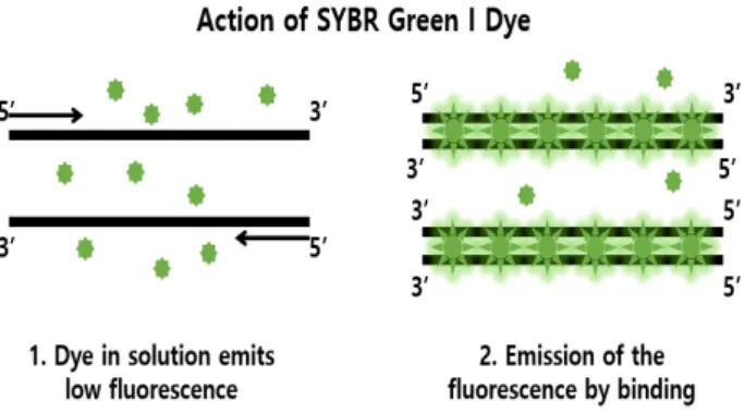
실시간 PCR 증폭 방법은 PCR 증폭이 수행되는 동안 실시 간으로 증폭 양상을 확인할 수 있으며, 그를 위하여 형광 표지 물질이 접목된 방법이다. 사용되는 표지 물질은 주형에 특이 적이거나 비 특이적인 물질 모두 가능하며 주로 사용되고 있 는 것은 이중 나선 DNA 사이에 끼어 형광 빛을 띠는 SYBR
®Green I [25], EvaGreen [76], SYBR
®Gold [72] 등의 비 특이적 형광 물질이나 형광 표지자가 부착되어 있는 주형 특이적 -probe, PNA probe [9]나 LNA
®[82] 등의 핵산 analogue [50]
등이다. SYBR
®Green I의 경우, 이중 가닥 DNA가 증폭되면서
그 사이에 끼어들어 형광 빛을 방출하며(Fig. 7) 어떠한 주형이
Fig. 8. TaqMan probe chemistry mechanism (photo by Brain- damaged).
라도 증폭함에 따라 사이로 끼어드는 형광 물질의 양이 증가 하는 것을 측정하여 실시간으로 증폭 결과를 확인할 수 있다 [25]. 주형에 특이적인 물질 중 주형-특이적 probe의 경우 주로 사용되는 방법은 TaqMan probe를 활용하는 방법으로, probe 에는 Fig. 8에 표현된 것처럼 한 쪽에는 형광 물질이, 한 쪽에는 Quencher이 위치하고 있다.
실시간 PCR 증폭 방법을 사용하면 증폭되는 RNA의 초기 양을 측정할 수 있기 때문에 주로 세포에 존재하는 mRNA의 발현 정도를 연구하기 위해 활용되었으나 2004년, Bloonham, N. 등에 의해 porato spindle tuber viroid (PSTVd)를 검출하 기 위해 도입된 이후로 바이로이드를 검출하기 위하여 활발하 게 활용되고 있다[3].
현재에도 실시간 PCR 증폭 방법은 바이로이드 검출을 위해 꾸준히 사용되고 있으나, PCR 증폭 과정을 수행하기 위한 온 도 변화 과정과 모니터링을 위한 값비싼 기기 및 복잡한 과정 에 따른 전문 인력이 요구되기 때문에 현장 진단 용으로 활용 하기에는 한계가 존재한다.
Loop-mediated isothermal amplification(LAMP)의 원리 및 활용
2000년, Notomi, T. 등에 의하여 온도의 변화 없이 단시간 내에 등온에서 핵산을 증폭시킬 수 있는 Loop-mediated iso- thermal amplification (LAMP) 방법이 개발되었다. 이 방법은 기존 PCR 방법에서 수행되었던 핵산 서열의 변성(denatura-
tion), 접합(annealing), 신장(extension)의 세 가지 단계를 수행 하지 않고도 단일 온도에서 핵산의 접합 및 신장이 가능하다 [45]. 또한 증폭 효율이 매우 높으며 특정한 기기가 요구되지 않아 현장에서의 활용도가 높으며, 현재 사람, 식물, 동물 모두 에서 병원성 세균, 바이러스, 곰팡이, 기생충만이 아니라 식물 에서만 병원성을 일으키는 바이로이드의 검출 등 광범위하게 활용되고 있다[5].
LAMP 방법에서 주로 활용되는 효소는 Bst polymerase로, 이는 Bacillus stearothermophilus의 DNA polymerase I을 인공 적으로 합성한 것으로 등온에서 핵산을 증폭시킨다. 또한 5‘→
3’ DNA polymerase 활성을 띠고 있으나 반대로 5’→3’나 3‘→
5’의 exconuclease의 활성은 띠고 있지 않아[22] 증폭 산물이 쭉 연결되어 생성되며 겔 전기영동 결과에서는 끌리는 band 양상이 나타나게 된다(Fig. 9).
LAMP을 수행하기 위해서는 2~3쌍의 primer가 필요하며, 각 primer들은 주형 가닥에 특이적으로 부착되어 증폭된다.
primer가 부착되는 부위는 총 6군데로, 주형 가닥의 B1, B2, B3 부위이며 상보적인 가닥의 F1, F2, F3이다. primer 쌍은 기본적으로 F3, B3 primer (inner primer) 쌍, FIP, BIP primer (outer primer) 쌍으로 구성되며 증폭 효율을 상승시키기 위해 Loop (LF, LB) primer 쌍을 추가하여 수행되기도 한다. 안쪽 (inner)에서 작용하는 primer인 FIP와 BIP primer는 각각 F1c, F2 부위와 B1c, B2 부위의 서열을 포함하고 있으며 후기의 증 폭 과정에서는 오로지 이 두 primer만이 사용된다. 각 primer 가 부착되는 위치와 증폭 원리는 Fig. 10에 나타나 있다[45].
Inner primer인 FIP primer가 주형 가닥의 F2c 부위에 결합 되어 상보적인 서열을 합성하며, outer primer인 F3 primer가 F3c 부위에 느리게 부착되어 FIP primer가 부착된 가닥이 방 출된 DNA 가닥의 합성이 시작된다. 여기서 방출된 FIP pri- mer 부착 DNA 가닥은 한 말단에서 환형의 구조를 형성할 수 있다. 이러한 단일 가닥의 DNA는 BIP primer에 의해 시작 되는 DNA 합성과 그 다음의 B3 primer가 부착된 가닥의 합성 을 위한 주형 가닥으로써 작용하며 dumb-bell 모양의 산물을 형성한다. 자가 부착된 DNA의 생성은 stem-loop 모양으로 빠르게 전환된다. 이러한 stem-loop 모양의 DNA는 LAMP cycle의 주요한 시작 물질로 활용된다.
이와 같이 LAMP 방법은 기존에 활용되던 PCR 증폭 방법 과는 다르게 온도 변화 없이 등온으로 증폭이 가능하다는 장 점으로 인해 현장 진단 형 검출 방법으로 각광받고 있다. 국내 에서 안 등에 의해서 수행된 salmonella spp.의 검출 관련 연구 에서는 일반적인 PCR 증폭과 실시간 PCR 증폭 방법과 LAMP 결과를 비교하였으며, 일반적인 PCR 증폭 방법보다 LAMP 방법의 검출 능력이 더 높으며, 실시간 PCR 증폭 방법과는 비슷한 양상을 보임을 검증하였다[2]. 그 밖에도 다양한 주형 에 대하여 LAMP 방법의 효율이 검증되었다[35, 69, 81].
RNA로만 구성되어 있는 바이로이드를 검출하기 위하여 기
Fig. 9. Agarose gel image of the included RNA LAMP Control Reaction.
The common “ladder” banding pattern within a HMW smear is customary of LAMP reactions. No dedicated RT step or use of addi- tional RT enzyme was used [photo by biocat].
Table 2. Summary of detection methods used in one-pot LAMP assays [16]
Hydroxynaphthol blue Calcein SYBR Green I EvaGreen Berberine
Substance Hydroxynaphthol blue (HNB)
Calcein AM +
MnCl2 SYBR Green I EvaGreen Berberine-SO4
Toxicity May cause eye
irritation
May be harmful to skin and eyes
Mutation
enhancer Possible carcinogen May be toxic in high concentrations Detection
mechanism Decrease of free Mg2+ Decrease of free Mn2
dsDNA intercalation
dsDNA intercalation
Small groove intercalation
Readout Absorption
Absorbance: 650 nm
Fluorescence Excitation: 495 nm
Emission: 515 nm
Fluorescence Excitation: 494 nm Emission: 521 nm
Fluorescence Excitation: 500 nm
Emission: 530 nm
Fluorescence Excitation: 450 nm
Emission: 530 nm Effect on
amplification None Manganese may
inhibit reaction
Not inhibiting when used 0.5–1×
Not inhibiting when used 0.5–1×
Not inhibiting (≤180 μM) One-pot real-time
assay - + + + + + + + + + + + + + + +
Equipment for
real-time detection - Fluorometer with
FAM filter
Fluorometer with FAM filter
Fluorometer with FAM filter
Fluorometer with FAM filter One-pot end point
assay + + + + + + + + - -
+ + + (UV light with low background signal) Equipment for
end point detection None UV lamp (optional) Not applicable Not applicable UV lamp
Field applicability (Depending on
visualization.)
+ + + + + + + + + + + + + + + +
존에는 식물체에서 RNA를 추출한 후 역전사 과정으로 cDNA 를 생성한 후 그를 활용해 LAMP 과정을 수행하였다[49]. 그러 나 최근 들어 RNA 주형을 그대로 도입하여 한 튜브 내에서 역전사와 LAMP 과정을 동시에 수행하는 방법이 고안되었고 [4], 이를 활용하여 Peach Latent Mosaic Viroid (PLMVd), Potato Spindle Tuber Viroid (PSTVd) 등이 검출되었다[34, 71].
LAMP 수행의 결과물을 확인하기 위해서 그 전 까지만 해 도 겔 전기 영동 방법이 사용되었으나 이는 현장 진단에서는 활용될 수 없기 때문에 최근에는 육안 관찰을 용이하게 하는 물질을 포함하여 부가적 과정을 생략하는 방법이 고안되었다.
그 종류는 필터 기반의 형광 관찰 기기(Fluormeter, Gel. Doc.
등)을 활용하여 관찰할 수 있는 Caseine, SYBR
®Green I, ber-
Fig. 10. Schematic representation of the mechanism of LAMP [45].
berine 등과 특정한 장비 없이 육안으로 색 변화를 관찰할 수 있는 hydroxynaphthol blue (HNB) 등이다. 각 물질들의 작용 원리는 Fig. 12와 같으며, Jens Fischbach 등은 각 검출 방법에 관한 장단점을 비교하였다(Table 2) [16].
Table 2에서도 나타나 있는 것처럼, 결과 확인을 위해 사용 될 수 있는 여러 물질들 중에서도 Calcein과 Hydroxynaph- thol blue (HNB)이 현장 진단용으로써 가장 높은 활용도를 보였다. 그러나 그 중 Calcein의 경우, 사용자의 피부와 눈에 독성을 나타내며 현장에서 활용하기 위해 따로 형광을 측정하 기 위한 기기가 필요하다. 그러므로 관찰을 위한 부가적인 장 비 없이 육안으로 결과를 확인할 수 있으며, 독성이 가장 약한 HNB가 현장 진단용으로 사용되기에 가장 적합하다고 사료된 다.
상기 서술된 이점으로 인하여 현장에서의 유효성이 크게 대두되고 있는 LAMP 방법이지만, 이 역시 한계점이 존재한 다. 기존 PCR처럼 핵산 기반으로 한 분자생물학적인 방법이 기 때문에 주형 가닥에 특이적으로 부착되는 primer의 역할이 상당히 중요하다. 그러나 LAMP의 경우, 특정한 조건 하에서 primer끼리의 자가 증폭 과정이 수행되어 비 특이적인 반응이
일어나게 된다[33, 66, 75]. 그러나 아직 그에 대한 정확한 연구 보고는 없으며, 바이로이드 자체가 GC%가 높기 때문이거나 primer들의 농도가 높아 primer dimer가 형성되면서 비 특이 적인 증폭이 유도되거나[33] primer들이 증폭의 민감도를 향 상 시키기 때문일 것이라고 추측되고 있다[66]. 이러한 자가 증폭 과정을 감소시키기 위해 Kazuya S. 등은 LAMP 반응 시간을 반으로 줄이는 방법을 도입했고[66], Lee, D. 등은 각 primer 쌍들을 실온에서 30분 동안 보관한 후 바로 LAMP 반 응을 수행 하였다[33]. 비 특이적인 증폭을 유도할 수 있는 MgSO
4나 MgCl
2의 농도를 낮추거나 반응 온도를 높여 비 특이 적인 결합을 줄일 수 있다[38].
고 찰
외피 단백질이 없고 오로지 RNA로만 구성된 바이로이드는
증폭과 감염과정에서 간단한 염기의 변화로도 수많은 변종,
즉 변이체의 생산이 쉽게 초래된다. 또한 바이로이드에 대한
특정한 매개체가 잘 알려져 있지 않아 매개체 방제를 통한
통제 수단은 존재하지 않고, 다른 방제 약제 또한 존재하지
A
B C
Fig. 11. Overview of indirect and direct detection methods used for monitoring loop-mediated isothermal amplification (LAMP) [16].
않는다. 바이로이드에 감염되면 초기에는 별다른 병징을 보이 지 않다가 수확기에 접어들어 발병하는 경우가 많아 초기에 발견하기가 어려운 편이다. 또한 토종 농작물 및 부가가치가 높은 화훼 작물에 질병을 유발해 상품적 가치를 떨어트려 심 각한 경제적 손실을 일으키기 때문에 바이로이드 질병에 감염 되지 않은 기주의 확보가 중요하며 그로 인해 조기 진단의 필요성이 크게 대두되고 있다. 이로 인하여 현재 식물체에서 바이로이드를 검출하기 위한 여러 방법이 고안되었으며 최근 에 주로 사용되는 방법은 역전사 PCR 증폭 방법과 실시간 검출 기법이 접목된 방법과 등온 증폭(LAMP) 방법이다. 전자 의 경우 민감도가 높고 반응 시간이 짧기 때문에 바이로이드 검출에 꾸준히 활용되고 있으나 현장 진단에서 활용되기에는 값비싼 기기와 전문 인력이 필요하다는 한계점이 존재한다.
그로 인하여 현재는 온도 변화를 위한 기기가 필요 없고 복잡 하지 않은 방법으로 실시간 검출 방법과 비슷하거나 높은 민 감도를 보이는 LAMP 방법이 현장 진단에서 활용될 수 있는 방법으로 크게 각광받고 있다. LAMP 방법은 아직 현장 진단 용으로 활용되기에는 여러 가지 해결할 점이 존재한다. 그러 나 상기 서술된 한계점이 극복된다면 LAMP 방법은 바이로이 드 진단 방법에서 가장 큰 효용성을 가질 것이며 이와 같은 현장 진단용 방법의 개발로 인하여 단기적으로 농가 경제적 손실을 최소화할 뿐 아니라, 장기적으로 선진형 무병묘목 선 발, 국가 주요 종자 관리 및 외래 병종 유입에 대한 관리에서도 주요한 작용을 할 것이라고 사료된다.
감사의 글
본 논문은 농촌진흥청 연구사업(세부과제번호: PJ011642) 의 지원에 의해 이루어진 결과이며, 이에 감사드립니다.
References
1. Adkar-Purushothama, C. R., Nagaraja, H., Sreenivasa, M.
Y. and Sano, T. 2013. First Report of Coleus blumei viroid Infecting Coleus in India. Plant Dis. 97, 149.
2. Ahn, Y. C., Cho, M. H., Yoon, I. K., Jung, D. H., Lee, E.
Y., Kim, J. H. and Jang, W. C. 2010. Detection of Salmonella Using the Loop Mediated Isothermal Amplification and Real-time PCR. J. Kor. Chem. Soc. 54, 215-221.
3. Boonham, N., Perez, L. G., Mendez, M. S., Peralta, E. L., Blockley, A., Walsh, K., Barker, I. and Mumford, R. A. 2004.
Development of a real-time RT-PCR assay for the detection of potato spindle tuber viroid. J. Virol. Methods 116, 139-146.
4. Boubourakas, I. N., Fukuta, S. and Kyriakopoulou, P. E.
2009. Sensitive and rapid detection of peach latent mosaic viroid by the reverse transcription loop-mediated isothermal amplification. J. Virol. Methods 160, 63-68.
5. Budziszewska, M., Wieczorek, P. and Obrepalska-Steplowska, A. 2016. One-step reverse transcription loop-mediated iso- thermal amplification (RT-LAMP) for detection of tomato torrado virus. Arch. Virol. 161, 1359-1364.
6. Bustin, S. A., Benes, V., Nolan, T. and Pfaffl, M. W. 2005.
Quantitative real-time RT-PCR--a perspective. J. Mol. Endoc- rinol. 34, 597-601.
7. Bustin, S. A. and Mueller, R. 2005. Real-time reverse tran- scription PCR (qRT-PCR) and its potential use in clinical diagnosis. Clin. Sci. (Lond). 109, 365-379.
8. Choi, J. J., Cho, M., Oh, M., Kim, H., Kil, M. S. and Park, H. 2010. PNA-mediated Real-Time PCR Clamping for Detection of EGFR Mutations. Bull. Kor. Chem. Soc. 31, 3525-3529.
9. Collmer, C. W., Hadidi, A. and Kaper, J. M. 1985. Nucleotide sequence of the satellite of peanut stunt virus reveals struc- tural homologies with viroids and certain nuclear and mi-
tochondrial introns. Proc. Natl. Acad. Sci. USA 82, 3110-3114.
10. De La Torre A, R., Téliz Ortiz, D., Pallás, V. and Sánchez Navarro, J. A. 2009. First report of avocado sunblotch viroid in avocado from Michoacán, México. Plant Dis. 93, 202.
11. Desvignes, J. C. 1999. Pear blister canker viroid: host range and improved bioassay with two new pear indicators, fieud 37 and fieud 110. Plant Dis. 83, 419-422.
12. Diener, T. O. 1989. Circular RNAs: relics of precellular evo- lution? Proc. Natl. Acad. Sci. USA 86, 9370-9374.
13. Elleuch, A., Marrakchi, M., Fakhfakh, H., Levesque, D., Bessais, N. and Perreault, J. P. 2003. Molecular Variability of Citrus Exocortis Viroid in a Single Naturally Infected Citrus Tree. Plant Protect. Sci. 39, 139-145.
14. Fadda, Z., Daros, J. A., Fagoaga, C., Flores, R. and Duran- Vila, N. 2003. Eggplant latent viroid, the candidate type spe- cies for a new genus within the family Avsunviroidae (hammerhead viroids). J. Virol. 77, 6528-6532.
15. Fawcett, H. S. and Klotz, L. J. Exocortis on trifoliate orange.
1948. http://ucce.ucdavis.edu/files/repositoryfiles/ca210p13- 71287.pdf.
16. Fischbach, J., Xander, N. C., Frohme, M. and Glökler, J. F.
2015. Shining a light on LAMP assays―A comparison of LAMP visualization methods including the novel use of berberine. BioTechniques 58, 189.
17. Flores, R., Hernandez, C., Llacer, G. and Desvignes, J. C.
1991. Identification of a new viroid as the putative causal agent of pear blister canker disease. J. Gen. Virol. 72, 1199-1204.
18. Fonseca, M. E., Marcellino, L. H. and Gander, E. 1996. A rapid and sensitive dot-blot hybridization assay for the de- tection of citrus exocortis viroid in Citrus medica with di- goxigenin-labelled RNA probes. J. Virol. Methods 57, 203-207.
19. Graca, J. V. da. and van Lelyveld, L. J. Peroxidase and in- dole3- acetic acid oxidase activities and isoenzymes in the mature bark of sunblotch-infected avocado (Persea ameri- cana). J. Phytopathol. 92, 143-149.
20. Hadidi, A., Flores, R., Randles, J. W. and Semancik, J. S.
2003. Viroids, Properties, Detection, Diseases and their Control. pp. 37-141: CSIRO Publishing, California. USA.
21. Hadidi, A. and Yang, X. 1990. Detection of pome fruit vi- roids by enzymatic cDNA amplification. J. Virol. Methods 30, 261-269.
22. Hafner, G. J., Yang, I. C., Wolter, L. C., Stafford, M. R. and Giffard, P. M. 2001. Isothermal amplification and multi- merization of DNA by Bst DNA polymerase. Biotechniques 30, 852-856, 858, 860.
23. Hashimoto, J. and Koganezawa, H. 1987. Nucleotide se- quence and secondary structure of apple scar skin viroid.
Nucleic Acids Res. 15, 7045-7052.
24. Hernández, C., Elena, S. F., Moya, A. and Flores, R. 1992.
Pear blister canker viroid is a member of the apple scar skin subgroup (apscaviroids) and also has sequence homology with viroids from other subgroups. J. Gen. Virol. 73, 2503.
25. Higuchi, R., Dollinger, G., Walsh, P. S. and Griffith, R. 1992.
Simultaneous amplification and detection of specific DNA sequences. Biotechnology (NY) 10, 413-417.
26. Horne, W. T. and Parker, E. R. 1930. The Avocado disease called sun-blotch. Phytopathology 20, 852.
27. Horst, R. K. and Kawamoto, S. O. 1980. Use of poly- acrylamide Gel electrophoresis for Chrysanthemum Stunt Viroid in Infected Tissues. Plant Dis. 64, 186-188.
28. Ito, T. and Yoshida, K. 1998. Reproduction of apple fruit crinkle disease symptoms by apple fruit crinkle viroid. Acta Horticulturae 472, 587-594.
29. Jo, Y., Yoo, S. H., Chu, H., Cho, J. K., Choi, H., Yoon, J.
Y., Choi, S. K. and Cho, W. K. 2015. Complete genome se- quences of peach latent mosaic viroid from a single peach cultivar. Genome Announc. 3, e01098-15.
30. Keese, P. and Symons, R. H. 1985. Domains in viroids: evi- dence of intermolecular RNA rearrangements and their con- tribution to viroid evolution. Proc. Natl. Acad. Sci. USA 82, 4582-4586.
31. Kim, D. H. Kim, H. R. Heo, S. Kim, S. H. Kim, M. Shin, I. S. Kim, J. H. Cho, K. H. and Hwang, J. H. 2010. Occurrence of Apple Scar Skin viroid and Relative Quantity Analysis Using Real-time RT-PCR. Res. Plant Dis. 16, 247-253.
32. Kim, W. S., Haj Ahmada, Y., Stobbsb, L. W. and Greigb, N. 2015. Evaluation of viroid extraction methods and appli- cation of a one-step reverse transcription real-time polymer- ase chain reaction assay (RT-qPCR) for the rapid detection of Chrysanthemum stunt viroid (CSVd) infection. Can. J.
Plant Pathol. 37, 221-229.
33. Lee, D., Kim, E. J., Kilgore, P. E., Kim, S. A., Takahashi, H., Ohnishi, M., Anh, D. D., Dong, B. Q., Kim, J. S., Tomono, J., Miyamoto, S., Notomi, T., Kim, D. W. and Seki, M. 2015.
Clinical evaluation of a loop-mediated isothermal amplifica- tion (LAMP) assay for rapid detection of Neisseria meningi- tidis in cerebrospinal fluid. PLoS One 10, e0122922.
34. Lenarčič, R., Morisset, D., Mehle, N. and Ravnikar, M. 2013.
Fast real-time detection of Potato spindle tuber viroid by RT-LAMP. Plant Pathol. 62, 1147-1156.
35. Lin, Z., Zhang, Y., Zhang, H., Zhou, Y., Cao, J. and Zhou, J. 2012. Comparison of loop-mediated isothermal amplifica- tion (LAMP) and real-time PCR method targeting a 529-bp repeat element for diagnosis of toxoplasmosis. Vet. Parasitol.
185, 296-300.
36. Marcelo, E., Maria Luisa, P. N.,Targon, T. V. M., Fajardo, R. F. and Elliot W. K. 2006. Citrus exocortis viroid and Hop Stunt viroid Doubly Infecting Grapevines in Brazil. Fitopatol.
bras. 31, 440-446.
37. Mishra, M. D., Hammond, R. W., Owens, R. A., Smith, D.
R. and Diener, T. O. 1991. Indian bunchy top disease of to- mato plants is caused by a distinct strain of citrus exocortis viroid. J. Gen. Virol. 72, 1781-1785.
38. Mousumi, D., Godavarthi, B. K. S. P. and Prakash, S. B. 2010.
Molecular Diagnostics: Promises and Possibilities, pp. 132:
Springer Science+Business Media LLC. Springer, New York, USA.
39. Mullis, K. B. 1990. The unusual origin of the polymerase chain reaction. Sci. Am. 262, 56-61, 64-55.
40. Nakahara, K., Hataya, T. and Uyeda, I. 1999. A simple, rapid method of nucleic acid extraction without tissue homoge-
nization for detecting viroids by hybridizaiton and RT-PCR.
J. Virol. Methods 77, 47-58.
41. Narayanasamy, P. 2001. Plant Pathogen Detection and Disease Diagnosis. pp. 197-257, 2nd ed. : CRC Press, Florida, USA.
42. Nathalie, A., Jose, F. M., Guy, M., Thierry, C. and Vicente, P. 1996. Studies on the diagnosis of hop stunt viroid in fruit trees: identification of new hosts and application of a nucleic acid extraction procedure based on non-organic solvents.
Eur. J. Plant Pathol. 102, 837-846.
43. Nikon, V., Oxana, K., Maria, P. and Christina, V. 2012.
Comparison of direct-RT-PCR and dot-blot hybridization for the detection of Potato spindle tuber viroid in natural host plant species. Eur. J. Plant Pathol. 134, 859-864.
44. Nome, C., Giagetto, A., Rossini, M., Di Feo, L. and Nieto, A. 2012. First report and molecular analysis of Apple scar skin viroid in sweet cherry. New Dis. Rep. 25, 3.
45. Notomi, T., Okayama, H., Masubuchi, H., Yonekawa, T., Watanabe, K., Amino, N. and Hase, T. 2000. Loop-mediated isothermal amplification of DNA. Nucleic Acids Res. 28, E63.
46. Ohtsuka, Y. 1987. On Manshu-sabika-byo of apple, graft transmission and symptom variation in cultivars. J. Jpn. Soc.
Hortic. Sci. 9, 282-286.
47. Owens, R. A. 2008. Identification of viroids by gel electrophoresis. Curr. Protoc. Microbiol. 16, 1.1-1.9.
48. Palukaitis, P., Hatta, T., Alexander, D. M. and Symons, R.
H. 1979. Characterization of a viroid associated with avoca- do sunblotch disease. Virology 99, 145-151.
49. Park, J., Jung, Y., Kil, E. J., Kim, J., Thi Tran, D., Choi, S.
K., Yoon, J. Y., Cho, W. K. and Lee, S. 2013. Loop-mediated isothermal amplification for the rapid detection of Chrysan- themum chlorotic mottle viroid (CChMVd). J. Virol. Methods 193, 232-237.
50. Petersson, B., Nielsen, B. B., Rasmussen, H., Larsen, I. K., Gajhede, M., Nielsen, P. E. and Kastrup, J. S. 2005. Crystal structure of a partly selfcomplementary peptide nucleic acid (PNA) oligomer showing a duplex–triplex network. J. Am.
Chem. Soc. 127, 1424-1430.
51. Pringle, C. R. 1998. The universal system of virus taxonomy of the International Committee on Virus Taxonomy (ICTV), including new proposals ratified since publication of the Sixth ICTV Report in 1995. Arch. Virol. 143, 203-210.
52. Puchta, H. and Sanger, H. L. 1988. An improved procedure for the rapid one-step-cloning of full-length viroid cDNA.
Arch. Virol. 101, 137-140.
53. Reuther, W., Calavan, E. C. and Carmen, G. E. 1989. The Citrus Industry, Vol. V., http://websites.lib.ucr.edu/ag- nic/webber/citrus_history.pdf.
54. Rizza, S., Nobile, G., Tessitori, M., Catara, A. and Conte, E. 2009. Real time RT-PCR assay for quantitative detection of Citrus viroid III in plant tissues. Plant Pathol. 58, 181-185.
55. Rodolfo, U., Clara, P., Juan, R. A., Fernando, R. and Gabriela, P. 2013. Evaluation of four viroid RNA extraction methods for the molecular diagnosis of CEVd in Citrus lem- on using RT-PCR, Dot blot and Northern blot. Biotecnología Aplicada. 30, 125-130.
56. Rohde, W. and Sanger, H. L. 1981. Detection of comple- mentary RNA intermediates of viroid replication by Nor- thern blot hybridization. Biosci. Rep. 1, 327-336.
57. Running, C. M. and Schnell, R. J. 1996. Detection of avocado sunblotch viroid and estimation of infection among ac- cessions in the national germplasm collection for avocado.
Proc. Fla. State Hort. Soc. 109, 235-237.
58. Sänger, H. L. 1988. Viroids And Viroid Diseases. Acta Horticulturae. 234. 10.17660/ActaHortic.1988.234.9.
59. Saiki, R. K., Gelfand, D. H., Stoffel, S., Scharf, S. J., Higuchi, R., Horn, G. T., Mullis, K. B. and Erlich, H. A. 1988. Primer- directed enzymatic amplification of DNA with a thermo- stable DNA polymerase. Science 239, 487-491.
60. Sano, T., Barba, M., Li, S. F. and Hadidi, A. 2010. Viroids and RNA silencing: mechanism, role in viroid pathogenicity and development of viroid-resistant plants. GM Crops. 1, 80-86.
61. Sano, T., Hataya, T. and Shikata, E. 1988. Complete nucleo- tide sequence of a viroid isolated from Etrog citron, a new member of hop stunt viroid group. Nucleic Acids Res. 16, 347.
62. Semancik, J. S. and Szychowski, J. A. 1994. Avocado sun- blotch disease: a persistent viroid infection in which variants are associated with differential symptoms. J. Gen. Virol. 75, 1543-1549.
63. Serra, P., Barbosa, C. J., Daros, J. A., Flores, R. and Duran- Vila, N. 2008. Citrus viroid V: molecular characterization and synergistic interactions with other members of the ge- nus Apscaviroid. Virology 370, 102-112.
64. Shamloula, A. M., Hadidia, A., Zhua, S. F., Singhb, R. P.
and Sagredoc, B. 1997. Sensitive detection of potato spindle tuber viroid using RT-PCR and identification of a viroid variant naturally infecting pepino plants. Can. J. Plant.
Pathol. 19, 89-96.
65. Shikata, E. 1990. New viroids from Japan. Sem. Virol. 1, 107-115.
66. Shirato, K., Yano, T., Senba, S., Akachi, S. Kobayashi, T., Nishinaka, T., Notomi, T. and Matsuyama, S. 2014.
Detection of Middle East respiratory syndrome coronavirus using reverse transcription loop-mediated isothermal ampli- fication (RT-LAMP). Virol. J. 11, 139.
67. Singh, R. P. 2014. The discovery and eradication of potato spindle tuber viroid in Canada. Virusdisease 25, 415-424.
68. Singh, R. P., Dilworth, A. D., Baranwal, V. K. and Gupta, K. N. 2006. Detection of Citrus exocortis viroid, Iresine vi- roid, and Tomato chlorotic dwarf viroid in New Ornamental Host Plants in India. Plant Dis. 90, 1457.
69. Sugiyama, H., Yoshikawa, T., Ihira, M., Enomoto, Y., Kawana, T. and Asano, Y. 2005. Comparison of loop-medi- ated isothermal amplification, real-time PCR, and virus iso- lation for the detection of herpes simplex virus in genital lesions. J. Med. Virol. 75, 583-587.
70. Baumstark, A. R. S. T. and Riesner, D. 1997. Viroid process- ing: switch from cleavage to ligation is driven by a change from a tetraloop to a loop E conformation. J. EMBO 16, 599.
71. Tsutsumi, N., Yanagisawa, H., Fujiwara, Y. and Ohara, T.
초록:식물체에 감염성 질병을 유발하는 바이로이드 검출 및 진단 방법
이세희․김양훈․안지영*
(충북대학교 자연과학대학 미생물학과)
바이로이드는 매우 작은 RNA 분자로 구성되어 있으며, 외피 단백질이 없고 오로지 식물에만 감염되어 질병을 유발한다. 바이로이드 감염 질병을 예방하거나 진단하는 것은 상당히 어려운 일이며, 이는 병징이 초기에는 발견 되지 않고 수확기에 접어들어서 발견되기 때문이다. 한편, 혈청학적인 방법은 식물 병원체를 검출하기 위해 주로 사용되었으나 바이로이드는 핵산인 RNA로만 구성되어 있기 때문에 이 방법으로 검출할 수가 없다. 때문에 바이 로이드를 검출하기 위해 주로 사용되는 방법은 분자 생물학적인 방법으로, 초기에는 바이로이드의 분자적인 크기 와 구조적 특징을 이용한 겔 전기 영동 방법이 주로 사용되었다. 그 후에는 역전사 반응과 중합효소 연쇄반응을 접목시킨 역전사 중합효소 연쇄반응(RT-PCR) 방법이 활용되었고, 그에 대한 효율적인 결과 확인을 위해 형광 물 질을 도입한 실시간 역전사 중합효소 연쇄반응(Real-time RT-PCR)이 도입되었다. 그러나 그들은 온도를 변화시키 기 위한 값비싼 기기와 전문적인 인력이 필요함으로 현장에서는 활용되기가 어렵다. 최근 개발된 고리 기반의 등온 증폭법(Loop-mediated isothermal amplification)의 경우, 온도의 변화가 필요 없어 비싼 온도 조절 기기가 필요하지 않다. 또한 매우 높은 증폭 효율을 지니며 반응 시간이 짧은 등의 여러 장점을 지니고 있기에 최근 현장 진단용 기술에 도입되고 있다. 이러한 배경으로, 이 총설에서는 바이로이드 유발 질병에 대하여 요약하고 그에 대한 검출 및 진단 방법에 대한 연구 동향에 대하여 기술하였다.
2010. Detection of potato spindle tuber viroid by reverse transcription loop-mediated isothermal amplification. Res.
Bull. Pl. Prot. Japan 46, 61-67.
72. Tuma, R. S., Beaudet, M. P., Jin, X., Jones, L. J., Cheung, C. Y., Yue, S. and Singer, V. L. 1999. Characterization of SYBR Gold nucleic acid gel stain: a dye optimized for use with 300-nm ultraviolet transilluminators. Anal. Biochem.
268, 278-288.
73. Visvader, J. E. and Symons, R. H. 1983. Comparative se- quence and structure of different isolates of citrus exocortis viroid. Virology 130, 232-237.
74. Visvader, J. E. and Symons, R. H. 1985. Eleven new se- quence variants of citrus exocortis viroid and the correlation of sequence with pathogenicity. Nucleic Acids Res. 13, 2907- 2920.
75. Wang, D. G., Brewster, J. D., Paul, M. and Tomasula, P.
M. 2015. Two methods for increased specificity and sensi- tivity in loop-mediated isothermal amplification. Molecules 20, 6048-6059.
76. Wang, W., Chen, K. and Xu, C. 2006. DNA quantification using EvaGreen and a real-time PCR instrument. Anal.
Biochem. 356, 303-305.
77. Weathers, L. G., Greer, F. C. J. and Harjung, M. K. 1967.
Transmission of exocortis virus of citrus to herbaceous
plants. Plant Dis. Rep. 51, 868-887.
78. Weidemann, H. and Buchta, U. 1998. A simple and rapid method for the detection of potato spindle tuber viroid (PSTVd) by RT-PCR. Potato Res. 41, 1-8.
79. Wu, Y. H., Cheong, L. C., Meon, S., Lau, W. H., Kong, L.
L., Joseph, H. and Vadamalai, G. 2013. Characterization of Coconut cadang-cadang viroid variants from oil palm af- fected by orange spotting disease in Malaysia. Arch. Virol.
158, 1407-1410.
80. Yamamoto, H., Kaqaml, Y., Kurokawa, M., Nishimura, S.
Ukawa, S. and Kubo, S. 1973. Studies on hop stunt disease in Japan. Report of the Research Laboratories of Kirin Brewery Co., Ltd. 16, 49-62.
81. Zhang, G., Brown, E. W. and Gonzalez-Escalona, N. 2011.
Comparison of real-time PCR, reverse transcriptase real- time PCR, loop-mediated isothermal amplification, and the FDA conventional microbiological method for the detection of Salmonella spp. in produce. Appl. Environ. Microbiol. 77, 6495-6501.
82. Zhu, L., Shen, D., Zhou, Q., Li, Z., Fang, X. and Li, Q. Z.
2015. A locked nucleic acid (LNA)-based real-time PCR as- say for the rapid detection of multiple bacterial antibiotic resistance genes directly from positive blood culture. PLoS One 10, e0120464.
![Fig. 2. Newly known symptom of Apple scar skin viroid (ASSVd) on apples [31].](https://thumb-ap.123doks.com/thumbv2/123dokinfo/5008192.548941/3.892.160.725.159.489/fig-newly-known-symptom-apple-viroid-assvd-apples.webp)
![Fig. 5. Sanguinella sweet orange tree showing severe bark scaling on the Poncirus trifoliata rootstock due to the exocortis viroid [photo by Kotra Experiment Station, Mazandaran province, Iran].](https://thumb-ap.123doks.com/thumbv2/123dokinfo/5008192.548941/4.892.84.435.160.293/sanguinella-poncirus-trifoliata-rootstock-exocortis-experiment-station-mazandaran.webp)

![Table 2. Summary of detection methods used in one-pot LAMP assays [16]](https://thumb-ap.123doks.com/thumbv2/123dokinfo/5008192.548941/7.892.84.817.671.1150/table-summary-detection-methods-used-pot-lamp-assays.webp)
![Fig. 10. Schematic representation of the mechanism of LAMP [45].](https://thumb-ap.123doks.com/thumbv2/123dokinfo/5008192.548941/8.892.142.748.599.1095/fig-schematic-representation-mechanism-lamp.webp)
![Fig. 11. Overview of indirect and direct detection methods used for monitoring loop-mediated isothermal amplification (LAMP) [16]](https://thumb-ap.123doks.com/thumbv2/123dokinfo/5008192.548941/9.892.176.715.218.460/overview-indirect-detection-methods-monitoring-mediated-isothermal-amplification.webp)
