41(9), 1326~1330(2012)
등온증폭법을 이용한
Clostridium difficile
검출⁃
연구노트⁃
인예원․하수정․양승국․오세욱†
국민대학교 식품영양학과
Detection of Clostridium difficile by Loop-Mediated Isothermal Amplification
Ye-Won In, Su-Jeong Ha, Seung-Kuk Yang, and Se-Wook Oh† Dept. of Food and Nutrition, Kookmin University, Seoul 136-702, Korea
Abstract
This study was conducted to develop a loop-mediated isothermal amplification (LAMP) method for the de- tection of Clostridium difficile. The tested target gene was 16S ribosomal RNA. Five different LAMP primer sets were designed, and LAMP was performed. All primer sets targeting the 16S rRNA gene (BIP, FIP, B3, F3, LF, PF) were determined as positive in tcdA-positive, tcdB-postive (A
+B
+) and tcdA-negative, tcdB-neg- ative (A
-B
-) Clostridium difficile strains. As the LAMP reaction took less than 80 min and did not require ex- pensive machine such as thermocycler, it can be used as a rapid and simple detection method for foodborne pathogens.
Key words: Clostridium difficile, LAMP, rapid detection, 16S rRNA gene
†
Corresponding author. E-mail: [email protected]
†
Phone: 82-2-910-5778, Fax: 82-2-910-5249
서 론
Clostridium difficile
은 혐기성 그람 양성 간균으로 아포 를 생성하며 설사 입원 환자의 주요한 원인균이다. 1978년 항생제와 연관된
C. difficile대장염(
Clostridium difficilecolitis, CDC)의 원인으로 처음 기술되었고 위막성 대장염이 나 항생제 관련 설사의 주 원인균으로 보고되었다(1). 항생 제 사용이 빈번해짐에 따라 최근
C. difficile에 의한 설사 (
Clostridium difficile-associated diarrhea, CDAD)의 발생 빈도 역시 증가하고 있으며 항생제와 연관된 설사의 15~
25%를 차지하고 있다(2). 미국과 캐나다, 유럽을 중심으로 2003년 이후 BI/NAP1/027형이 유행하여 재발률과 사망률 이 높은 중증
C. difficile감염이 증가하였으며 국내에서도 2009년 고병원성 BI/NAP1/027형 환자가 최초 보고되었다 (3-5). 고병원성인 BI/NAP1/027형은 endonuclease analy- sis group(BI), pulse-field gel electrophoresis type(NAP1), 그리고 polymerase chain reaction ribotype(027)로써 bina- ry toxin을 생산하며
tcdC 유전자의 18염기의 결실이 있고
in vitro결과에서, control 균주보다 16~23배 많은 독소를 생산한다고 알려져 있다(2,6).
한편, 소매점 분쇄육에서 분리한 12균주 중 8균주가 환자 에게 발견되는 ribotype 027과 80%의 유사성이 있다고 하였 으며, 또한 소고기에서 분리한 31균주는 8개의 ribotype을 가지고 있었는데 7종은 사람에게 발견되는 것이었다고 하였
다(7). 또한 애리조나 주에서 식육에 대한 조사 결과 88 시료 중 37개 시료에서
C. difficile이 검출되었다고 하였으며 분리 균주 중 25균주는 ribotype 078에 속하였으며 ribotype 027에 속하는 것도 4균주라고 하였다(8). 이와 같이
C. difficile이 식품을 통한 감염이 가능한 식중독 균이라는 증거들이 계속 적으로 제시되고 있다. 원인 식품으로는 고기, 야채, 즉석식 품 등 다양한 식품이 제시되고 있다(9,10). 또한
C. difficile은 열 저항을 가지는 포자를 형성하기 때문에 햄버거 패티 등을 조리 시 일반적인 조리시간이나 온도에 사멸되지 않고 존재 할 가능성이 있으며 조리된 즉석편의 식품이나 냉장 식품, 샐러드에서도 포자 상태로 존재하고 있다가 germinate 되어 CDAD 식중독의 원인이 될 수 있다(11,12).
염기서열 분석은 미생물 동정의 최종적인 방법이 되며 이 때 이용되는 유전자는 16S rRNA, 16S-23S rRNA ITS(in- ternally transcribed spacer), 65 kDa heat-shock protein gene(hsp65), RNA polymerase-subunit gene(rpoB)이다 (13). 이 중 16S rRNA 유전자는 모든 박테리아에 존재하며 보존부위 및 변이부위를 가지고 있어 미생물 계통분류에 이 상적인 유전자로 미생물 동정에도 많이 연구되었다(14).
C. difficile
의 검출에 이용되고 있는 유전자는 house-
keeping 유전자인
tpi유전자와 병원성을 나타내는 주요 독
소 유전자인 toxin A(enterotoxin,
tcdA)와 toxin B(cyto-
toxin,
tcdB)이다(15). 일부 균주에 대하여 binary toxin
(CDT, actin-specific ADP-ribosyltransferase)이 생성되고
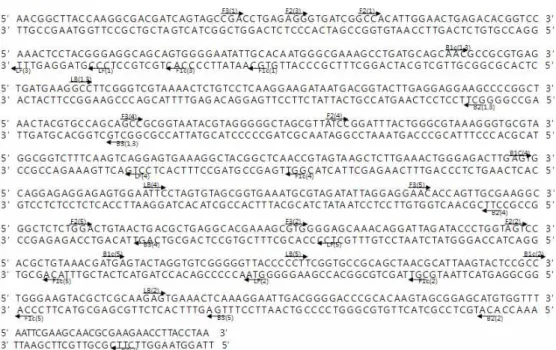
Fig. 1. Oligonucleotide pri- mers used for amplification of 16S rRNA gene, single- underlined and double-un- derlined letter indicate the sequences of primers for LAMP, respectively.
pathogenic locus(PaLoc)에는 주요 독소 유전자인
tcdA,
tcdB외에 accessory 유전자로
tcdR, tcdE, tcdC이 함께 존 재하고 있어 이 독소 유전자를 타깃으로 한 검출방법 등도 연구되고 있다(16-18).
C. difficile
검출방법으로 전 세계적으로 공인된 시험법은 없으며 식품에서
C. difficile에 대한 분리법도 현재 몇 종류 의 배지와 polymerase chain reaction(PCR)과 ribotyping, loop-mediated amplification 방법 등이 제시될 뿐 최적화된 방법은 없다.
최근 loop-mediated isothermal amplification(LAMP)이 식중독 균 분리기술로서 이용되고 있는데 기존 polymerase chain reaction(PCR) 방법과 유사하나 기존의 PCR 방법이 변성(denaturation), 접합(annealing), 신장(extension) 세 가 지 단계를 거치면서 온도의 변화를 주어야 하는 반면, 일정 한 온도에서 접합 및 신장이 가능하다는 특징이 있다(19).
별도의 온도변화를 주지 않고 63~65
oC 조건에서 주형 DNA 의 증폭이 가능하게 됨에 따라 PCR 방법보다 소요시간을 줄일 수 있으며 고가의 PCR 장비가 아닌 항온 수조나 오븐, 온장고 등 저가의 장비에서도 증폭이 가능하며 증폭산물을 전기영동 과정 없이 눈으로 확인할 수 있다(20).
본 연구에서는 최근 식중독 균으로 인정되고 있는
C. dif-ficile
에 대하여 LAMP에 의한 검출가능성을 타진하고자
16S rRNA 유전자를 타깃으로 하는 서로 다른 primer set를 구성하여 검출실험을 실시하였으며 이에 그 결과를 보고하 고자 한다.
재료 및 방법
사용균주 및 배지
Clostridium difficile
(KCTC 5009, NCCP 10868, NCCP
10898)와
C. difficile32(isolation from beef)를 Brain Heart Infusion(BHI; Oxoid, Hampshire, England)을 이용하여 37
o
C에서 48시간 혐기적으로 배양한 후 CCFA(Cefoxitin- Cycloserine-Fructose Agar; Oxoid)에 도말하여 형성된 단 일 콜로니를 실험에 사용하였다.
DNA 분리
DNA는 BHI에 배양한
C. difficile배양액을 AccuPrep
ⓇGenomic DNA Extraction kit(Bioneer Co., Daejeon, Korea) 를 이용하여 추출하였다. 공급자가 제시한 방법에 따라 DNA 를 분리하였다.
LAMP primer set 구성
C. difficile
의 16S rRNA를 target region으로 하는 염기서 열을 National center for biotechnology information(NCBI) 에서 검색하였다. 이 염기서열을 이용하여 LAMP primer set(BIP, FIP, B3, F3, LF, and PF)를 구성하기 위하여 Primer Explorer V4 software program and Primer3 Input(version 0.4.0, DNA STAR, Inc., Madison, WI, USA) program을 이용하였으며 설계한 primer set를 Bioneer사 (Bioneer Co.)에 의뢰하여 합성하였다(Table 1, Fig 1).
LAMP 반응 조건
Loopamp DNA amplification kit(Eiken Chemical, Tokyo,
Japan)를 사용하여 수행하였으며 총 반응용액은 반응튜브
당 2×reaction mix.(Tris-HCl; pH 8.8 40 mM, KCl 20 mM,
MgSO
416 mM, (NH4)
2SO
420 mM, Tween 20 0.2%, betaine
1.6 M, dNTPs 2.8 mM each) 12.5 μL, FIP 40 ρmol, BIP
40 ρmol, F3 5 ρmol, B3 5 ρmol, 1 μL의
BstDNA polymer-
ase와 distilled water를 포함하여 23 μL가 되도록 혼합하였
으며 한 번에 제조하여 분주하였다. 여기에 sample DNA
Table 1. Primer sequence sets LAMP detection of Clostridium difficile
Target ID Primer Sequence (5'-3')
16S rRNA 1
F3 B3 FIP BIP LF LB
CGACCTGAGAGGGTGATCG CTGCTGGCACGTAGTTAGC
TGCAATATTCCCCACTGCTGCC-GCCACATTGGAACTGAGACA AACGCCGCGTGAGTGATGAAG-GCTTCCTCCTCAAGTACCGT CCCGTAGGAGTTTGGACCG
GCCTTCGGGTCGTAAAACTCT
2
F3 B3 FIP BIP LF LB
GTGGGGAGCAAACAGGATT TCTTCGCGTTGCTTCGAATT
CGTTAGCTGCGGCACCGAAG-AGTCCACGCTGTAAACGATG CCTGGGAAGTACGCTCGCAA-ACATGCTCCGCTACTTGTG GTAACCCCCGACACCTAGTAG
GAGTGAAACTCAAAGGAATTGACG
3
F3 B3 FIP BIP LF LB
CGATCAGTAGCCGACCTGA CTGCTGGCACGTAGTTAGC
CACTGCTGCCTCCCGTAGGA-AGGGTGATCGGCCACATT AACGCCGCGTGAGTGATGAAG-GCTTCCTCCTCAAGTACCGT TGGACCGTGTCTCAGTTCC
GCCTTCGGGTCGTAAAACTC
4
F3 B3 FIP BIP LF LB
GCCGCGGTAATACGTAGG AGTTACAGTCCAGAGAGCCG
GGTTGAGCCGTAGCCTTTCACT-ATCCGGATTTACTGGGCGTA AGTGCAGGAGAGGAGAGTGGA-CTTCGCAACTGGTGTTCCT CTGACTTGAAAGACCGCCTAC
ATTCCTAGTGTAGCGGTGAAATG
5
F3 B3 FIP BIP LF LB
AACACCAGTTGCGAAGGC TGAGTTTCACTCTTGCGAGC
ACAGCGTGGACTACCAGGGTAT-GGACTGTAACTGACGCTGAG ATGAGTACTAGGTGTCGGGGGT-CCAGGCGGAGTACTTAATGC TCCCCACGCTTTCGTGC
CCTTCGGTGCCGCAGCTAA
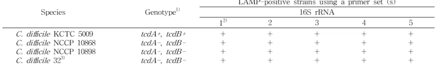
Table 2. Results of LAMP assays for detection of the 16S rRNA gene
Species Genotype
1)LAMP-positive strains using a primer set (s) 16S rRNA
1
2)2 3 4 5
C. difficile KCTC 5009 C. difficile NCCP 10868 C. difficile NCCP 10898 C. difficile 32
3)tcdA+ , tcdB+
tcdA- , tcdB- tcdA- , tcdB- tcdA- , tcdB-
+ +
+ +
+ +
+ +
+ +
+ +
+ +
+ +
+ +
+ +
1)
+: toxin positive, -: toxin negative.
2)ID of primer sets tested.
3)Strain was isolated from beef.
2 μL를 첨가하여 최종 volume이 25 μL가 되도록 하고 62
oC 에서 1시간, 80
oC에서 5분 thermal cycle로 반응시켰다.
검출 한계치 산출
C. difficile
를 사용하여 BHI에 혐기적 조건으로 37
oC에서 24시간 배양한 배양액을 연속적으로 희석하여 10
1~10
5cfu/
mL의 농도로 조정하였다. 각 배양액과 희석액에서 1 mL를 취하여 DNA를 분리하고 LAMP를 수행하여 검출 한계 값을 구하였다. 또한 시간 별로 CCFA에 분주하여 성장시킨 후 균수를 측정하여 검출 한계 값을 보정하였다.
증폭 산물 확인
1.5%(w/v) agarose gel로 Mupid-one(Advance Co., Ltd., Tokyo, Japan)을 이용하여 전기영동 하여 증폭산물을 분석
하였다. UV transilluminator 위에 agarose gel을 올려 Gel print system(Core Bio Corp., Seoul, Korea)으로 최종 확인 하였다.
결과 및 고찰
LAMP를 이용한
C. difficile검출
C. difficile
3종과 고기에서 분리한 분리균주
C. difficile1종에서 추출한 genomic DNA를 16S rRNA 유전자 se-
quence를 이용하여 구성한 5종의 primer set의 LAMP 수행
결과를 Table 2에 나타내었다. 실험에 사용된 primer set 중
16S rRNA를 타깃으로 하여 구성된 primer set 5개 중 5종
모두에 대해서
C. difficile4종이 증폭되었다(Fig. 2). 증폭된
(a) (b)
Fig. 2. The results of LAMP reactions detected by naked eye (a) and gel electrophoresis (b). Primer set 1: 1~4, primer set 2: 5~8, primer set 3: 9~12, primer set 4: 13~16, primer set 5: 17~20. 1, 5, 9, 13, 17: Clostridium difficile KCTC 5009, 2, 6, 10, 14, 18: Clostridium difficile NCCP 10868, 3, 7, 11, 15, 19: Clostridium difficile NCCP 10898, 4, 8, 12, 16, 20: Clostridium difficile 32 (isolated from beef), PC: positive control, NC: negative control.
C. difficile
은 TcdA positive, TcdB positive genotype과 TcdA negative, TcdB negative genotype을 가지는 균주였 다. 그리고 detection limit를 관찰하기 위하여 균주를 대상으 로 10
1-5cfu/mL 수준으로 십진 희석하여 각각 분리한 DNA 를 대상으로 16S rRNA primer set로 LAMP를 수행하였다.
그 결과, detection limit는 6.0×10
1cfu/mL로 측정되었으며 tube 상으로 관찰하였을 때도 증폭산물을 확인할 수 있었다.
Kato 등(21)은 toxin B 유전자를 타깃으로 하여 대변검체에 서 toxin A(TcdA) positive, toxin B(TcdB) positive
C. dif- ficile과 toxin A(TcdA) negative, toxin B(TcdB) positive
C. difficile을 검출하였다고 하였으며, Norén 등(22)은
tcdA유전자를 타깃으로 구성하여 대변 검체로부터
C. difficile을 검출하였다고 보고하였다. 그러나 아직까지 16S rRNA 유전 자를 타깃으로 하여 LAMP를 수행한 보고는 없는 실정으로 16S rRNA 유전자는 모든 미생물에 존재하고 미생물 계통분 류에 이상적인 유전자로 미생물 동정에도 많이 연구되고 있 다(14).
본 연구에서 16S rRNA 유전자를 바탕으로 구성된 5개의 target sequence에서
C. difficile의 TcdA-positive, TcdB- positive(A
+B
+)와 TcdA-negative, TcdB-negative(A
-B
-)의 genotype 균주와 분리 균주 모두에서 양성으로 나타났으며 detection limit는 10
1cfu/mL로 매우 낮게 측정되었다. 따라 서 16S rRNA를 타깃으로 하는 LAMP는 toxin 생성 여부에 상관없이 모든
C. difficile을 분리할 수 있을 것으로 생각되 며, 검출한계도 realtime PCR 수준으로 매우 낮으므로 신속 검출 기술로 활용이 가능할 것으로 생각되었다. 또한 분석 시 고가의 장비를 필요로 하지 않으며 검출 결과를 눈으로 직접 확인할 수 있으며 고도의 분석기술을 요구하지 않기 때문에 식품 생산 현장에서 활용될 수 있을 것이라고 생각되 었다.
요 약
본 연구는 loop-mediated isothermal amplification(LAMP) 을 이용하여
Clostridium difficile을 검출하고자 하였다.
LAMP 수행을 위하여 선택적인 타깃 유전자로
C. difficile의 16S ribosomal RNA를 타깃으로 하여 primer set를 구성하 였다. 5개의 primer set(BIP, FIP, B3, F3, LF, PF)를 이용하 여 TcdA와 TcdB toxin이 모두 양성인 균주, TcdA와 TcdB toxin이 모두 음성인 균주와 식품 분리균주를 효과적으로 검출할 수 있었다. LAMP는 80분 이내의 시간이 필요하며 thermocycler와 같은 장비를 필요로 하지 않고 또한 결과를 직접 눈으로 확인할 수 있기 때문에 식품 생산 현장에서 활 용될 수 있을 것이라고 생각되었다.
감사의 글
이 논문은 2012년도 정부(교육과학기술부)의 재원으로 한 국연구재단의 지원을 받아 수행된 연구임(2012-0005363).
문 헌