ISSN 1225-6552, eISSN 2287-7630 http://dx.doi.org/10.7853/kjvs.2013.36.2.79
< Original Article >
Veterinary Service
Available online at http://kjves.org
*Corresponding author: Ba-Ra-Da Koh, Tel. +82-62-613-7673, Fax. +82-62-613-7649, E-mail. [email protected]
Mycobacterium bovis 와 M. tuberculosis 감별을 위한 등온증폭법
고바라다
1*ㆍ김재명
2ㆍ성창민
1ㆍ지태경
1ㆍ나호명
1ㆍ박성도
1ㆍ김용환
1ㆍ김은선
11광주광역시보건환경연구원, 2농림축산검역본부 세균질병과
Loop-mediated isothermal amplification assay for differentiation of Mycobacterium bovis and M. tuberculosis
Ba-Ra-Da Koh
1*, Jae-Myung Kim
2, Chang-Min Sung
1, Tae-Kyung Ji
1, Ho-Myung Na
1, Seong-Do Park
1, Yong-Hwan Kim
1, Eun-Sun Kim
11
Health & Environment Research Institute of Gwangju, Gwangju 500-210, Korea
2
Bacterial Disease Division, Animal and Plant Quarantine Agency, Anyang 430-757, Korea (Received 1 May 2013; revised 4 June 2013; accepted 12 June 2013)
Abstract
Mycobacterium (M.) bovis, a member of the M. tuberculosis complex (MTC), is a re-emerging, zoonotic agent of bovine tuberculosis whose prevalence probably depends on variations in direct exposure to cat- tle and ingestion of raw milk. Accurate species differentiation of M. bovis and M. tuberculosis is needed to distinguish between human and zoonotic tuberculosis. This study successfully developed a loop-medi- ated isothermal amplification (LAMP) assay for rapid detection and differentiation of M. bovis and M.
tuberculosis, however showed negative reactions in eight non-tuberculous mycobacteria (NTM) samples and ten other bacterial species. Sensitivity of this assay for detection of genomic M. bovis DNA was 10 fg/ μ l. And this assay successfully detected M. bovis in bovine clinical specimens. In conclusion, the LAMP assay is a simple and powerful tool for rapid detection of M. bovis in both pure bacterial culture and in clinical samples.
Key words : M. bovis, M. tuberculosis, LAMP, Bovine tuberculosis, Differentiation
서 론
소에서 결핵은 M. bovis 감염에 의한 만성 소모성 질 병이며 인수공통전염병으로써 축산업에서 경제적 손실 이 크다는 점에서 중요한 질병으로 취급되고 있다 (Grange와 Collins, 1987; Thoen 등, 2006). 한·육우에서 결핵발생 두수는 우리나라 동물방역통합시스템(http://
kahis.nvrqs.go.kr)에 따르면 2007년 228두에서 2010년 1,129 두 , 2011년 1,113두 그리고 2012년 1,127두가 발생하였다.
소결핵은 대부분 임상증상이 나타나지 않기 때문
에 도축장에 출하되어 도축과정에서 결핵 감염소가 발견되기도 한다(Koh 등, 2011). 소의 임상 시료로부 터 M. bovis DNA를 polymerase chain reaction (PCR)과 같은 분자생물학적인 방법으로 신속하게 직접 검출 할 수 있었다(Koh 등, 2011; Parra 등, 2008). PCR은 변성(denaturation), 접합(annealing), 신장(extension) 세 가지 단계를 거치면서 온도의 변화를 주어야 한다.
Loop-mediated isothermal amplification (LAMP)은
Bst DNA polymerase와 같은 strand displacement 활성
을 가진 효소에 의해 일정한 온도에서 세 가지 단계
의 온도 변화를 거치지 않고도, 유전자를 증폭할 수
있는 새로운 핵산 증폭방법으로 Notomi 등(2000)에
의해서 개발되었다. LAMP는 일정한 온도조건하에서 높은 특이성, 효율성 그리고 신속성을 지닌 DNA 증 폭 기술이다. LAMP assay로 MTC를 검출하거나 동정 하기 위해서 IS6110 (Aryan 등, 2010), hspX (Bi 등, 2012), 16S rRNA (Pandey 등, 2008), gyrB (Iwamoto 등, 2003), mpt83 (Zhang 등, 2011) 및 rimM 유전자 (Zhu 등, 2009) 등이 사용되었다.
국내에서 M. bovis에 의한 사람 감염은 외국 이주 민에서 발생한 사례 1건이 보고되었다(Lim 등, 2011).
따라서 사람 결핵 감염에서 M. bovis를 감별하여 신 속한 의학적 처지에 도움이 되고 도축장에서 발견되 는 결핵 병변 시료에 대한 신속ㆍ정확한 실험실 진단 방법이 필요하다.
Tomita 등(2008)이 보고한 LAMP assay는 여러 가지 시약을 다양한 농도로 사용하는 복합한 실험절차, Zhang 등(2011)은 증폭 후 결과판독을 위해 반응용기 개방에 의한 실험실 오염문제, Zhu 등(2009)의 육안판 독에 따른 객관성 부족 그리고 Bi 등(2012)은 복잡한 primer에 의한 비특이 반응과 실험실 오염에 의한 의 양성 결과 등 실험방법을 개선할 여지가 있다.
따라서 이번 연구의 목적은 이런 단점을 개선하기 위해서 Genie
ⓇII (OptiGene, UK) 장비를 이용하여 M.
tuberculosis에만 존재하는 12.7-kb 절편부위로 M. bo- vis와 M. tuberculosis를 감별할 수 있는 LAMP assay를 개발하였으며, 소의 림프절 시료와 분리된 결핵균주 에 적용하였다.
재료 및 방법
공시재료
공시 균주: 이번 연구에 이용한 10종의 결핵균은 M.
bovis AN5, M. tuberculosis, M. avium, M. intra- cellulare, M. fortuitum, M. phlei, M. peregrinum, M.
scrofulaceum, M. smegmatis, M. kansasii이며, 이들 균 주는 농림축산검역본부에서 보유하고 있는 DNA를 사용하였으며, 이들 균주에 대한 LAMP 특이성 검사 는 농림축산검역본부에서 수행하였다. 다른 병원성 세균 10종은 E. coli, Salmonella spp., Staphylococcus (Sta.) aureus, Clostridium (Cl.) perfringnes, Listeria (L.) monocytogenes, L. innocua, Campylobacter (Cam.) coli, Cam. jejuni, Enterobacter (Ent.) faecium, Ent. faecalis이 다.
임상시료: 2010년부터 2012년까지 광주지역 도축장
에서 발견된 소의 육아종성 림프절 병변 24건과 정상 림프절 32건을 LAMP assay 실험에 사용하였다.
분리배양: 2010년부터 2012년까지 광주지역 도축장
에서 발견된 소의 육아종성 림프절 병변 24개 시료로 부터 결핵균 분리 배양을 위해서 Koh 등(2011) 방법 에 따라 균질화된 시료 100 μl를 Lowenstein-Jensen (LJ) 배지에 접종하여 37
oC에서 3∼6주 동안 배양하 면서 균 발육을 관찰하였다. 조직처리 방법을 간략하 게 요약하면 다음과 같다. 림프절 조직 1∼3 g 정도 를 멸균 생리식염수 5 ml과 혼합하여 균질화하였다.
균질액에 4% NaOH 용액 5 ml를 첨가하여 상온에서 15분간 액화시킨 후 2% HCl 6.8 ml를 첨가하여 중화 시켰다. 중화된 균질액은 3,000 ×g에서 15분 동안 원 심분리를 한 후 상층액은 버리고 침전물에 멸균된 PBS (pH 7.2) 2 ml를 첨가하여 재균질화시킨 후 300 μl를 eppendorf tube에 채취하여 DNA 추출과 결핵균 분리배양에 사용하였다.
Genomic DNA 추출
E. coli 등 병원성 세균 10종은 균주별 배양액을 끓 는 물에 15∼20분 처리한 후 원심분리하여 상층액을 실험에 사용하였다. 림프절 시료와 LJ 배지에서 분리 배양한 결핵균의 DNA 추출은 Koh 등(2011)의 방법 에 따랐다. Proteinase K 처리 후 결핵균 사멸을 위해 95
oC에서 5분간 처리하였다.
추출된 DNA는 NanoDrop
ⓇND-1000 UV-Vis Spec- trophotometer (NanoDrop Technologies, USA)를 이용하 여 260 : 280 nm에서 정량하여 실험 전까지 −20
oC에 보관하였다.
DNA oligonucleotides
M. bovis와 M. tuberculosis 감별을 위한 oligonucleo-
tide 디자인은 Zumárraga 등(1999)이 보고한 M. bovis의
고유한 연속된 229-bp의 염기서열을 분석하여 M. bovis
AF2122/97 전체 유전자서열(accession No. BX248339)
과 M. tuberculosis에 특이적인 12.7-kb 절편 부위
(accession no. BX842576)를 분석하여 Tomita 등(2008)
의 방법과 PrimerExplorer V4 프로그램(http://primer-
explorer.jp/elamp4.0.0/index. html)을 이용하여 primer
(F3, B3, FIP 및 BIP)를 설계하였으며, 2개의 loop pri-
mer (FLP, BLP)는 직접 설계하였다(Table 1). Primer는
Fig. 1. The 229-bp contiguous sequences of M. bovis (accession No. BX248339) was used to design the six primers. The sequences of the LAMP primer sites are in rectangles. Right and left arrows indicate sense and complementary sequences were used for the primer. Forward inner primer (FIP) contains the sequence (F1c) complementary to F1 and F2, backward inner primer (BIP) contains B1 and the sequence (B2c) complementary to B2. FLP: forward loop primer, BLP: backward loop primer.
Fig. 2. Amplification curves of gradient LAMP for M. bovis AN5.
Using the GenieⓇ II, a gradient of 60 to 67°C was set. M.
bovis was amplified faster at 67
oC than at 65°C recom- mended by the manufacturer of the Isothermal Master Mix.M. bovis DNA detection time (min:sec) was 60°C, 15:54;
61°C, 15:09; 62°C, 13:56; 63°C, 13:15; 64°C, 11:53; 65°C, 11:19; 66°C, 10:27; 67°C, 10:11.
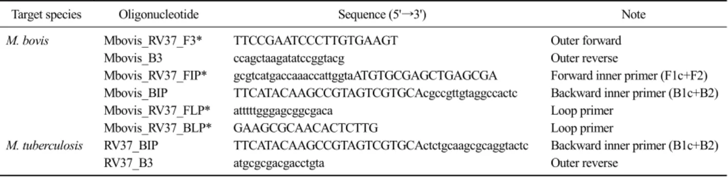
Table 1.Nucleotide sequences used in this study to detect M. bovis (229-bp contiguous sequences) and M. tuberculosis (12.7-kb fragment)
Target species Oligonucleotide Sequence (5'→3') Note
M. bovis
Mbovis_RV37_F3* TTCCGAATCCCTTGTGAAGT Outer forwardMbovis_B3 ccagctaagatatccggtacg Outer reverse
Mbovis_RV37_FIP* gcgtcatgaccaaaccattggtaATGTGCGAGCTGAGCGA Forward inner primer (F1c+F2) Mbovis_BIP TTCATACAAGCCGTAGTCGTGCAcgccgttgtaggccactc Backward inner primer (B1c+B2)
Mbovis_RV37_FLP* atttttgggagcggcgaca Loop primer
Mbovis_RV37_BLP* GAAGCGCAACACTCTTG Loop primer
M. tuberculosis
RV37_BIP TTCATACAAGCCGTAGTCGTGCActctgcaagcgcaggtactc Backward inner primer (B1c+B2)RV37_B3 atgcgcgacgacctgta Outer reverse
*Consensus between M. bovis and M. tuberculosis.
Bioneer (Korea)에 의뢰하여 합성하였으며, FIP와 BIP 는 HPLC 정제를 하였다. Primer는 표적 DNA에 대해 서 Fig. 1과 같이 loop primer (FLP, BLP)를 제외한 네 개의 primer가 각기 다른 여섯 개 부위와 특이적으로 결합한다. 이번 연구에 사용된 F3, FIP, FLP, BLP는 M. bovis와 M. tuberculosis와 공통으로 사용되도록 설 계하였다.
LAMP PCR 반응 조건
LAMP 반응액은 1.6 μM의 FIP와 BIP, 0.2 μM의 F3 와 B3, 0.4 μM의 FLP와 BLP, 1 μl의 template DNA 그리고 Isothermal Master Mix (OptiGene, UK) 12 μl를 첨가하고 총반응액은 20 μl가 되도록 증류수로 조절 하였다. 모든 template DNA는 96
oC에서 3분간 변성과 정을 거친 후 LAMP 반응액에 첨가하여 실험하였다.
최적의 온도조건을 찾기 위하여 60∼67
oC 범위에 서 1
oC 차이로 7개 온도구역을 설정하였고, 사용된 균주는 M. bovis AN5를 template DNA로 이용하였다.
LAMP는 Genie
ⓇII (OptiGene, UK)을 이용하여 이중 나선 DNA에 형광물질 결합여부를 여기파장 488 nm, 방출파장 520 nm에 측정하였다. 반응시간은 30분이며,
이후 PCR 반응액을 95
oC로 상승시킨 후 80
oC까지 낮 추면서 증폭산물의 re-annealing 온도를 측정하였다. 증 폭결과는 Genie
ⓇII software (Ver. 0.99, OptiGene, UK) 프로그램을 이용해 확인하였다 .
M. bovis에 대한 LAMP의 검출한계 검사를 위해서
Table 2.Comparison of the detection time between 65°C and 67°C using GenieⓇ II for M. bovis
Lane in Fig. 3 and Fig. 4
DNA concentration
Peak position (min:sec)
65°C 67°C
1 10.0 ng/μl 08:44 07:46
2 1.0 ng/μl 09:44 08:31
3 0.1 ng/μl 10:59 09:31
4 10.0 pg/μl 13:14 11:01
5 1.0 pg/μl 14:44 12:16
6 0.1 pg/μl 14:29 17:16
7 10.0 fg/μl 17:14 -*
8 1.0 fg/μl - -
*Not detected.
Fig. 5. Melting curves for M. bovis LAMP product as monitored in GenieⓇ II. The cruves were obtainded after LAMP amplification for 30 min. M. bovis had a melting temperature (Tm) of 87.3±0.1°C.
A B
Fig. 3.A minimum of 10.0 pg/μl of DNA could be detected. (A) Real-time fluorescence curves of M. bovis DNA dilution series were amplified by LMAP at 65°C for 40 min using the GenieⓇ II. (B) 2% agarose gel shows LAMP amplicon as a ladder of bands. Lanes M) 100 bp DNA ladder, 1) 10.0 ng/μl 2) 1.0 ng/μl, 3) 0.1 ng/μl, 4) 10.0 pg/μl, 5) 1.0 pg/μl, 6) 0.1 pg/μl, 7) 10.0 fg/μl, 8) 1.0 pg/μl.
A B
Fig. 4. A minimum of 10.0 pg/μl of DNA could be detected. (A) Real-time fluorescence curves of M. bovis DNA dilution series were amplified by LMAP at 67°C for 40 min using the GenieⓇ II. (B) 2% agarose gel shows LAMP amplicon as a ladder of bands. Lanes M) 100 bp DNA ladder, 1) 10.0 ng/μl 2) 1.0 ng/μl, 3) 0.1 ng/μl, 4) 10.0 pg/μl, 5) 1.0 pg/μl, 6) 0.1 pg/μl, 7) 10.0 fg/μl, 8) 1.0 pg/μl.
DNA를 10 ng/ μl로 정량한 후 10배 계단 희석하여 실 험하였다.
결 과
Gradient LAMP assay
60°C에서 67°C까지 온도구배에 따른 최단시간에 결핵균을 검출할 수 있는 온도는 67°C이었고, 검출시 간은 10분 11초였다. 제조사에서 제시한 LAMP 실험 의 최적 온도조건은 65°C로 이때 우결핵 검출된 시간 은 11분 19초로 67°C일 때보다 약 1분 정도 지연되었 다(Fig. 2).
M. bovis의 LAMP assay 검출한계
M. bovis의 DNA를 10 ng/μl부터 10배 계단 희석하 여 65°C와 67°C 조건에서 LAMP assay의 검출한계를 실험하였다. 65°C에서 검출한계는 10 fg/μl까지 검출 이 가능하였지만(Fig. 3A), 67°C에서 검출한계는 0.1 pg/μl까지 검출되었다(Fig. 4A). 이들 두 가지 온도조 건에 대한 전기영동 결과 사다리 모양의 LAMP assay 증폭산물이 확인되었으며, 검출한계도 실시간 그래프 와 일치하였다(Fig. 3B, 4B). 그리고 LAMP assay를 이 용한 M. bovis의 검출시간은 20분 이내에 확인이 가 능하였다(Fig. 3, Table 2). 따라서 이번 연구에서 림프 절 시료에 대한 온도조건은 65°C에서 LAMP assay를 수행하였다.
M. bovis에 대한 LAMP assay 증폭산물에 대한 melt-
ing temperature (Tm)를 측정한 결과 87.3±0.1
oC이었다
(Fig. 5).
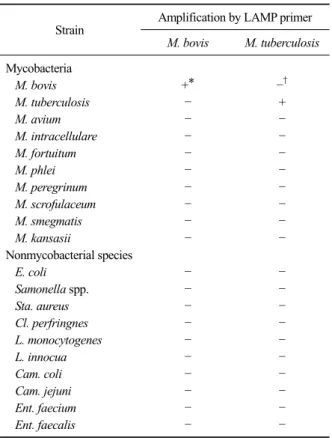
Table 3. Specificity of LAMP primers for identification of M.
bovis and M. tuberculosis
Strain Amplification by LAMP primer
M. bovis M. tuberculosis
MycobacteriaM. bovis
+* †M. tuberculosis
+M. avium
M. intracellulare
M. fortuitum
M. phlei
M. peregrinum
M. scrofulaceum
M. smegmatis
M. kansasii
Nonmycobacterial species
E. coli
Samonella spp.
Sta. aureus
Cl. perfringnes
L. monocytogenes
L. innocua
Cam. coli
Cam. jejuni
Ent. faecium
Ent. faecalis
*Positive result. †Negative result.
Table 4. Comparison of LJ medium and LAMP assay for the detection of M. bovis
Pathological status LJ medium LAMP assay Positive Negative Positive Negative
VL* (n=24) 21 3 24 0
NVL† (n=32) Not tested 0 32
*Visible lesions. †Non visible lesions.
LAMP assay 특이성
소 결핵균(M. bovis)을 포함한 항산성 균주 10주와 E. coli 등 병원성 세균 10주에서 추출된 DNA를 대상 으로 LAMP assay의 특이성 실험은 65°C에서 수행하 였다. M. bovis와 M. tuberculosis에 특이적인 LAMP primer는 해당 결핵균에만 반응하였고, 다른 결핵균 과 병원성 세균에서는 교차반응이 관찰되지 않았다 (Table 3).
분리균주와 임상시료에 대한 LAMP assay
도축장에서 발견된 소의 육아종성 림프절 병변 24 개 시료 중 21건에서 결핵균이 분리되었으며, M. bo- vis와 M. tuberculosis에 대한 LAMP assay 실험결과 21 개 분리주 모두 M. bovis로 확인되었다. 그리고 소의 육아종성 림프절 24건은 LAMP assay에서 모두 M.
bovis가 검출되었으며, 정상 림프절 32건은 모두 음성 이었다(Table 4).
고 찰
Notomi 등(2000)이 일정한 온도 조건에서 높은 특 이성, 효율성 그리고 신속성을 지닌 LAMP라 불리는 DNA 증폭 기술을 개발하였다. 이는 기존의 PCR에서 사용되는 Taq DNA polymerase 대신 Bst DNA poly- merase를 사용함으로써 일정한 온도에서 접합 및 신 장을 할 수 있게 되었다. Bst DNA polymerase는 일반 PCR의 Taq DNA polymerase와는 달리 5'→3' exonu- clease의 성격을 가지고 있어 DNA의 이중나선 구조 를 열에 의한 변성을 거치지 않고 Tm값에 가까운 접 합 온도에서 접합 및 신장이 수행된다(Nagamine 등, 2002b).
LAMP 실험에서는 DNA를 변성시킬 필요가 없으
며(Nagamine 등, 2001), 네 개 primer가 표적 DNA의
각기 다른 여섯 개 부위를 인식하기 때문에 특이성이
매우 높을 뿐만 아니라 loop primer가 고리형성의 속
도를 증가시키기 때문에 신속하게 결과를 도출할 수
있다(Nagamine 등, 2002a). 이번 연구에서도 loop pri-
mer를 사용하여 실험결과를 10분 정도 단축할 수 있
었다(자료 미제시). LAMP assay의 다른 특징은 상당
량의 DNA가 합성되기 때문에 dNTP에서 방출된 py-
rophosphate 이온(P
2O
74-)이 magnesium 이온과 결합하
여 생성된 부산물인 magnesium pyrophosphate
(Mg
2P
2O
7)에서 유래한 탁도를 측정함으로써 실시간
으로 표적 DNA를 정량할 수 있게 되었다(Mori 등,
2001; 2004). Pandey 등(2008)과 Bi 등(2012)은 tur-
bidimeter 기기를 이용하여 M. tuberculosis를 검출하였
다. 또한, Mori 등(2006)은 FITC와 ROX와 같은 형광
물질로 표지된 probe를 첨가하여 LAMP 증폭산물에
양이온 물질인 polyethylenimine (PEI)를 첨가하여 두
개의 표적 DNA를 동시에 시각화하였다. 이번 연구에
서는 DNA 증폭 부산물에서 유래한 탁도 형성을 억
제하는 상품화된 premix 제품과 Genie
ⓇII (OptiGene,
UK) 기기로 결핵균 증폭 여부를 실시간으로 확인할
수 있었다.
LAMP는 PCR과 대등한 실험결과를 나타내지만, primer 설계에서 까다로운 조건 때문에 응용범위가 제한적이다(Gandelman 등, 2011). Tomita 등(2008)은 형광물질로써 calcein을 사용하여 특별한 장비없이 LAMP 증폭산물을 시각화할 수는 실험방법과 적절한 primer 설계기준을 제시하였다. LAMP assay는 현재 다양한 방법으로 개선되고 있다. 첫 번째 Gandelman 등(2011)이 제시한 stem-LAMP에서 stem primer를 loop primer와 병용하거나 loop primer 설계가 곤란할 때 대체하여 사용함으로써 속도, 민감도 그리고 재현 성이 동시에 향상됨을 입증하였다. 두 번째 다른 실 험법과 병용한 신속한 검출과 multiplex 기능으로 진 화되어 가고 있다(Jung 등, 2010; Liang 등, 2012). 세 번째 LAMP assay을 위한 spectrofluorimeter 등 다양한 기기들이 소개되고 있다(Ahmed 등, 2010; Gandelman 등, 2010; Tong 등, 2008). 그리고 이번 연구에서는 이 전 Bst DNA polymerase보다 증폭속도가 개선된 Gsp DNA polymerase가 포함되어 있으면서 일반 PCR처럼 제품화된 premix 제품을 사용하였기 때문에 여러 가 지 시약을 제조해야 되는 불편한 점이 개선되었다.
또한, 이 제품은 LAMP assay 후 증폭용기를 개방하 여 발색시약을 첨가할 필요가 없으므로 실험실 오염 방지에 매우 유용하였다.
Zhang 등(2011)은 LAMP assay으로 1시간 동안 증 폭 후 SYBR Green I를 첨가하여 MTC 세포 표면의 lipoprotein인 mpt83 염기서열 부위로 M. bovis를 10 copy/reaction까지 검출하였다. 그리고 이들 방법을 이 용한 Iwamoto 등(2003)은 MTC를 검출하기 위해서 gyrB 유전자를 사용하였으며, 검출한계는 50 copy 수 준이었다. Hong 등(2012)은 mpt83 염기서열 부위로 M. tuberculosis와 M. bovis를 감별하였고, 일반 PCR의 검출한계 10 copy/ μl보다 뛰어난 1 copy/μl로 민감도 가 우수하였다. Bi 등(2012) 등은 hspX 염기서열 부위 로 MTC를 10 copy까지 검출하였다. Hwang 등(2011) 은 IS6110 유전자 부위로 MTC를 200 copy까지 검출 하였다. 이번 연구에서 M. bovis의 검출한계는 10 fg/μl 즉, 2 genome에 상당하는 DNA 농도로 이들 연 구결과와 대등하거나 보다 우수하였다.
M. bovis 유전자가 M. tuberculosis에 존재하는 12.7-kb 절편이 결여된 부위는 RD 7 부분에 해당하고 이번 연구에서는 LAMP assay로 이들 결핵균을 감별 하기 위해서 BIP와 B3는 각각의 primer를 사용하였 다. 그리고 F3, FIP, FLP, BLP는 M. bovis와 M. tuber-
culosis와 공통으로 사용하였지만, loop primer (FLP, BLP)를 제외한 네 개의 primer가 각기 다른 여섯 개 부위와 특이적으로 결합하였기 때문에 이들 두 가지 결핵균에서 교차반응은 관찰되지 않았다.
Nagamine 등(2001)은 LAMP 반응에 있어서 초기 단계의 template DNA 변성 과정없이 증폭이 가능하 다고 보고하였지만, Geojith 등(2011)은 Zhu 등(2009) 이 보고한 LAMP 반응의 민감도를 증가시키기 위해 서는 template DNA의 최초 변성이 필수적이며 반응 시간 연장을 통해 민감도가 증가한다고 보고하였다.
Aryan 등(2010)도 IS6110 유전자로 MTC DNA를 검출 하기 위해서 template DNA를 96
oC에서 3분간 초기 변성 과정을 실시하였고, LAMP assay의 반응시간이 검출한계에 영향을 끼치기 때문에 최적반응 시간은 90분으로 하였다. 그리고 이들은 초기 변성과정 없이 실험하면 검출한계는 200 copy이었지만, 열변성 과정 을 거친 후에는 1 copy까지 검출할 수 있었다. 따라 서 이번 연구에서 사용된 모든 template DNA는 96
oC 에서 3분간 변성과정을 거친 후 LAMP 반응액에 첨 가하여 실험하였고, M. bovis의 검출한계는 10 fg/μl 즉, 2 genome에 상당하는 DNA 농도였다.
M. bovis의 표준균주에 대한 LAMP assay에서 검출 시간은 20분 이내이었지만, 육아종성 림프절 시료에 서 추출한 template DNA에는 우결핵과 조직의 유전 자가 혼재되어 있기 때문에 반응시간이 다소 지연되 었다. 다만 이번 실험에 사용된 Isothermal Master Mix (OptiGene, UK)는 40분을 초과하여 실험하면 비특이 적인 증폭산물이 생성되기 때문에 LAMP assay 반응 시간을 30분으로 제한하였다. 그리고 임상시료의 template DNA는 다양한 유전자가 혼재되어 있기 때 문에 LAMP peak가 표준균주에 비해 정상적인 증폭 곡선(Fig. 2)처럼 보이지 않거나 높이가 낮으면 반드 시 Tm값을 확인할 것을 권장한다.
이번 연구에서, 도축장에서 발견된 육아종성 림프
절 시료 24건 중 21건에서 결핵균을 분리하는데 4주
이상의 시간이 필요하였고 분리율은 87.5%이었지만,
LAMP assay에서는 신속하게 모든 시료에서 M. bovis
DNA가 검출되었다. 기존의 PCR은 한번에 많은 시료
를 검사할 수 있으며, 동시에 두 개 이상의 표적 유전
자를 검출할 수 있는 장점이 있다. 이번 연구에 사용
된 LAMP assay에는 값비싼 시약과 고가의 장비가 필
요하고 1회 처리건수가 16건으로 다소 제약은 있었
지만, 기기를 이용한 객관적인 결과 판정, 간소화된
실험방법, 검사시간 단축 그리고 전기영동을 할 필요
가 없다는 장점이 있었다.
국내에서 2010년 이후 매년 1,100여 두 이상 한․육 우에서 우결핵이 검색되고 있으며, 이들 감염개체 중 일부분은 도축장에서 발견되고 있기 때문에, 이번 연 구에서 개발된 LAMP assay가 도축장에서 결핵병을 검사하는데 유용하게 활용될 수 있다. 그리고 M. bo- vis와 M. tuberculosis를 감별할 수 있으며, 동물 임상 시료에 존재하는 M. bovis를 신속하게 검출해낼 수 있었을 뿐만 아니라 사람 결핵 진단에도 매우 유용하 게 사용될 것으로 생각한다.
결 론
이번 연구에서 M. bovis와 M. tuberculosis를 감별할 수 있는 LAMP assay를 개발하였고, 이를 이용하여 육안적으로 확인된 소의 육아종성 림프절 24건과 정 상 림프절 32건의 시료와 육아종성 림프절에서 분리 된 결핵균 21주에 대해서 진단 기법의 활용성을 평가 하였다. M. bovis와 M. tuberculosis에 대한 LAMP as- say의 특이성은 100%이었고, M. bovis 검출한계는 10 fg/μl이었다. 결핵병변을 보이는 조직시료에 적용한 결과 균 분리율은 87.5% (21/24)이었으나, LAMP as- say 적용결과 M. bovis 유전자를 100% 검출하였다.
따라서 이번 연구에서 개발된 LAMP assay는 소 림프 절에 존재하는 M. bovis를 신속․정확하게 진단할 수 있을 뿐만 아니라 의학적 진단에 매우 유용하게 사용 될 것으로 생각한다.
참 고 문 헌
Ahmed MU, Hasan Q, Hossain MM, Saito M, Tamiya E. 2010.
Meat species identification based on the loop mediated isothermal amplification and electrochemical DNA sensor. Food Control 21: 599-605.
Aryan E, Makvandi M, Farajzadeh A, Huygen K, Bifani P, Mousavi SL, Fateh A, Jelodar A, Gouya MM, Romano M. 2010. A novel and more sensitive loop-mediated iso- thermal amplification assay targeting IS6110 for de- tection of Mycobacterium tuberculosis complex.
Microbiol Res 165: 211-220.
Bi A, Nakajima C, Fukushima Y, Tamaru A, Sugawara I, Kimura A, Kawahara R, Hu Z, Suzuki Y. 2012. A rapid loop-mediated isothermal amplification assay targeting
hspX for the detection of Mycobacterium tuberculosis
complex. Jpn J Infect Dis 65: 247-251.Gandelman O, Jackson R, Kiddle G, Tisi L. 2011. Loop-mediated
amplification accelerated by stem primers. Int J Mol Sci 12: 9108-9124.
Gandelman OA, Church VL, Moore CA, Kiddle G, Carne CA, Parmar S, Jalal H, Tisi LC, Murray JAH. 2010. Novel bioluminescent quantitative detection of nucleic acid am- plification in real-time. PLoS ONE 5: e14155.
Geojith G, Dhanasekaran S, Chandran SP, Kenneth J. 2011.
Efficacy of loop mediated isothermal amplification (LAMP) assay for the laboratory identification of
Mycobacterium tuberculosis isolates in a resource lim-
ited setting. J Microbiol Methods 84: 71-73.Grange JM, Collins CH. 1987. Bovine tubercle bacilli and disease in animals and man. Epidemiol Infect 99: 221-234.
Hong M, Zha L, Fu W, Zou M, Li W, Xu D. 2012. A modified visual loop-mediated isothermal amplification method for diagnosis and differentiation of main pathogens from
Mycobacterium tuberculosis complex. World J Microbiol
Biotechnol 28: 523-531.Hwang ES, Lee TU, Jung DY, Cho HS. 2011. Development of loop-mediated isothermal amplification method for the rapid and sensitive detection of bovine tuberculosis in Korea native cattle. Korean J Vet Serv 34: 333-339.
Iwamoto T, Sonobe T, Hayashi K. 2003. Loop-mediated iso- thermal amplification for direct detection of Mycobacte-
rium tuberculosis complex, M. avium, and M. intra- cellulare in sputum samples. J Clin Microbiol 41:
2616-2622.
Jung C, Chung JW, Kim UO, Kim MH, Park HG. 2010.
Isothermal target and signaling probe amplification method, based on a combination of an isothermal chain amplification technique and a fluorescence resonance en- ergy transfer cycling probe technology. Anal Chem 82:
5937-5943.
Koh BRD, Jang YB, Ku BK, Cho HS, Bae SY, Na HM, Park SD, Kim YH, Mun YU. 2011. Development of real-time PCR for rapid detection of Mycobacterium bovis DNA in cattle lymph nodes and differentiation of M. bovis and
M. tuberculosis. Korean J Vet Serv 34: 321-331.
Liang C, Chu Y, Cheng S, Wu H, Kajiyama T, Kambara H, Zhou G. 2012. Multiplex loop-mediated isothermal amplifica- tion detection by sequence-based barcodes coupled with nicking endonuclease-mediated pyrosequencing. Anal Chem 84: 3758-3763.
Lim SK, Park JY, Park SD, Chang HK. 2011. Localized empye- ma due to Mycobacterium bovis. Korean J Med 81:
792-796.
Mori Y, Hirano T, Notomi T. 2006. Sequence specific visual de- tection of LAMP reactions by addition of cationic polymers. BMC Biotechnol 6: 3.
Mori Y, Kitao M, Tomita N, Notomi T. 2004. Real-time tur- bidimetry of LAMP reaction for quantifying template DNA. J Biochem Biophys Methods 59: 145-157.
Mori Y, Nagamine K, Tomita N, Notomi T. 2001. Detection of loop-mediated isothermal amplification reaction by tur- bidity derived from magnesium pyrophosphate forma-
tion. Biochem Biophys Res Commun 289: 150-154.
Nagamine K, Hase T, Notomi T. 2002a. Accelerated reaction by loop-mediated isothermal amplification using loop primers. Mol Cell Probes 16: 223-229.
Nagamine K, Kuzuhara Y, Notomi T. 2002b. Isolation of sin- gle-stranded DNA from loop-mediated isothermal ampli- fication products. Biochem Biophys Res Commun 290:
1195-1198.
Nagamine K, Watanabe K, Ohtsuka K, Hase T, Notomi T. 2001.
Loop-mediated isothermal amplification reaction using a nondenatured template. Clin Chem 47: 1742-1743.
Notomi T, Okayama H, Masubuchi H, Yonekawa T, Watanabe K, Amino N, Hase T. 2000. Loop-mediated isothermal am- plification of DNA. Nucleic Acids Res 28: E63.
Pandey BD, Poudel A, Yoda T, Tamaru A, Oda N, Fukushima Y, Lekhak B, Risal B, Acharya B, Sapkota B, Nakajima C, Taniguchi T, Phetsuksiri B, Suzuki Y. 2008.
Development of an in-house loop-mediated isothermal amplification (LAMP) assay for detection of Mycobacte-
rium tuberculosis and evaluation in sputum samples of
Nepalese patients. J Med Microbiol 57: 439-443.Parra A, García N, García A, Lacombe A, Moreno F, Freire F, Moran J, Hermoso de Mendoza J. 2008. Development of a molecular diagnostic test applied to experimental abat- toir surveillance on bovine tuberculosis. Vet Microbiol
127: 315-324.
Thoen C, Lobue P, de Kantor I. 2006. The importance of
Mycobacterium bovis as a zoonosis. Vet Microbiol 112:
339-345.
Tomita N, Mori Y, Kanda H, Notomi T. 2008. Loop-mediated isothermal amplification (LAMP) of gene sequences and simple visual detection of products. Nat Protoc 3:
877-882.
Tong Y, Tang W, Kim HJ, Pan X, Ranalli TA, Kong H. 2008.
Development of isothermal TaqMan assays for detection of biothreat organisms. Biotechniques 45: 543-557.
Zhang J, Zhang GH, Yang L, Huang R, Zhang Y, Jia K, Yuan W, Li SJ. 2011. Development of a loop-mediated iso- thermal amplification assay for the detection of
Mycobacterium bovis. Vet J 187: 393-396.
Zhu RY, Zhang KX, Zhao MQ, Liu YH, Xu YY, Ju CM, Li B, Chen JD. 2009. Use of visual loop-mediated isothermal amplification of rimM sequence for rapid detection of
Mycobacterium tuberculosis and Mycobacterium bovis. J
Microbiol Methods 78: 339-343.Zumárraga M, Bigi F, Alito A, Romano MI, Cataldi A. 1999. A 12.7 kb fragment of the Mycobacterium tuberculosis ge- nome is not present in Mycobacterium bovis. Microbiol- ogy 145: 893-897.