15 ISSN 1225-6552, eISSN 2287-7630
https://doi.org/10.7853/kjvs.2017.40.1.15
< Original Article >
Veterinary Service
Available online at http://kjves.org
*Corresponding author: Choi-Kyu Park, Tel. +82-53-950-5973, Fax. +82-53-950-5973, E-mail. [email protected]
Reverse transcription loop-mediated isothermal amplification assay for the rapid and simultaneous detection of H5 and other
subtypes of avian influenza viruses
Yu-Ri Park1, Eun-Mi Kim1, Do-Hyun Han1,2, Dae-Young Kang1,2, Sang-Geon Yeo1, Choi-Kyu Park1
*
1College of Veterinary Medicine&Animal Disease Intervention Center, Kyungpook National University, Daegu 41566, Korea
2Animal and Plant Quarantine Agency, Gimcheon 39660, Korea
(Received 23 January 2017; revised 13 March 2017; accepted 15 March 2017)
Abstract
A two-tube reverse transcription loop-mediated isothermal amplification (RT-LAMP) assay was designed for the rapid visual detection of the M gene of all subtypes of avian influenza virus (AIV) and the H5 gene of the H5 subtype of highly pathogenic AIV (HPAIV). The reaction carried out in two tubes in a single step at 58°C for 40 min, and the assay results could be visually detected by using hydrox- ynaphthol blue dye. Using M or H5 gene-specific primers, the assay successfully detected all subtypes or H5 subtypes of AIVs, including the Korean representative H5N1 and H5N8 HPAIVs. The detection limit of the assay was approximately 102.0 EID50/reaction for the M and H5 genes of H5N1 HPAIV, respectively, and was more sensitive than that of previously reported RT-LAMP and comparable to that of real-time RT-PCR. These results suggest that the present RT-LAMP assay, with its high specificity, sensitivity, and simplicity, will be a useful diagnostic tool for surveillance of currently circulating H5 HPAIVs and other subtypes of AIV in bird population, even in under-equipped laboratories.
Key words : Avian influenza virus, matrix gene, H5 gene, Loop-mediated isothermal amplification (LAMP)
INTRODUCTION
The H5 subtype highly pathogenic avian influenza vi- ruses (HPAIVs) have caused considerable economic losses in the poultry industry, and continue to pose po- tential threats to animal and human health worldwide.
Furthermore, these viruses are continually evolving be- cause of their extensive genetic diversity and reassort- ment with other subtypes of influenza viruses (WHO, 2015). Since 2003, periodical outbreaks of H5N1 or H5N8 HPAI have occurred in Korea, and despite ex- tensive national control strategies, various subtypes of AIV, including H5 subtype HPAIVs, have been sporadi- cally detected in domestic poultry and wild birds (Kim et al, 2012; Lee et al, 2014). Rapid and accurate diag-
nostic methods are essential for the surveillance, out- break management, and early infection control of emerg- ing HPAIVs. Currently, molecular diagnostic methods, including the reverse transcription polymerase chain re- action (RT-PCR) and real time RT-PCR (RRT-PCR), have been successfully applied for rapid and reliable AIV detection (Kim et al, 2013; WHO, 2014). However, these techniques require sophisticated and expensive in- strumentation and specialized technicians, thereby limit- ing their effectiveness in smaller and under-equipped laboratories in developed or developing countries.
Since the development of loop-mediated isothermal amplification (LAMP) in 2000 (Notomi et al, 2000), the assay has been considered as a valuable tool for de- tection of various pathogens, with high sensitivity, spe- cificity, rapidity, and simplicity (Mori and Notomi, 2009).
Recently, some reverse transcription-LAMP (RT-LAMP)
assays have been successfully applied to detect all sub- types of AIV with matrix (M) gene-specific primer sets (Kim et al, 2015; Shivakoti et al, 2010; Yoshida et al, 2011) or H5 HPAIVs (Dhin et al, 2011; Imai et al, 2007;
Jayawardena et al, 2007; Postel et al, 2010) However, considering that various subtypes of AIV and H5 HPAIV co-circulate among wild and domestic bird populations in HPAI epidemic situation, a RT-LAMP assay that is able to detect H5 HPAIVs and other subtypes of AIV simultaneously is required for rapid virus diagnosis and surveillance. Therefore, in this study, we describe a two-tube RT-LAMP assay with M gene-specific and H5 gene-specific primers for the simultaneous visual de- tection of the H5 and other subtypes of AIV.
MATERIALS AND METHODS
The M gene-specific RT-LAMP primers for the de- tection of all subtypes of AIV were taken from our pre- vious research (Kim et al, 2015), and the primers for M gene are F3: 5’-AGTCTTCTAACCGAGGTCGA-3’ (20 nt); B3; 5’-TGCAGTCCTCGCTCACTG-3’ (18 nt); FIP:
5’-ACATCTTCAAGTCTCTGCGCGATC-ACGTTCTCT CTATCRTCCCG-3’ (44 nt); LF: 5’-GCAAAGACATC- TTCAAGTCTCTGC-3’ (24 nt); LB: 5’-GACTAARGGG- ATTTTGGGATTTGT-3’ (24 nt). The H5 gene-specific primers for detecting H5 subtypes of HPAIV were new- ly designed using the Web-based primer design software Primer Explorer V4 (Eiken Chemical, Japan; http://www.
primerexplorer.jp/e/) and were based on the available H5 gene sequences of H5 subtype AIVs (including H5N1 and H5N8 HPAIVs) deposited in the Influenza Sequence Database (http://www.ncbi.nlm.nih.gov/genomes/FLU/FLU.
html) in April 2016. The primers for H5 gene are F3:
5’-TTATAGAGGGAGGATGGCAR (20 nt); B3: 5’-A- AYCCGTCTTCCATCTTCTTR-3’ (21 nt); FIP: 5’-TTG- TCTGCAGCGTAYCCAC-AATGGTAGAYGGTTGGTA TGG-3’ (40 nt); BIP: 5’-GGCAATAGAYGGAGTYAC- CAAT-GCCTCAAAYTGRGTGTTCA (42 nt); LF: 5’-GC- TCRTTGCTGTGGTGRTA-3’ (19 nt); LB: 5’-AAGGTCA- ACTCRATCATTGACA-3’ (22 nt). Some of these pri- mers were modified as degenerate primers for sequence matching with all of the targeted gene sequences of
AIVs. The RT-LAMP with M or H5 gene-specific pri- mer sets was carried out in two reaction tubes, as pre- viously described (Kim et al, 2015). Briefly, 5 L of viral RNA was added to each RT-LAMP premix con- taining 40 pmol each of the inner primers FIP and BIP, 5 pmol each of the outer primers F3 and B3, 20 pmol each of the loop primers LF and LB, 8 U of Bst DNA polymerase (New England Biolabs, USA), 10 U of AMV reverse transcriptase (Promega, USA), 10 mM dNTPs (Takara, Japan), 250 mM betaine, 150 mM MgSO4, and 3 mM hydroxynaphthol blue (HNB) (Lemon-Green, China), and the final volume was adjusted to 25 L with diethylpyrocarbonate (DEPC)-treated water. The RT-LAMP was performed at 58°C for 40 min, followed by heating at 80°C for 2 min to terminate the reaction. Results of the RT-LAMP were visually confirmed by color change of the HNB dye (from initial purple to positive sky blue) in the reaction tubes, and detected by 1.5% agar- ose gel electrophoresis with NEO green dye staining (Neoscience, Korea) and observation of the LAMP-spe- cific ladder-like DNA bands under an ultraviolet light transilluminator (Bio-Rad, USA).
RESULTS AND DISCUSSION
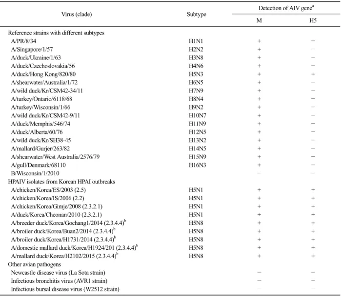
The specificity of the RT-LAMP assay was evaluated using viral RNAs extracted from 16 reference AIV strains with different H subtypes, one human influenza B virus, four H5N1 and five H5N8 Korean representa- tive HPAIV strains, and three other avian respiratory vi- ral pathogens (viz., Newcastle disease virus, infectious bronchitis virus, and infectious bursal disease virus vac- cine strains) (Table 1). The RT-LAMP with M gene-spe- cific primers successfully amplified the M genes of all tested AIVs, and the assay with H5 gene-specific pri- mers also successfully amplified H5 genes of all tested H5 subtype AIVs, including strains H5N3, H5N1, and H5N8, but not the influenza B virus and other avian pathogens. Furthermore, for the H5N1 and H5N8 Korean representative HPAIVs, the M and H5 genes were simultaneously detected by the RT-LAMP assay in a one-step reaction, as expected (Table 1). Although some researchers have previously described RT-LAMP
Table 1. Virus strains used for validation of the reverse transcription loop-mediated isothermal amplification (RT-LAMP) assay and its specificity
Virus (clade) Subtype Detection of AIV genea
M H5
Reference strains with different subtypes
A/PR/8/34 H1N1 + −
A/Singapore/1/57 H2N2 + −
A/duck/Ukraine/1/63 H3N8 + −
A/duck/Czechoslovakia/56 H4N6 + −
A/duck/Hong Kong/820/80 H5N3 + +
A/shearwater/Australia/1/72 H6N5 + −
A/wild duck/Kr/CSM42-34/11 H7N9 + −
A/turkey/Ontario/6118/68 H8N4 + −
A/turkey/Wisconsin/1/66 H9N2 + −
A/wild duck/Kr/CSM42-9/11 H10N7 + −
A/duck/Memphis/546/74 H11N9 + −
A/duck/Alberta/60/76 H12N5 + −
A/wild duck/Kr/SH38-45 H13N2 + −
A/mallard/Gurjer/263/82 H14N5 + −
A/shearwater/West Australia/2576/79 H15N9 + −
A/gull/Denmark/68110 H16N3 + −
B/Wisconsin/1/2010 − −
HPAIV isolates from Korean HPAI outbreaks
A/chicken/Korea/ES/2003 (2.5) H5N1 + +
A/chicken/Korea/IS/2006 (2.2) H5N1 + +
A/chicken/Korea/Gimje/2008 (2.3.2.1) H5N1 + +
A/duck/Korea/Cheonan/2010 (2.3.2.1) H5N1 + +
A/breeder duck/Korea/Gochang1/2014 (2.3.4.4)b H5N8 + +
A/broiler duck/Korea/Buan2/2014 (2.3.4.4)b H5N8 + +
A/broiler duck/Korea/H1731/2014 (2.3.4.4)b H5N8 + +
A/domestic mallard duck/Korea/H1924/201 (2.3.4.4)b H5N8 + +
A/mallard duck/Korea/H2102/2015 (2.3.4.4)b H5N8 + +
Other avian pathogens
Newcastle disease virus (La Sota strain) − −
Infectious bronchitis virus (AVR1 strain) − −
Infectious bursal disease virus (W2512 strain) − −
aViral RNA amplification was evaluated bythe developed two-tube RT-LAMP using M and H5 gene-specific primers (+, RT-LAMP positive; −, RT-LAMP negative).
bThis group of hemagglutinin gene segments was re-designated as clade 2.3.4.4 from the original clade of 2.3.4.6 according to a recommendation of the WHO/OIE/FAO H5 working group.
assays for the detection of the H5 gene of HPAIVs, these assays targeted for the gene of H5N1 but not H5N8 HPAIV, and never evaluated for the current cir- culating H5N8 subtype (Dhin et al, 2011; Imai et al, 2007; Jayawardena et al, 2007). The RT-LAMP assay developed in this study was capable of detecting both H5N1 and H5N8 HPAIVs, and expected to be more useful than previously reported RT-LAMP assays in countries where avian populations are infected by differ- ent H5 subtype HPAIVs, including H5N1 and H5N8 subtypes.
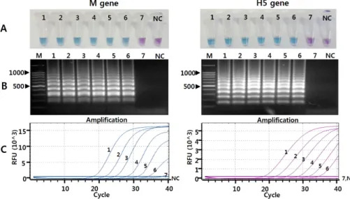
The sensitivity of the RT-LAMP assay was evaluated with Korean representative H5N1 [A/chicken/Korea/Gim- je/2008 (H5N1)] HPAIVs (Kim et al, 2012). The viruses were propagated in specific pathogen-free (SPF) em- bryonated chicken eggs (ECEs), and the 50% egg in- fectious dose (EID50) titers were determined in 11-day-old SPF ECEs using the Reed and Muench method (Reed and Muench, 1938). To calculate the limit of detection (LOD) of the assay, viral RNAs were extracted with tit- ers of 107.0 EID50/0.1 mL, serially 10-fold diluted in DEPC-treated water, and tested by the RT-LAMP assay,
Fig. 1. Comparison of the sensitivity of reverse transcription loop-mediated isothermal amplification (RT-LAMP) and real time reverse tran- scription polymerase chain reaction (RRT-PCR) assays with serially diluted RNAs of H5N1 HPAIV. (A) Detection of the RT-LAMP results by the hydroxynaphthol blue dye-mediated colorimetric method. Tubes 1∼7, 10-fold dilutions of viral RNAs from 107.0 to 101.0 EID50/0.1 mL; tube NC, negative control. (B) Detection of the RT-LAMP results by agarose gel electrophoresis. Lane M, 100 bp DNA marker; lanes 1∼7, 10-fold dilutions of viral RNAs from 107.0 to 101.0 EID50/0.1 mL; lane NC, negative control. (C) Results of RRT-PCR. Lines 1∼7, 10-fold dilutions of viral RNAs from 107.0 to 101.0 EID50/0.1 mL; line NC, negative control. The Ct values for the detection of M gene were determined as 17.3, 21.06, 23.92, 27.37, 30.35 and 35.51 for each serial viral diluent (from 107.0 to 101.0 EID50/0.1 mL), respectively. The Ct values for the detection of H5 gene were de- termined as 18.72, 22.11, 25.45, 28.58, 31.87 and 36.27 for each serial viral diluent (from 107.0 to 10 EID /0.1 mL), respectively.
and the results were compared with those of the pre- viously reported RRT-PCR assay (Kim et al, 2013). The RRT-PCR for detection of M or H5 gene of HPAIV was performed using a one-step PrimeScript RT-PCR kit (Takara Bio, Inc., Shiga, Japan) and a CFX96 TouchTM Real-Time PCR Detection System (Bio-Rad, USA) as previously described (Kim et al, 2013). For interpre- tation of the RRT-PCR results, samples producing a cy- cle threshold (Ct) of less than 37 were considered to be positive, and a high Ct value (>37) was considered to be negative. The LOD of the RT-LAMP assay for the M and H5 genes of H5N1 HPAIV was determined to be about 102.0 EID50/0.1 mL, and consistent with the LOD of the RRT-PCR assay (Fig. 1). Previously, some RT-LAMP assays were described for the detection of all AIV subtypes using M gene-specific primers (Shivakoti et al, 2010; Yoshida et al, 2011) or H5 HPAIVs using H5 gene-specific primers (Dhin et al, 2011; Imai et al, 2007; Jayawardena et al, 2007; Postel et al, 2010). In those studies, the sensitivity of each RT-LAMP assay was different according to the target gene or tested virus. Shivakoti et al, (2010) reported the sensitivity of the RT-LAMP assay for detection of the AIV M gene
to be 103.0 EID50/reaction (Shivakoti et al, 2010). Postel et al, (2010) reported the sensitivity of the RT-LAMP assay for the AIV H5 gene to be 1,000-fold lower than that of RRT-PCR assay (Postel et al, 2010). Considering these earlier results, the sensitivity of the RT-LAMP as- say in this study should be sufficient for the monitoring and surveillance of H5 and other AIV subtypes in bird populations with suspected HPAI (Lee et al, 2005; Zhao et al, 2013).
To evaluate the clinical performance of the developed RT-LAMP assay, a total of 627 fecal samples of domes- tic chickens (n=252) and wild birds (n=375) were col- lected from domestic chicken farms and migratory bird habitats in 2014∼2015 (Table 2), and tested by the RT-LAMP with M gene or H5 gene-specific primer set.
For further evaluation of virus-positive samples, the HA protein was subtyped as previously described (Kim et al, 2012). The RT-LAMP assay with M or H5 gene-specif- ic primers detected 15 samples as IAV-positive or one sample as H5 IAV-positive, respectively, which was con- sistent with the RRT-PCR results, Subtypes of IAV-pos- itive cases were confirmed as H5 (n=1), H6 (n=3), H8 (n=10) and H11 (n=1), respectively (Table 2).
Table 2. Results of virus detection in clinical samples by the M or H5 gene-specific reverse transcription loop-mediated isothermal amplification (RT-LAMP) and real time reverse transcription polymerase chain reaction (RRT-PCR)
Fecal samples No. of tested
No. of virus positive
HA subtype (No. of virus)
RRT-PCR RT-LAMP
M gene H5 gene M gene H5 gene
Domestic chicken 252 8 0 8 0 H9 (8)
Wild bird 375 7 1 7 1 H5 (1), H6 (3), H9 (2), H11 (1)
Total 627 15 15 1
With regard to the detection methods for the ampli- cons, previously described RT-LAMP assays for the de- tection of the AIV M or H5 gene determined the ampli- fication results by gel electrophoresis, real-time turbidity monitoring (derived by phosphate precipitates), and/or fluorescence dye-mediated colorimetric detection (Shivakoti et al, 2010; Yoshida et al, 2011; Dhin et al, 2011; Imai et al, 2007; Jayawardena et al, 2007; Postel et al, 2010).
However, these detection methods require complicated, bulky, and specialized instrumentations, which diminish the point-of-care testing capability of RT-LAMP, and may increase the potential risk for contamination of am- plified RT-LAMP products by opening the lid of the re- action tube. Therefore, to take full advantage of the RT-LAMP assay, the detection methodology should be simple, rapid, and cost-effective (Mori and Notomi, 2009). In this study, we were able to visually detect RT-LAMP results with the naked eye by adding HNB into the pre-reaction solution without opening the tubes, and without any additional detection process or appara- tus after amplification (Fig. 1), thereby reducing the chance of carryover contamination by the amplified product (Mori and Notomi, 2009; Goto et al, 2009; Cardoso et al, 2010).
Recently, some multiplex RT-LAMP (mRT-LAMP) assays were developed for the simultaneous detection of more than two specific genes in a single reaction tube (Kouguchi et al, 2010; Jung et al, 2015; Mahonya et al, 2013). However, further processing and additional equip- ment are required for these methods including restriction enzyme digestion (Kouguchi et al, 2010), immunoch- romatographic detection system (Jung et al, 2015), or
real time fluorometer (Mahonya et al, 2013), making it more laborious, time and cost consuming. Moreover, the high sensitivity of the RT-LAMP assay raises the con- cern of carry-over contamination during post-amplifica- tion analysis (Kouguchi et al, 2010; Jung et al, 2015).
Despite the inconvenience of using two tubes, the two-tube RT-LAMP assay that can visually detect the result of the assay and prevent a carry-over contamination thought to be more preferable than mRT-LAMP for si- multaneous detection of M and H5 genes of AIV. The highly specific and sensitive RT-LAMP assay developed in the study can serve as a rapid, cost-effective, and user-friendly diagnostic tool for currently circulating H5 subtype of HPAIVs, and other subtypes of AIV in avian populations, even in under-equipped laboratories.
ACKNOWLEDGMENTS
We thank Dr. Y.J. Lee (Animal and Plant Quarantine Agency, Korea) for providing the influenza viruses. This research was supported by Animal Disease Management Technology Development (Project No. 313060-03-1-HD020) and Golden Seed Project (Project No. PJ009921), Rural Development Administration (RDA), Ministry of Agri- culture, Food and Rural Affairs (MAFRA), Republic of Korea.
CONFLICT OF INTEREST
The authors declare no conflict of interest.
REFERENCES
Cardoso TC, Ferrari HF, Bregano LC, Silva-Frade C, Rosa ACG, Andrade AL. 2010. Visual detection of turkey coronavi- rus RNA in tissues and feces by reverse-transcription loop-mediated isothermal amplification (RT-LAMP) with hydroxynaphthol blue dye. Mol Cell Probes 24: 415-417.
Dinh DT, Le MTQ, Vuong CD, Hasebe F, Morita K. 2011. An updated loop-mediated isothermal amplification method for rapid diagnosis of H5N1 avian influenza viruses.
Trop Med Health 39: 3-7.
Goto M, Honda E, Ogura A, Nomoto A, Hanaki KI. 2009.
Colorimetric detection of loop-mediated isothermal am- plification reaction by using hydroxynaphthol blue.
Biotechniques 46: 167-172.
Imai M, Ninomiya A, Minekawa H, Notomi T, Ishizaki T, Van Tu P, Tien NTK, Tashiro M, Odagiri T. 2007. Rapid di- agnosis of H5N1 avian influenza virus infection by new- ly developed influenza H5 hemagglutinin gene-specific loop-mediated isothermal amplification method. J Virol Methods 141: 173-180.
Jayawardena S, Cheung CY, Barr I, Chan KH, Chen H, Guan Y, Peiris JSM, Poon LLM. 2007. Loop-mediated isothermal amplification for influenza A (H5N1) virus. Emerg Infect Dis 13: 899-901.
Jung JH, Oh SJ, Kim YT, Kim SY, Kim WJ, Jung J, Seo TS.
2015. Combination of multiplex reverse-transcription loop-mediated isothermal amplification with an im- munochromatographic strip for subtyping influenza A virus. Anal Chim Acta 853: 541-547.
Kim EM, Jeon HS, Kim JJ, Shin KK, Lee YJ, Yeo SG, Park CK.
2015. Evaluation of reverse-transcription loop-mediated isothermal amplification assay for screening influenza A viruses from different animal species. J Anim Vet Adv 14: 155-160.
Kim HR, Lee YJ, Park CK, Oem JK, Lee OS, Kang HM, Choi JG, Bae YC. 2012. Highly pathogenic avian influenza (H5N1) outbreaks in wild birds and poultry, South Korea. Emerg Infect Dis 18: 480-483.
Kim HR, Oem JK, Bae YC, Kang MS, Lee HS, Kwon YK. 2013.
Application of real-time reverse transcription polymerase chain reaction to the detection the matrix, H5 and H7 genes of avian influenza viruses in field samples from South Korea. Virol J 10: 85.
Kouguchi Y, Fujiwara T, Teramoto M, Kuramoto M. 2010.
Homogenous, real-time duplex loop-mediated isothermal amplification using a single fluorophore-labeled primer and an intercalator dye: Its application to the simulta- neous detection of Shiga toxin genes 1 and 2 in Shiga toxigenic Escherichia coli isolates. Mol Cell Probes 24:
190-195.
Lee C, Suarez DL, Tumpey TM, Sung H, Kwon Y, Lee Y, Choi
J, Joh S, Kim M, Lee E, Park J, Lu X, Katz JM, Spackman E, Swayne DE, Kim J. 2005. Characterization of highly pathogenic H5N1 avian influenza A viruses isolated from South Korea. society 79: 3692-3702.
Lee YJ, Kang HM, Lee EK, Song BM, Jeong J, Kwon YK, Kim HR, Lee KJ, Hong MS, Jang I, Choi KS, Kim JY, Lee HJ, Kang MS, Jeong OM, Baek JH, Joo YS, Park YH, Lee HS. 2014. Novel reassortant influenza A (H5N8) vi- ruses, South Korea, 2014. Emerg Infect Dis 20:
1087-1089.
Mahony J, Chong S, Bulir D, Ruyter A, Mwawasi K, Waltho.
2013. Multiplex loop-mediated isothermal amplification (M-LAMP) assay for the detection of influenza A/H1, A/H3 and influenza B can provide a specimen-to-result diagnosis in 40min with single genome copy sensitivity.
J Clin Virol 58: 127-131.
Mori Y, Notomi T. 2009. Loop-mediated isothermal amplification (LAMP): A rapid, accurate, and cost-effective diagnostic method for infectious diseases. J Infect Chemother 15:
62-69.
Notomi T, Okayama H, Masubuchi H, Yonekawa T, Watanabe K, Amino N, Hase T. 2000. Loop-mediated isothermal am- plification of DNA. Nucleic Acids Res 28: 63-69.
Postel A, Letzel T, Frischmann S, Grund C, Beer M, Harder T.
2010. Evaluation of two commercial loop-mediated iso- thermal amplification assays for detection of avian influ- enza H5 and H7 hemagglutinin genes. J Vet Diagn Invest 22: 61-66.
Reed LJ, Muench H. 1938. A simple method of estimating fifty percent endpoints. American J Epidemiol 27: 493- 497.
Shivakoti S, Ito H, Murase T, Ono E, Takakuwa H, Yamashiro T, Otsuki K, Ito T. 2010. Development of reverse tran- scription-loop-mediated isothermal amplification (RT-LAMP) assay for detection of avian influenza vi- ruses in field specimens. J Vet Med Sci 72: 519-523.
WHO. 2014. WHO information for molecular diagnosis of influ- enza virus - update 56.
WHO. 2015. Evolution of the influenza A (H5) haemagglutinin:
WHO/OIE/FAO H5 Working Group reports a new clade designated 2.3.4.4.
Yoshida H, Sakoda Y, Endo M, Motoshima M, Yoshino F, Yamamoto N, Okamatsu M, Soejima T, Senba S, Kanda H, Kida H. 2011. Evaluation of the reverse transcription loop-mediated isothermal amplification (RT-LAMP) as a screening method for the detection of influenza viruses in the fecal materials of water birds. J Vet Med Sci 73:
753-758.
Zhao K, Gu M, Zhong L, Duan Z, Zhang Y, Zhu Y, Zhao G, Zhao M, Chen Z, Hu S, Liu W, Liu X, Peng D, Liu X.
2013. Characterization of three H5N5 and one H5N8 highly pathogenic avian influenza viruses in China. Vet Microbiol 163: 351-357.