67 Copyright © 2015 The Korean Society of Cardiology
Korean Circulation Journal
Introduction
Stent fractures have recently become an important concern in the medical community due to their potential association with se- rious conditions such as in-stent restenosis (ISR) and stent throm- bosis after drug-eluting stent (DES) implantation.
1) Most currently reported stent fractures are found in lesions implanted with siroli- mus-eluting stents (SES), likely due to the inherent platform material and design of such stents.
2) Furthermore, while the exact timing of fractures following stent implantation remains unclear, nearly all reports identify fractures after a time period of follow-up for patients after stenting, suggesting a delayed complication timeline.
1)3-5) In this report, we present two unusual cases of immediate stent fracture that occurred after implantation of new-generation DES, a zotarolimus-
Case Report
http://dx.doi.org/10.4070/kcj.2015.45.1.67
Print ISSN 1738-5520 • On-line ISSN 1738-5555
Two Cases of Immediate Stent Fracture after Zotarolimus-Eluting Stent Implantation
Pil Hyung Lee, MD, Seung-Whan Lee, MD, Jong-Young Lee, MD, Young-Hak Kim, MD, Cheol Whan Lee, MD, Duk-Woo Park, MD, Seong-Wook Park, MD, and Seung-Jung Park, MD
Department of Cardiology, University of Ulsan College of Medicine, Asan Medical Center, Seoul, Korea
Drug-eluting stent (DES) implantation is currently the standard treatment for various types of coronary artery disease. However, previous reports indicate that stent fractures, which usually occur after a period of time from the initial DES implantation, have increased during the DES era; stent fractures can contribute to unfavorable events such as in-stent restenosis and stent thrombosis. In our present report, we describe two cases of zotarolimus-eluting stent fracture: one that was detected six hours after implementation, and the other case that was detected immediately after deployment. Both anatomical and technical risk factors contributed to these unusual cases of imme- diate stent fracture. (Korean Circ J 2015;45(1):67-70)
KEY WORDS: Drug-eluting stents; Percutaneous coronary intervention; Complications.
Received: February 23, 2014 Revision Received: May 17, 2014 Accepted: June 9, 2014
Correspondence: Seung-Jung Park, MD, Department of Cardiology, Uni- versity of Ulsan College of Medicine, Asan Medical Center, 86 Asanbyeong- won-gil, Songpa-gu, Seoul 138-736, Korea
Tel: 82-2-3010-3152, Fax: 82-2-486-5918 E-mail: [email protected]
• The authors have no financial conflicts of interest.
This is an Open Access article distributed under the terms of the Creative Commons Attribution Non-Commercial License (http://creativecommons.
org/licenses/by-nc/3.0) which permits unrestricted non-commercial use, distribution, and reproduction in any medium, provided the original work is properly cited.
eluting stent (ZES).
Cases
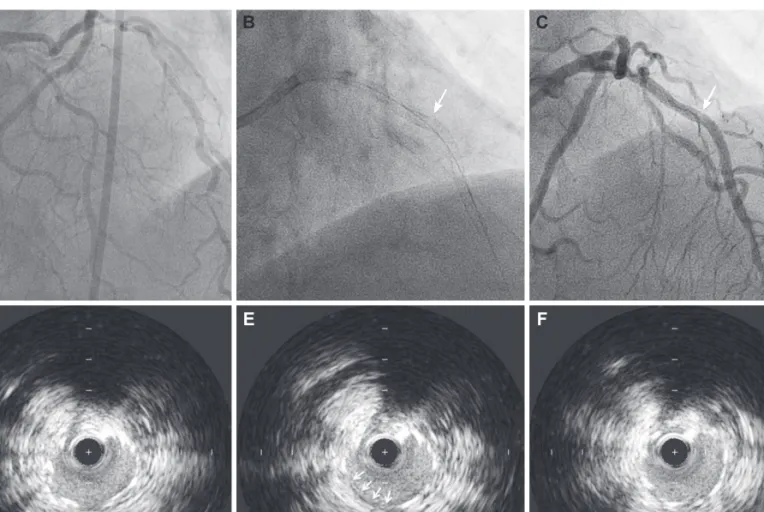
Case 1
A 62-year-old man, who had undergone cardiac transplantation
for advanced heart failure, was referred to our catheterization labo-
ratory for a regular surveillance coronary angiogram and concomi-
tant intravascular ultrasound (IVUS) imaging. After identifying a
normal right coronary angiogram (Fig. 1), the clinician attempted to
insert a 0.014-inch BMW (Abbott Vascular, Melno Park, CA, USA)
wire for IVUS evaluation. However, resistance was quickly met, and
the wire failed to pass through the proximal portion of the right
coronary artery (RCA). A subsequent angiogram revealed a spiral lu-
minal filling defect from the proximal to the distal RCA indicating
coronary dissection and Thrombolysis in Myocardial Infarction (TIMI)
2 flow (Fig. 1). The ST segment elevation appeared in lead II and III
on electrocardiography (ECG) monitoring, while hemodynamic pa-
rameters were stable. Urgent bailout stenting was performed: the
RCA was engaged with 8 Fr guiding catheter (JR 4.0, Cordis, Bridge-
water, NJ, USA); a BMW wire was inserted using a 1.8 Fr Finecross
(Terumo Medical, Tokyo, Japan) micro-guiding catheter; the true lu-
men was confirmed by IVUS-virtual histology; and three ZES stents
(Resolute Integrity 2.75×30 mm, 3.0×38 mm, 3.5×30 mm, Medtron-
ic, Santa Rosa, CA, USA) were deployed in order from distal to proximal
at 12, 16, and 16 atm, respectively, with overlap between adjacent