Yonsei Med J http://www.eymj.org Volume 50 Number 4 August 2009 585
Cholesterol granuloma (CG) of the middle ear typically presents as a conductive hearing loss and a blue eardrum, whereas those at the petrous apex either manifest as the side effects of bony erosion (with sensorineural hearing loss, tinnitus, vertigo, or cranial nerve impairment), or are incidentally identified.1CG can be a perfectly localized and isolated mass in any pneumatized area in temporal bone, the middle ear cavity,2,3mastoid antrum,4external auditory canal,5or the petrous apex,1as has been found in as many as 12 - 20% of temporal bones with chronic otitis media.2-4 According to a histopathologic study, CG may be present in 12%
of patients with chronic otitis media with an intact tympanic membranes, and in 21% of those with a perforated tympanic membrane.6However, intramembranous tympanic membrane CG occurs less frequently, and only one case has so far been reported in the English literature.7Here, we report our experiences with a case of CG in the tympanic membrane and review relevant medical literature.
A 42-year-old man presented with a history of childhood coalescent otitis media and, as consequent, hearing loss in the right ear. He complained of middle ear fullness without otalgia. A blue eardrum was identified at the local clinic. The patient was referred to our department. Otomicroscopic findings of the right tympanic membrane demonstrated a bulging blue eardrum. Two weeks after presentation, otomicroscopic findings showed a pinhole perforation in the anterior area with a brown discharge. A
Case Report
DOI 10.3349/ymj.2009.50.4.585pISSN: 0513-5796, eISSN: 1976-2437 Yonsei Med J 50(4): 585-587, 2009
Cholesterol Granuloma of the Tympanic Membrane Presenting as a Blue Eardrum
Chul Ho Jang,
1,2Jun Sung Kim,
1and Yong Bum Cho
11Department of Otolaryngology, Chonnam National University Medical School, Gwangju; 2Research Center for Resistant Cells, Chosun Medical School, Gwangju, Korea.
Intramembranous tympanic membrane cholesterol granuloma (CG) occurs infrequently. Here, the authors report a case of CG in the tympanic membrane presenting as a blue eardrum in the right ear. In addition, a pinhole perforation noted in the anterosuperior area revealed a brown discharge. High-resolution temporal bone CT showed a bulging mass shadow in the middle ear and a soft tissue dense lesion that filled both the epitympanum and mastoid cavity. Tympanomastoidectomy was performed under general anesthesia. New bone formation was confirmed in the mastoid antrum and epitympanum, and the epitympanum was blocked by new bone. The tympanic membrane revealed a round, brownish mass with a glistening surface and a severely thickened pars tensa.
We herein report this case and review pertinent medical literature.
Key Words : Cholesterol granuloma, tympanic membrane
Received: May 15, 2007 Revised: November 15, 2007 Accepted: November 15, 2007 Corresponding author: Dr. Chul Ho Jang, Department of Otolaryngology,
Chonnam National University Medical School, 8 Hak-dong, Gwangju 501-757, Korea.
Tel: 82-62-220-6774, Fax: 82-62-220-6776 E-mail: [email protected]
∙The authors have no financial conflicts of interest.
© Copyright:
Yonsei University College of Medicine 2009
INTRODUCTION
CASE REPORT
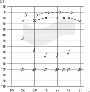
pure tone audiogram showed a dead ear on the right side (Fig.
1). High resolution temporal bone CT showed a bulging mass shadow in the middle ear and a dense soft tissue lesion, which filled both the epitympanum and mastoid cavity. New bone formation was noted between the epitympanum and middle ear (Fig. 2A), and bulging soft tissue in the middle ear (Fig. 2B). No definite ossicular shadow was identified.
Tympanomastoidectomy was performed under general anesthesia, and new bone formation was confirmed in the mastoid antrum and epitympanum, the latter of which was blocked by the new bone. No ossicular structure was identified in the epitympanum. The dead ear finding is believed to have been caused by a complication of childhood coalescent otitis media. After elevation of the tympanomeatal flap, the tympanic membrane revealed a round, brownish mass with a glistening surface and a severely thickened pars tensa (Fig. 3). The incus and stapes were not identified. After removing the mass from the tympanic membrane, it was sent
for pathological examination, which confirmed CG (Fig. 4).
Postoperatively, the patient had an uneventful course without any sign of blue eardrum recurrence.
Three factors are considered to play an important role in the development of CGs: interference with drainage, hemorrhage, and ventilation obstruction,8although remain- ing mesenchyme9and hematopoietic bone marrow10,11have also been suggested to be associated with CG formation.
Clinically, CG of the temporal bone appears in three forms:12in association with chronic otitis media (especially cholesteatoma), idiopathic hemotympanum, or a localized lesion in the middle ear, mastoid antrum, external auditory canal, and petrous apex. CG can also occur without any previous history of infection.2,13
In the present case, the patient had a history of otitis media from childhood. Moreover, the bulging tympanic Chul Ho Jang, et al.
Yonsei Med J http://www.eymj.org Volume 50 Number 4 August 2009 586
DISCUSSION
125 130 120 110 100 90 80 70 60 50 40 30 20 10 0 -10 (dB)
250 500 1 k 2 k 4 k 8 k (Hz)
Fig. 1. The pure tone audiogram showing that the hearing level in the right ear had scaled out at all frequencies.
Fig. 3. After elevation of the tympanomeatal flap, the inner surface of the tympanic membrane showed this round, brownish mass which had a glistening surface (arrow indicates) with severely thickened pars tensa at the posteroin- ferior quadrant.
Fig. 4. Microscopic examination showing typical foreign body giant cells (arrows indicate) surrounding cholesterol crystals (Hematoxylin & Eosin stain, ×100) Fig. 2. High-resolution temporal bone CT findings of the middle ear and mastoid.
(A) Temporal bone CT showing a dense soft tissue lesion filling both the epitympanum and mastoid cavity on the right side. New bone formation as a complication of childhood coalescent mastoiditis was noted. (B) Shows the bulging soft tissue in the middle ear.
A
Rt Rt
B
Cholesterol Granuloma of Tympanic Membrane
Yonsei Med J http://www.eymj.org Volume 50 Number 4 August 2009 587
membrane had a pinhole perforation with brown discharge in the anterior pars tensa. CGs contain hemosiderin, lipids, and cholesterol crystals. Microscopically, these crystals are surrounded by foreign body giant cells in fibrous connec- tive tissue, which is infiltrated by round cells.2
Jaisinghani et al.14correlated pathologic findings of the tympanic membrane with pathologic changes in the middle ear cleft in cases with chronic otitis media in 150 temporal bones. They found a significant correlation between CG and tympanic membrane retraction and a mild additive effect when retraction was associated with perforation, however, the described case showed no tym- panic membrane retraction. CG in the tympanic membrane has been rarely reported. In a recently published case, Haginomori et al.7reported histopathological charac- teristics of CG in the tympanic membrane, and speculated that, when hemorrhage occurs in the tympanic membrane, it is likely due to the absence of drainage or air exchange systems. On the other hand, when a CG develops in the tympanic membrane, it may promote its own growth in a vicious circle, by becoming another source of hemorrhage from small vessels whose numbers continue to increase.
Park15suggested that blue eardrum is an independent clinical entity, and found no bone changes with temporal bone CT or surgical findings. In the present case, however, temporal bone CT showed new bone formation in the epitympanum. Moreover, intraoperative findings indicated a complete attic block by new bone formation.
This finding together with the resolution of blue eardrum indicate that the main reason for blue eardrum in the present case was CG at the tympanic membrane.
In conclusion, we report a case of CG of the tympanic membrane, which presented as blue eardrum.
This work was supported by the National Research Foundation (NRF) grant funded by the Korea government (MEST) through the Research Center for Resistant Cells (R13-2003-009).
1. Brackmann DE, Toh EH. Surgical management of petrous apex cholesterol granulomas. Otol Neurotol 2002;23:529-33.
2. Miglets AW, Booth JB. Cholesterol granuloma presenting as an isolated middle ear tumor. Laryngoscope 1981;91:410-5.
3. Hoffman RA. Cholesterol cyst manifesting a middle ear vascular tumor. Am J Otolaryngol 1984;5:68-70.
4. Palva T, Lehto VP, Johnsson LG, Virtanen I, Mäkinen J. Large cholesterol granuloma cysts in the mastoid. Clinical and histopa- thologic findings. Arch Otolaryngol 1985;111:786-91.
5. Matt BH, Myer CM 3rd, Bellet PS. Cholesterol granuloma present- ing the ear canal. Ann Otol Rhinol Laryngol 1990;99:672-3.
6. da Costa SS, Paparella MM, Schachern PA, Yoon TH, Kim- berley BP. Temporal bone histopathology in chronically infected ears with intact and perforated tympanic membranes. Laryn- goscope 1992;102:1229-36.
7. Haginomori S, Takamaki A, Ito K, Takenaka H, Kurisu Y, Tsuji M. Cholesterol granuloma in the tympanic membrane. Otol Neurotol 2006;27:1201-2.
8. Nager GT, Vanderveen TS. Cholesterol granuloma involving the temporal bone. Am Otol Rhinol Laryngol 1976;85:204-9.
9. Nager GT. Cholesterol granuloma. In: Nager GT, Hyams VJ, editors. Pathology of the ear and temporal bone. Baltimore:
Williams & Wilkins; 1993. p.914-39.
10. Jackler RK, Cho M. A new theory to explain the genesis of petrous apex cholesterol granuloma. Otol Neurotol 2003;24:96-106.
11. Miura M, Sando I, Orita Y, Hirsch BE. Histopathologic study of the temporal bones and Eustachian tubes of children with cho- lesterol granuloma. Ann Otol Rhinol Laryngol 2002;111:609-15.
12. Alaminos D, Gamboa J, Prades J. Cholesterol granuloma in the middle ear. In: Sade J, editor. Basic aspects of the Eustachian tube and middle ear disease. Conference on the Eustachian tube and middle ear disease. Genova: Kugler and Ghedini Publi- cations; 1991. p.103-9.
13. Leonetti JP, Shownkeen H, Marzo SJ. Incidental petrous apex findings on magnetic resonance imaging. Ear Nose Throat J 2001;80:200-2, 205-6.
14. Jaisinghani VJ, Paparella MM, Schachern PA, Le CT. Tympanic membrane/middle ear pathologic correlates in chronic otitis media.
Laryngoscope 1999;109:712-6.
15. Park K. Cholesterol granuloma of the middle ear. In: Park KH, editor. Middle ear diseases. Seoul: Academy’A; 2002. p.53-9.