정중흉골절개술(median sternotomy)은 종격동 종괴의 수술 이나 대부분의 심장수술에 가장 많이 사용되는 개흉방법이다.
정중흉골절개술 후 합병증은 0 . 5 % - 5 %로 매우 빈도가 낮으며, Serry 등( 1 )은 합병증의 스펙트럼을 (a) 무균성 장액혈액성 배농(sterile serosanguineous discharge)이 있는 안정성 흉골 (stable sternum); (b) 무균성 배농이 동반되거나 동반되지 않 은 불안정성 흉골(unstable sternum); (c) 종격동염( m e d i a s- t i n i t i s )이 동반되지 않은 흉골열개(sternal dehiscence); (d) 종 격동염이 동반되지 않은 표재성 창상 감염(superficial wound infection); (e) 피하감염(subcutaneous infection), 불안정성 흉 골과 흉골후방침범; (f) 흉골열개가 동반되거나 동반되지 않 은 종격동염 등 6가지로 분류하였다. 그들의 연구에서 초기 감염을 가지지 않은 환자의 경우(a, b) 적절한 치료로 모두 생존하였으나, 종격동염 없이 흉골열개나 표재성 감염이 있었 던 환자의 경우(c, d) 사망률이 2 4 %였으며, 심부 감염이 있었 던 환자의 경우(e, f) 70% 이상의 사망률을 보였다(1). 낮은 합병증의 빈도에도 불구하고 높은 사망률은 합병증의 조기 발견과 즉각적인 치료를 요구한다. 이러한 합병증을 가지고 있는 환자는 임상 소견상 국소적으로는 통증, 압통, 발적( e r y- thema), 수술 후 배농 등을 일으키며, 전신적으로는 발열, 패 혈증, 백혈구증가증(leukocytosis) 등을 자주 일으킨다. 이러한 임상적 소견만으로 정중흉골절개술 후 감염 부위의 깊이를 판단하는 것은 매우 어렵다. 또한 임상 소견과 기존의 영상기 법으로도 감염 부위의 범위를 진단하는 것은 어렵다. 단순흉 부사진 역시 정중흉골절개술의 합병증의 유무와 병변의 범위 를 평가하는 데는 별로 도움이 되지 않는다(7). 따라서, 정중
흉골절개술의 합병증의 유무와 병변의 범위를 평가하는 데는 C T가 중요한 역할을 한다(5). 또한 C T를 이용하여 배액 가능 한 농양과 미만성 종격동염을 구별할 수 있고, 농양과 농흉 (empyema) 사이의 교통여부를 알아내어 불필요한 수술을 줄 일 수 있다( 6 ) .
정중흉골절개술 후에 흉골후방에 감염이 있거나 흉골열개 가 있는 경우에는 종격동 감염의 배액과 흉골 재결찰( r e s u t u r- i n g )을 위해 재수술이 필요하며, 이보다 표면에 국한된 감염 은 국소적인 처치만으로 치료가 가능하다(2). 널리 알려져 있 는 재수술 방법은 근치적인 변연절제술(debridement), 대흉근 (pectoralis major muscle)이나 복직근(rectus abdominis muscle) 을 이용한 근육판(muscle flap) 전위( t r a n s p o s i t i o n )와 대망판 (omental flap) 전위이다.
흉골주위조직의 합병증
흉골주위조직은 흉골 앞쪽으로는 피부와 피하조직, 흉골 뒤 쪽으로는 종격동 지방으로, 흉골 양 옆으로는 늑연골( c o s t a l c a r t i l a g e )과 늑간근육(intercostal muscle)으로 이루어져 있다.
수술 후 피부결손과 부종이 정상적으로 보일 수 있으며, 대부 분 흉골앞 연부조직은 빠르게 정상화되고 조직내에 보이던 공기음영 또한 일주일이 지나면 대개 보이지 않는다. 수술 후 흉골은 좀처럼 완벽하게 근사( a p p r o x i m a t i o n )되어 있지는 않 으며 전종격동에 경도의 부종이나 국소적 액체음영이 있을 수 있다(3). 대부분의 흉골주위조직 합병증은 염증 내지 감염, 농양형성, 배액누관(draining sinus tract)에 의한 것이다. 표재 성 감염의 경우, 수술 중에 피하조직의 오염( c o n t a m i n a t i o n )이 일어나는 것으로 생각되고 수술 후 5일 내지 7일 이내에 감염 소견이 명확해진다. 환자의 면역체계가 감염을 국소화( l o c a l- 정중흉골절개술(median sternotomy)은 종격동 종괴의 수술이나 대부분의 심장수술에 가장
많이 사용되는 개흉방법이다. 정중흉골절개술 후 합병증은 0 . 5 % - 5 %로 매우 빈도가 낮으나, 실제 발생할 경우 매우 위험하며 높은 사망률을 보이기 때문에 합병증의 빠른 발견과 즉각 적인 치료를 요구한다. 단순흉부촬영과 임상적 소견만으로 정중흉골절개술 후 합병증의 존 재와 침범 정도를 판단하는 것은 매우 어렵다. 이에 비해 C T는 정중흉골절개술의 합병증의 유무와 병변의 범위를 평가하는데 중요한 정보를 제공한다. 이 임상화보에서는 정중흉골절 개술 후 생긴 합병증들의 임상적 소견과 C T소견을 기술하고자 한다.
1서울대학교 의과대학 방사선과학교실, 방사선의학연구소
2서울시립 보라매병원 진단방사선과
이 논문은 1 9 9 9년 1월 6일 접수하여 1 9 9 9년 3월 2일에 채택되었음.
정중흉골절개술의 합병증: CT 소견1
최영호・구진모・서준범・송재우2・이동경・한대희・임정기
i z a t i o n )시킬 수 없을 경우 감염은 흉골을 통해 종격동까지 범 위가 확대되고 피부를 통해 배농된다. 대부분의 정상면역체계 를 유지하고 있는 환자는 표재성 감염의 종격동 침윤을 막기 위해 5일 내지 7일 이내에 흉골 뒤 연부조직에 효과적인 방어 선을 구축한다. 더 심각한 문제는 수술 중 종격동에 일차적인 오염이 있을 때 발생한다. 이런 경우 감염은 흉골을 통해 피 하조직으로, 그리고 농성배액(pus drainage)으로 나타난다(2).
표재성 창상감염의 발견은 대개 봉와직염(cellulitis), 농성배 액, 또는 만져지는 농양에 의한 임상적 소견에 기초를 두고 있다. 감염의 흉골이나 종격동으로의 확장을 밝혀내는 것이 중요한데, 이는 높은 질병 이환율과 높은 사망률이 신속하고 공격적인 수술적 처치를 요구하기 때문이다(1). CT는 염증변 화의 정도, 농양의 크기, 주위조직과의 관계를 잘 보여준다.
농성배액이 있다면 CT sinography가 종격동으로의 확장을 확 인하는데 도움을 줄 수 있다. 드물게 늑연골염이 올 수 있는 데 C T에서 염증의 정도와 주위조직과의 관계를 잘 볼 수 있 다(Fig. 1). 흉골주위조직의 문제없이 수술 창상부위의 심한 통증만이 있을 수 있는데 이는 잠재적인 심각한 합병증의 하 나로 고려되어져야 한다( 9 ) .
흉골열개
수술 후 흉골열개를 일으킬 수 있는 요인은 감염, 재수술, 심폐소생술, 지속적인 기계적 호흡술, 그리고 부적절한 절개 또는 봉합 등이다. 국소적인 상처 감염은 흉골의 유합( u n i o n ) 을 방해하고 결과적으로 흉골의 분리를 일으킨다. 반대로 일 차적인 흉골열개가 이차적으로 감염을 일으키기도 한다( 3 ) . 흉골의 분리는 대개 방사선학적 소견이 있기 이전에 임상적 으로 명확해진다. 흉골의 불안정( i n s t a b i l i t y )은“c l i c k”으로 나 타나거나 기침에 의해 불안정이 강조되는 것으로 나타난다.
흉골열개의 가장 믿을 만한 방사선학적 소견은 주변조직과 봉합물질의 상대적 위치가 변하는 것이다(Fig. 2). 흉골에서 보이는 얇은 방사선투과도( r a d i o l u c e n c y )는 수술 후에 정상적 으로 보일 수 있고 흉골열개를 의미하는 것은 아니다. 점진적 으로 넓어지는 방사선투과도가 대개 흉골의 분리를 의미하지
만, 흉골열개가 있다고 하여 이것이 모두 의미 있는 임상적 문제를 가지는 것은 아니다. CT상에서 흉골열개는 쉽게 찾아 지지만, 다른 소견들이 모호하다면 임상적 소견과 9 9 m T c - p h o s p h a t e와 6 7 G a - c i t r a t e스캔 등의 추가적인 검사가 도움을 줄 수 있다. 스캔에서 불규칙한 모양으로 흉골 또는 종격동의 넓은 부위에서 강한 활동성( a c t i v i t y )을 보인다면 동반된 감염 을 시사한다. 동반된 감염이 없다면 방사핵종( r a d i o n u c l i d e )의 흡수( u p t a k e )는 미미하거나 절개가 가해진 골과 연부조직의 좁은 부위에서 미약하게 보일 수 있다(8). 임상적으로 합병증 이 없는 경우도, 수술 후 3주 이내에 실시된 C T에서 흉골이 2 mm 이하로 분리되어 보이거나, 어긋남(step-off), 감입( i m- paction) 등의 소견이 보고되어 있다( 3 ) .
흉골골수염
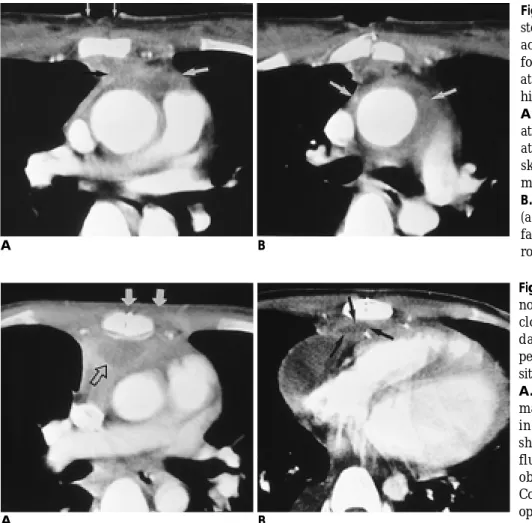
수술적 절개의 직접적 효과로부터 흉골골수염을 구분해내 는 것은 어렵다. 경미한 정도의 흉골의 분리나 어긋남은 증상 이 없고 합병증이 없는 수술에서도 흔하다. 단순흉부촬영에서 흉골에 골막거상(periosteal elevation)이 보일 수 있는데 이는 창상감염의 증거가 없는 환자에서도 보일 수 있어 수술 후 흉 골에 생긴 골수염의 특징적인 소견이라고 할 수는 없다. 불행 하게도 분명한 골파괴(bone destruction) 소견만이 골수염의 믿을만한 방사선학적 소견으로 알려져 있다(7). CT상에서 골 수염 초기의 흉골소견은 수술 중 톱질에 의한 약간 불규칙해 보이는 소견 혹은 정상변이(normal variation)와 구별되기 힘 드나, 진행하면서 흉골에 분명한 골파괴, 심한 광물이탈( d e m- ineralization), 열개 등이 보이게 되며, 미란(erosion), 골막성 신생골형성(periosteal new bone formation), 골경화( s c l e r o s i s ) , 주변의 연부조직부종 등도 보일 수 있다(5) (Fig. 3, 4).
종격동염
종격동에서의 합병증은 생명을 위협할 만큼 심각한 것이며 농양, 종격동염, 종격동 혈종, 심낭삼출 또는 혈종, 흉수, 농흉 등이 있다. 그 중 가장 흔히 접하고 수술 후 사망률을 높이는
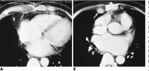
Fig. 1. 40-year-old man with acute peri- chondritis. He had had mitral valve re- placement(MVR) 40 days before and was suffering from substernal pain and discharge at wound site. Postoperative pathologic diagnosis was acute peri- c h o n d r i t i s .
A . Postcontrast CT scan shows a bulging low-attenuation lesion (arrow) surrounding the costal cartilage. Right half of the sternum (open arrow) shows bony destruction.
B . A scan at slightly upper level shows an enlarged ipsilateral internal mam- mary lymph node(arrow).
A B
C D
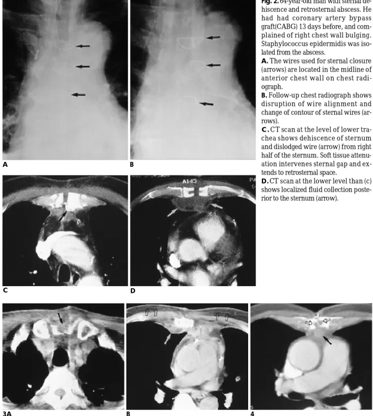
Fig. 2. 64-year-old man with sternal de- hiscence and retrosternal abscess. He had had coronary artery bypass graft(CABG) 13 days before, and com- plained of right chest wall bulging.
Staphylococcus epidermidis was iso- lated from the abscess.
A. The wires used for sternal closure (arrows) are located in the midline of anterior chest wall on chest radi- o g r a p h .
B. Follow-up chest radiograph shows disruption of wire alignment and change of contour of sternal wires (ar- r o w s ) .
C . CT scan at the level of lower tra- chea shows dehiscence of sternum and dislodged wire (arrow) from right half of the sternum. Soft tissue attenu- ation intervenes sternal gap and ex- tends to retrosternal space.
D . CT scan at the lower level than (c) shows localized fluid collection poste- rior to the sternum (arrow).
A B
Fig. 3. 64-year-old man with osteomyelitis of the sternum. The patient had had CABG 40 days before, and was suffering from fever, discharge, and substernal pain. No drainage procedure was performed between the initial operation and the CT exam.
A . The precontrast CT scan shows bony destruction of sternum (arrow). Soft tissue density replaces anterior portion of the ster- n u m .
B . CT scan at 5cm lower level than (a) shows posteriorly bulging peristernal soft tissue lesion (arrow) in anterior chest wall. Note al- so inflammatory thickening of retromammary fascia (open arrows).
Fig. 4. 61-year-old man with osteomyelitis of the sternum and retrosternal abscess. He had had mitral valve replacement 79 days b e f o r e .
CT scan shows concave smooth bony defect at the sternotomy site (open arrows). The anterior mediastinal fat is partially obliterat- ed by infiltration of soft tissue (arrow).
3 A B 4
것이 종격동염이다. 종격동염의 일반적인 임상소견은 백혈구 증가증, 적혈구침강속도(erythrocyte sedimentation rate)의 증 가, 열, 빈맥, 그리고 호흡곤란 등이다. 하지만 이러한 소견들 이 나타나지 않는 경우도 있으며 무기폐, 수술후 스트레스, 또 는 원발성 심장질환(primary heart disease) 등의 다른 질환에 서 나타나는 소견으로 오인되기도 한다. 종격동염의 주된 방 사선학적 소견은 종격동 확장, 종격동기흉( p n e u m o m e d i- astinum), 종격동 지방층의 소실, 국소 액체저류(localized fluid collection), 그리고 농양형성이고, 일측 혹은 양측 흉수의 동반 이 흔하다. 수술 후 시행한 단순흉부촬영은 빈번히 비정상적 으로 보이며, 상부 종격동의 확장, 종괴효과(mass effect), 이소 성 기체(ectopic gas), 관련된 흉막질환을 보여준다. 그러나, 이 러한 단순흉부촬영의 소견은 비특이적인 것으로 알려져 있다 (6). 단순흉부촬영을 포함한 기존의 영상기법은 감염과 수술 후의 정상변화를 감별해 내는 데 적합치 못하여 잘못된 진단 을 이끌어 낼 수도 있다.
C T에서 종격동염의 진단은, 흉골주위의 부종이나 흉골이 벌어지는 소견 등과 함께 종격동 내의 공기음영이나 액체음영 등의 소견들이 있을 때 가능하다(2, 3, 5, 6). 그러나, 수술 후 2 - 3주까지는 부종이나 출혈로 인한 경도의 연부조직 침윤( s o f t tissue infiltration), 액체음영이나 국소적인 공기음영 등의 소견
들이 보일 수 있으므로, 이러한 시기에는 임상적으로 아무런 이상이 없는 환자들에서도 종격동염과 같은 소견을 보일 수 있다(2). 흉골 앞 공간과 흉골 뒤 공간사이의 명백한 교통이 있는 경우를 포함한 불완전한 흉골의 봉합의 C T소견이 표재 성감염으로부터 심부감염을 구분하는데 도움을 주지는 못하 지만, 종격동내 연부조직의 C T소견은 수술 후 생긴 종격동염 을 진단하는데 도움을 준다(2) (Fig. 5). 많은 양의 공기음영이 나 공기음영의 증가소견이 또한 종격동염을 시사한다. 흉수나 심낭삼출액, 종격동 림프절종대, 종격동 지방내의 부종이나 선 상의 고밀도음영, 경도의 심막비후, 그리고 폐실질의 이상소견 은 비특이적으로 특별히 종격동염을 시사하지는 않는다(3, 4).
Goodman 등( 3 )은 흉골이 완전히 아물지 않아보이는 것이 수술 후 4개월 이내의 대조군에서도 흔히 보이는 소견으로서 합병증을 시사한다고 보기는 어렵다고 주장하였으나, Jolles 등 ( 4 )은 종격동염에서 흉골의 분리 혹은 미란의특이도가 9 3 %로 보고 하고 있어, 흉골분리 소견이 어떤 임상적 의미를 갖는지 에 대해서는 논란이 있다. Jolles 등의 보고에 의하면, 종격동 내의 액체저류나 공기음영의 존재를 근거로 종격동염을 진단 할 경우, 수술 후 2주 이후에는 위양성율이 매우 낮으나, 그 이 전에는 위양성율이 매우 높았다. 이러한 이유로, 수술 후 수 일 내에 CT 소견상 종격동염을 시사하는 소견이 보일 때 환
Fig. 6. 22-year-old man with postster- notomy abscess. The patient had had closure of ventricular septal defect 32 days before. He was suffering from persistent pus drainage at sternotomy site and high fever.
A . Postcontrast CT shows diffuse ede- matous change (arrows) of soft tissue in chest wall. The sternotomy site shows complete union. Note localized fluid collection (open arrow) totally obliterating anterior mediastinal fat.
Collection of pus was confirmed on re- o p e r a t i o n .
B . The fluid collection (arrows) extends to anterior mediastinum at the level of cardiac ventricular chamber.
A B
Fig. 5. 24-year-old female with post- sternotomy mediastinitis. She had had aortic valve replacement 12 days be- fore, and was suffering from postoper- ative persistent fever and wound de- h i s c e n c e .
A . Postcontrast CT scan shows obliter- ation of mediastinal fat with soft tissue attenuation (large arrows). Note also skin defect in anterior chest wall (s- mall arrows).
B . CT scan obtained at 2cm upper than (a) shows obliteration of mediastinal fat surrounding ascending aorta (ar- r o w s ) .
A B
자의 임상상과 면밀히 맞추어 봐야 함이 강조되어왔다(4, 6).
종격동염이 진단되면 창상 변연절제술과 이물질의 제거가 필수적이다. 감염의 원인은 한 보고에 의하면 9 0 %에서 녹농 균(Pseudomonas aeruginosa)이었고, 나머지는 황색포도상구균 (staphylococcus aureus)이었다( 1 0 ) .
종격동내 다른 합병증 (한국성농양, 석회화루, 종격동혈종)
수술 후 생긴 감염성 종격동 병변의 치료방침을 결정하는 데 있어 미만성 종격동염과 한국성농양을 구별할 필요가 있
다(6) (Fig. 6). 일단, 흉골후방 농양으로 진단되면, 종격동 배 액이 필수적이다. 병변의 치료를 위해 배양의 결과를 기다리 거나 또는 항생제 효과의 평가결과를 기다리는 것은 어떠한 이점도 없다(2). CT의 주된 장점은 종격동내 한국성 농양의 존재여부를 빠르게, 정확하게, 그리고 안전하게 알려준다는 것이다. CT상에서 특이소견이 없다는 것은 환자와 담당의사 에게 심각한 문제가 존재하지 않는다는 것을 확신시켜 줄 수 있다. 또한 환자의 증상이 지속된다면 C T로 추적검사를 하는 것이 도움을 준다. 농양이 만성화된 경우 드물게 흉골 뒤를 따라 석회화루(calcified tract)를 보이기도 한다(Fig. 7). 농양 과는 달리 종격동혈종은 C T상에서 감염에 의한 종격동 연부
A B
F i g .8 . 50-year-old woman with huge mediastinal hematoma. She had had mitral valve replacement 30 days before, and was suf- fering from dyspnea.
A . Chest radiograph shows widening of the mediastinum (arrows).
B . Precontrast CT scan shows no abnormality in subcutaneous fat, and the sternum. Hematoma appears as a huge, well-defined heterogeneously hyperdense soft tissue mass (arrows) in the anterior mediastinum. Despite the huge mass, anteiror mediastinal fat is only partially obliterated. Note intact portions of anterior mediastinal fat (open arrows). Multiple air bulbbles are probably due to previous tube drainage.
F i g .7 .56-year-old man with retroster- nal calcified tract. He had had aortic valve replacement 1-year before, and was suffering from oozing from an ori- fice on the anterior suface of his left lower chest wall. He also had leukocy- t o s i s .
A . Precontrast CT scan shows no abr- mality in skin or subcutaneous fat ex- cept postoperative scar. The sternum shows complete union. The anterior mediastinal fat is partially obliterated by soft tissue (open arrow), in which a calcified area (arrow) is seen.
B . The calcified lesion was seen at mul- tiple contiguous levels (not shown), and at the upper level than (A), the lesion directs to the posterior surface of the sternum (ar- rows). The lesion was confirmed as calcified fistulous tract on re-operation.
A B
조직음영에 비하여 높은 감쇄계수(>30 HU)를 보인다(Fig. 8).
하지만 매우 농축된 농양의 경우도 이론적으로는 유사한 정 도의 높은 감쇄계수를 보일 수 있다(3).
참 고 문 헌
1 . Serry C, Bleck P, Javid H, et al. Sternal wound complications. J Thorac Cardiovasc Surg 1980; 80:861-867
2 . Kay HR, Goodmann LR, Teplick SK, Mundith ED. Use of comput- ed tomography to assess mediastinal complications after median sternotomy. Ann Thorac Surg 1983; 36:706-714
3 . Goodman LR, Kay HR, Teplick SK, Mundth ED. Complications of median sternotomy: computed tomographic evaluation. AJR 1 9 8 3 ; 1 4 1 : 2 2 5 - 2 3 0
4 . Jolles H, Henry DA, Roberson JP, Cole TJ, Spratt JA. Mediastinitis following median sternotomy: CT findings. Radiology 1 9 9 6 ; 2 1 0 : 4 6 3 - 4 6 6
5 . Templeton PA, Fishman EK. CT evaluation of poststernotomy complications. AJR 1992; 159:45-50
6 . Carrol CL, Jeffrey RB, Federle MP, Vernacchia FS. CT evaluation of mediastinal infections. J Comput Assist Tomogr 1987; 11:449-454 7 . Carter AR, Sostman HD, Curtis AM, Swett HA. Thoracic alter-
ations after cardiac surgery. AJR 1983; 140:475-483
8 . Smith PW, Peterson RJ, Ferlic RM. Gallium scan in sternal os- teomyelitis. AJR 1979; 132:840-841
9 . Makada K, Oda Y, Terai T, Fujimori M. Chest pain in a patient af- ter median sternotomy. Masui 1995; 44:272-274
1 0 . Topalov I, Lazarov Z, Chalukov P, Vasilev B. Supprative complica- tions following sternotomy. Khirurgiia (Sofiia) 1990; 43(6):4-7
J Korean Radiol Soc 1999;40:1 1 47- 1 1 5 2
Complications of Median Sternoto my: CT Findings1
Young Ho Choi, M.D., Jin Mo Goo, M.D., Joon Beom Seo, M.D., Jae Woo Song, M.D.2, Dong Kyung Lee, M.D., Daehee Han, M.D., Jung-Gi Im, M.D.
1Department of Radiology, and the Institute of Radiation Medicine, Seoul National University College of Medicine
2Department of Radiology Seoul City Boramae Hospital
In heart surgery or surgery for mediastinal mass, median sternotomy is the most common surgical approach.
The frequency of complications after sternotomy is very low (0.5%-5%), but once a complication develops, the high risk involved results in high mortality; early detection and treatment are thus essential. In determining the presence and extent of complications after sternotomy, chest radiography and clinical findings are of limit- ed usefulness, though for detecting the existence of complications and evaluating the extent of lesions, CT is very useful. We illustrate the clinical characteristics and CT findings of complications after median sternoto- my.
Index words :Sternum, abnormalities Mediastinum, CT
Address reprint requests to : Jung-Gi Im, M.D., Department of Radiology, Seoul National University College of Medicine.
# 28, Yongon-dong, Chongno-gu, Seoul 110-744, Korea.
Tel. 82-2-760-2584 Fax. 82-2-743-6385