논문접수일 :2015년 8월 17일 논문수정일 :2015년 10월 7일 심사완료일 :2015년 11월 6일
교신저자 :김성희, 41199 대구광역시 동구 아양로 99 대구파티마병원 이비인후과
전화 :(053) 940-7350・전송:(053) 954-7417 E-mail:[email protected]
Introduction
Actinomycosis is a chronic suppurative infection by Actinomyces species, which are anaerobic, gram-posi- tive filamentous bacteria.1) The presentations of most patients with middle ear actinomycosis are similar to those of patients with chronic suppurative otitis media, that is, these nonspecific clinical features result in long delays to diagnose.2) Furthermore, it is difficult to find Actinomyces through microbiologic culture because Actinomyces are anaerobic and frequently coexist with other bacteria.2)
Actinomyces rarely involves the middle ear and sel- dom diagnosed prior to tympanomastoidectomy. Un-
like actinomycosis of the other body part, surgical removal is generally conducted in middle ear actino- mycosis. We present the case of a 72-year-old female with middle ear actinomycosis who was treated non surgically.
Case Report
A 72-year-old woman presented with an 1-month history of otorrhea and aggravating otalgia in her left ear. There was neither history of trauma to ear, dental manipulation nor otological surgery. The external au- ditory canal was filled with grayish polypoid soft tis- sue with serous otorrhea (Fig. 1). The pure tone audio- gram revealed four frequency average (500, 1,000, 2,000 and 3,000 Hz) of 85 dB flat mixed hearing loss with poor speech discrimination (0%) in the left ear and 35dB in the right ear (Fig. 2). The hearing loss in the left ear was worsening gradually, but she hadn’t had any work-up for her ear by then. On initial micro- biologic study, Streptococcus viridans, multi-drug sen- sitive, was cultured. Bone window computed tomo-
A Case of Middle Ear Actinomycosis Treated by Non-Surgical Method
Eun Jung Lim, MD, Ki Hwan Kwak, MD, Tae Hoon Kim, MD and SungHee Kim, MD, PhD Department of Otolaryngology-Head and Neck Surgery, Daegu Fatima Hospital, Daegu, Korea - ABSTRACT -
Actinomycosis is a chronic suppurative infection by Actinomyces species, which rarely involves the middle ear and seldom diagnosed prior to tympanomastoidectomy. Unlike actinomycosis of the other body part, surgical removal is generally conducted in middle ear actinomycosis. We present the case of a 72-year-old female with 1-month history of otalgia and otorrhea in the left ear. Otoscopic examination revealed voluminous granulation tissue obstructing the ear canal with serous discharge. Excisional biopsy of the granulation tissue revealed acti- nomycosis. Tympanomastoid surgery was advised to treat presumed residual mastoid disease, but it was refused by the patient. 2 weeks of intravenous antibiotics and 6 months of penicillin was administered, and with a fol- low-up of 2 years she is free of symptom. Early diagnosis of actinomycosis and proper medication with antibi- otics could control the symptoms and disease without surgery in some selected cases. (J Clinical Otolaryngol 2015;26:248-252)
KEY WORDS:ActinomycosisㆍMastoidㆍTemporal bone.
J Clinical Otolaryngol 2015;26:248-252 증 례
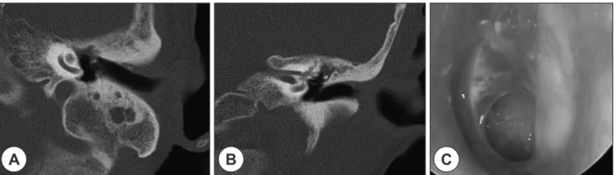
graphic scan showed soft tissue density replacing the middle ear cavity and external auditory canal and total opacification of sclerotic mastoid air cells without evi- dence of bony destruction (Fig. 3).
The polypoid granulation tissue had a broad stalk on middle ear mucosa and was protruding through the perforated ear drum, thus occupying the ear canal. Ex- cisional biopsy was done under the microscope in the outpatient clinic. Histopathologic examination showed
middle ear mucosa infiltrated by lymphocytes, plasma cells, and also adjacent Actinomycoces colonies char- acterized by coccoid and filamentous forms surround- ed by polymorphonuclear leukocytes (Fig. 4). Tympa- nomastoid surgery was advised to treat presumed residual mastoid disease, but it was refused by the pa- tient due to old age and rapid improvement in symp- tom after polypectomy. Intravenous antibiotics (amox- icillin sodium 1 g, clavulanate potassium 0.2 gm three times a day) for 2 weeks and 6-month course of anti- biotics with Amoxicillin/Clavulanate potassium (Amocla duo (7 : 1)® 875/125 mg, twice a day, Kuhnil
Fig. 1. Initial otoscopic finding of the left ear. Tympanic membrane is replaced by polypoid granulation tissue along with serous otorrhea. The polypoid granulation tis- sue had a broad stalk on middle ear mucosa and was protruding through the perforated ear drum, thus occu- pying the ear canal.
Fig. 3. Temporal bone CT showed soft tissue density occupying the whole middle ear and medial part of ear canal.
There is a total opacification of sclerotic mastoid. Ossicle erosion is suspected (A : axial view, B : coronal view).
A B
Fig. 2. Pure tone audiometry showed four frequency av- erage (500, 1,000, 2,000 and 3,000 Hz) of 90 dB flat mixed hearing loss in the left ear and 35dB in the right ear.
0 10 20 30 40 50 60 70 80 90 100 110
250 500 1,000 2,000 4,000 8,000 Frequency (Hz)
dB HL
pharm, Seoul, Korea) were implemented. Follow-up computed tomographic scan after 3 months showed an improvement in middle ear and mastoid air cell (Fig. 5A, B). There was no recurrence of otorrhea and otalgia with a follow-up of 2 years. Tympanic mem- brane perforation (Fig. 5C) and hearing loss remained.
Discussion
Actinomycosis is a chronic suppurative infection of the cervicofacial region caused by Actinomyces spe- cies, which are anaerobic, gram-positive filamentous bacteria.1) It is an uncommon infection of the middle ear and is seldom diagnosed prior to tympanomastoid- ectomy. It was once a fatal disease, however in the an- tibiotic era, it responds to the antibiotic treatment well.
The presentations of most patients with middle ear actinomycosis are similar to those of patients with
chronic suppurative otitis media, that is, these infections are characterized by a prolonged, indolent course.2,21) Even though middle ear actinomycosis tends to be re- fractory to conventional treatment, these nonspecific clinical features result in long delays to diagnose.
Actinomyces are known to be a commensal flora of the oral cavity; hence, it is postulated that entry to the middle ear is most likely the result of seeding from the nasopharynx via the Eustachian tube.1,2) Many cases of middle ear actinomycosis report intact bulging ear drum with middle ear granulation tissue, and these are supporting the idea of pathogens are coming upward through Eustachian tube.3-6) Direct infections of the external auditory canal or haematogenous spread are possible but less likely alternatives.1) In this patient, the patient didn’t have any history of ear infection pre- viously, but according to poor pneumatization of mas- toid and gradual aggravation of hearing loss in the left
Fig. 4. Histopathologic finding. Histopathologic examination shows middle ear mucosa infiltrated by lymphocytes, plasma cells, and also adjacent Actinomycoces colonies characterized by coccoid and filamentous forms surround- ed by polymorphonuclear leukocytes (sulfur granule : arrow) (A : H&E stain ×40, B : H&E stain ×100).
A B
Fig. 5. Follow-up temporal bone CT scan (A and B) and ear drum finding (C) at 3 months post-presentation. Temporal bone CT scan showed well aerated middle ear space, but soft tissue density in sclerotic mastoid cavity (A : axial view, B : coronal view). Otoscopic exam shows dry medium sized perforation of the tympanic membrane.
A B C
ear, it is presumed that she had a chronic history of ear infection in the left ear. Also considering the old age unlike most reported cases, it can be plausible that Ac- tinomyces could have been introduced in the middle ear through the external auditory canal and preexisted ear drum perforation rather than infection through the Eustachian tube.
Since 1980s, 19 cases in available English literature and 2 cases in Korean literature about middle ear and temporal bone actinomycosis were reported.1-18) Among them, 13 cases are children below 15 years old1-3,5,9-16,18)
and 9 cases under 10 years old.1,2,11-16,18) There is a ten- dency of middle ear actinomycosis being related to somewhat younger age than actinomycosis in the other part of body. It is presumed that the children in those ages are prone to infection through the Eustachian tube. Intact bulging ear drum is frequently reported in middle ear actinomycosis, so if middle ear actinomy- cosis occurs frequently in young children, it would be difficult to differentiate from regular bacterial acute suppurative otitis media.3,5)
Furthermore, it is difficult to find Actinomyces through microbiologic culture because Actinomyces are anaerobic and frequently coexist with other bacte- ria.2,7) Therefore, all the case reports except 2 cases are confirmed by pathologic result during operation.7,10) In pathologic exam, Actinomyces form basophilic and eosinophilic colonies with club-shaped filaments radi- ating into rosette patterns, so called ‘Sulfur granules’.1) So, the identification of Gram-positive filamentous or- ganisms with Sulphur granules is strongly suggestive of actinomycosis.1) Newer diagnostic techniques such as polymerase chain reaction, 16S rRNA sequencing and mass spectrometry hold promise, but are not rou- tinely used in clinical practice.1,8)
In our case, luckily the patient had protruding pol- ypoid granulation occupying middle ear space with stalk, so it could be removed quite easily, and actino- mycosis could be confirmed by pathologic reports.
However, in several reports, excisional biopsy through polypectomy showed just inflammatory polyp not ac- tinomycosis, so the patients needed an operation to get
the diagnosis.1,8,9) Even though excision of granulation is not enough for mastoid ventilation, persistent tym- panic perforation could be a mean for producing aero- bic conditions in the middle ear. However, at 3 months post- presentation, the temporal bone CT scan shows mastoid haziness, thus long term follow up will be needed because there is a tendency to recur after insuf- ficient treatment.
Among 21 cases reported so far, 2 cases were diag- nosed as actinomycosis by biopsy examination, which preceded surgical extraction, and they all had non-sur- gical treatment with antibiotics and reported good re- sult.7,10) So far, in middle ear actinomycosis, surgical debridement and proper antibiotics treatment were recommended.1-4) Especially the need for surgical de- bridement has been brought up because thick granula- tion tissue can provide anaerobic environment for the Actinomyces. However in non-severe case with con- firmation of actinomycosis through culture or tissue biopsy, medical treatment can be optional. In actino- mycosis of other body part, for example in lung, medi- cal treatment alone gives good result.19) And in cervi- cofacial actinomycosis, full recovery occurs in most of patients managed by antimicrobial treatment alone.20) Surgical debridement is often necessary for complete resolution, as this anaerobic species can survive for long periods within poorly vascularized tissues, where antibiotics may not reach therapeutic concentrations.12)
However, in selected cases with already confirmed actinomycosis and expected good aeration and drain- age of inflammation, surgery would not be mandatory.
It is possible that early diagnosis may facilitate suc- cessful non-surgical treatment of actinomycosis. It is hoped that alert awareness and clinical suspicion in patients with culture-negative infections or intractable inflammation might lead to earlier diagnosis and more success with non-surgical treatment regimens in se- lected patients with middle ear actinomycosis.
REFERENCES
1) Kullar PJ, Yates P. Actinomycosis of the middle ear. J Lar- yngol Otol 2013;127(7):712-5.
2) Gazzano E, Chanteret C, Duvillard C, Folia M, Romanet P. A case of actinomycosis of the middle ear and a review of the literature. Int J Pediatr Otorhinolaryngol Extra 2010;5:70-3.
3) Budenz CL, Tajudeen BA, Roehm PC. Actinomycosis of the temporal bone and brain: case report and review of the literature. Ann Otol Rhinol Laryngol 2010;119(5):313-8.
4) Subha ST, Raman R, Cheah PL, Soo Hoo TS. Actinomy- ces infection of the mastoid: a rare entity. Med J Malay- sia 2004;59(5):680-1.
5) Sobol SE, Samadi DS, Wetmore RF. Actinomycosis of the temporal bone: A report of a case. Ear Nose Throat J 2004;
83(5):327-9.
6) Shishegar M, Hosseini SH, Varedi P, Varedi P, Mahmoodi S, Ashraf MJ, et al. Actinomycosis of the Middle Ear. Otol Neurotol 2009;30(5):686-7.
7) Hoshino T, Amano H, Tanaka K. Actinomycosis of the middle ear and mastoid. Eur Arch Otorhinolaryngol 1996;
253(6):378-80.
8) Kakuta R, Hidaka H, Yano H, Miyazaki H, Suzaki H, Nakamura Y, et al. Identification of Actinomyces meyeri actinomycosis in middle ear and mastoid by 16S rRNA analysis. J Med Microbiol 2013;62:1245-8.
9) Mehta D, Statham M, Choo D. Actinomycosis of the tem- poral bone with labyrinthine and facial nerve involve- ment. Laryngoscope 2007;117(11):1999-2001.
10) Lester FT, Juhasz E. Actinomycosis of the ear. Ethiop Med J 1990;28(1):41-4.
11) Böör A, Jurkovic I, Friedmann I, Benický M, Dudriková K, Krajcár R. Actinomycosis of the middle ear. J Laryngol
Otol 1998;112(8):800-1.
12) Lezcano C, Simons JP, Colman KL, Cohen MS, Lin PL, Reyes-Múgica M. Actinomycotic mastoiditis complicated by sigmoid sinus thrombosis and labyrinthine fistula. Pe- diatr Dev Pathol 2014;17(6):478-81.
13) Ajal M, Turner J, Fagan P, Walker P. Actinomycosis oto- mastoiditis. J Laryngol Otol 1997;111(11):1069-71.
14) Tarabichi M, Schloss M. Actinomycosis otomastoiditis. Arch Otolaryngol Head Neck Surg 1993;119(5):561-2.
15) Olson TS, Seid AB, Pransky SM. Actinomycosis of the middle ear. Int J Pediatr Otorhinolaryngol 1989;17(1):51-5.
16) Shelton C, Brackmann DE. Actinomycosis otitis media.
Arch Otolaryngol Head Neck Surg 1988;114(1):88-9.
17) Lee SS, Park HJ, Cho HH. Middle ear actinomycosis in- volving facial nerve and lateral semicircular canal. Kore- an J Otorhinolaryngol-Head Neck Surg 2015;58(1):57-60.
18) Suh HK, Yoo KM, Hwang KS, Yoo DHm, Lim HH, Hwang SJ. Clinical experience of acute mastoiditis in recent 10 years.
Korean J Otorhinolaryngol-Head Neck Surg 1998;41(4):
461-6.
19) Kolditz M, Bickhardt J, Matthiessen W, Holotiuk O, Höffken G, Koschel D. Medical management of pulmo- nary actinomycosis: data from 49 consecutive cases. An- timicrob Chemother 2009;63(4):839-41.
20) Smego RA Jr, Foglia G. Actinomycosis. Clin Infect Dis 1998;26(6):1255-61.
21) Cho HP, Lee SW, Kim SH, Lim EJ. A case of malignant otitis externa caused by aspergillus in hemodialysis pa- tient. J Clinical Otolaryngol 2013;24(1):94-9.