INTRODUCTION
Congenital cholesteatoma appears as a whitish mass in the middle ear with an intact tympanic membrane in children, and it is a rare disease accounting for approximately 2% of all choles- teatoma cases (1).
Both computed tomography (CT) and magnetic resonance imaging (MRI) are used in the diagnosis of cholesteatoma and MR diffusion weight imaging or delayed enhancement imaging are evaluated for the differential diagnosis (2, 3). However, the cost of MRI scans is high and contrast media has complications such as allergic reaction and anaphylactic shock. In addition, CT is superior to MRI for delineating fine bone structure. Therefore,
temporal bone CT is the imaging technique of choice for clini- cally suspected cholesteatoma. Although temporal bone CT has high sensitivity of detection, its specificity is low because it is not a contrast-enhanced image, and occasionally the findings are similar to those of granulation tissue, secretions, cholesterol granuloma, or neoplasm, particularly when the lesion is not ac- companied by changes in bone (4). Congenital cholesteatoma re- quires a complete resection to prevent recurrence, whereas treat- ment for cholesterol granuloma or secretions is marsupialization (5, 6). Therefore, the preoperative diagnosis of cholesteatoma is important.
According to a recent study (7), Hounsfield unit (HU) mea- surements may aid in the diagnosis of cholesteatoma. Choleste-
J Korean Soc Radiol 2014;70(2):153-158 http://dx.doi.org/10.3348/jksr.2014.70.2.153
Received August 29, 2013; Accepted October 16, 2013 Corresponding author: Yong-Woo Kim, MD Department of Radiology, Pusan National University Yangsan Hospital, 20 Geumo-ro, Mulgeum-eup, Yangsan 626-770, Korea.
Tel. 82-55-360-1840 Fax. 82-55-360-1848 E-mail: [email protected]
This is an Open Access article distributed under the terms of the Creative Commons Attribution Non-Commercial License (http://creativecommons.org/licenses/by-nc/3.0) which permits unrestricted non-commercial use, distri- bution, and reproduction in any medium, provided the original work is properly cited.
This study was supported by a 2012 research grant from Pusan National University Yangsan Hospital.
Purpose: To evaluate the usefulness of Hounsfield unit (HU) measurements for di- agnosing of congenital cholesteatoma.
Materials and Methods: A total of 43 patients who underwent surgery due to middle ear cavity lesions were enrolled. Twenty-one patients were confirmed to have congenital cholesteatoma by histopathological results and the other 22 pa- tients were confirmed to have otitis media (OM) by operation. Their computed to- mography images were retrospectively reviewed. We measured HU of the soft tis- sue mass in the middle ear cavity. In addition, we evaluated the largest diameter and location of the mass, the presence of bony erosion in the ear ossicle, and the status of the tympanic membrane in the cholesteatoma group.
Results: The mean HU was 37.36 ± 6.11 (range, 27.5–52.5) in the congenital choles- teatoma group and 76.09 ± 8.74 (range, 58.5–96) in the OM group (p < 0.001). The cut-off value was 55.5. The most common location for congenital cholesteatoma was the mesotympanum, and ear ossicle erosion was present in 24%. All patients had an intact tympanic membrane.
Conclusion: HU measurement may be useful as an additional indicator to diagnose congenital cholesteatoma.
Index terms
Congenital Cholesteatoma Temporal Bone CT
Hounsfield Unit Measurement
Usefulness of Computed Tomography Hounsfield Unit Measurement for Diagnosis of Congenital Cholesteatoma
1Congenital Cholesteatoma의 진단에 있어 CT Hounsfield Unit 측정의 유용성1
Sang Hyuk Ahn, MD
1, Yong-Woo Kim, MD
1, Seung Kug Baik, MD
1, Jae-Yeon Hwang, MD
1, Il-Woo Lee, MD
2Departments of 1Radiology, 2Otolaryngology, Medical Research Institute, Pusan National University Yangsan Hospital, College of Medicine, Pusan National University, Yangsan, Korea
clusion of a history of OM.
The OM group was used as the control group to compare the measured HU values with the congenital cholesteatoma group.
We reviewed medical records including patient age, gender and symptoms in both groups.
Imaging Protocol
Temporal bone CT was performed in the axial plane, and re- formatted coronal images were obtained in all cases. CT imag- ing parameters were as follows: a Somatom Definition Flash (Siemens, Forchheim, Germany), 0.6 mm section thickness, 100 KVp, variable mAs protocol, CT dose index range 2–3 mGy, and 55–68 slices performed per examination.
CT Image Analysis
Two radiologists (Y.W.K and S.H.A) retrospectively reviewed the CT images and independently measured HU of the soft tis- sue mass in axial and reformatted coronal images. HUs were measured on axial and reformatted coronal image slices that showed the largest diameter of soft tissue mass for each case.
The region of interest (ROI) was placed within the mass in the middle ear cavity as large as possible, because a large ROI is gen- erally preferred for measuring HU to decrease the variation of its value. Therefore, the ROI circles were centered on the mass and lay 1 mm inside from the margin of the mass to eliminate potential artifacts from the adjacent bony structure and the pneumatized middle ear cavity (Figs. 1, 2). The lower HU of the atoma can be divided into congenital and acquired types, which
differ in their location, clinical history, and the status of the tym- panic membrane but are indistinguishable histologically (4).
Therefore, we hypothesized that the HU measurement in tem- poral bone CT would be useful for diagnosing congenital cho- lesteatoma.
MATERIALS AND METHODS
The Institutional Review Board approved this retrospective study and waived informed consent.
Patients
Among the patients who underwent temporal bone CT be- tween September 2010 and December 2012, 43 who underwent surgery due to middle ear cavity lesions were enrolled. Twenty- one patients were confirmed to have congenital cholesteatoma by histopathological results and the other 22 patients were con- firmed to have otitis media (OM) by operation.
Twenty-one patients in the congenital cholesteatoma group were consistent with the diagnostic criteria suggested by Leven- son et al. (8), including the following: 1) a whitish mass in the middle ear cavity with a normal tympanic membrane; 2) a nor- mal pars flaccida and pars tensa of the tympanic membrane; 3) no history of otorrhea or perforation; 4) no history of otological surgery; 5) exclusion of an occluded external canal, intramem- branous cholesteatoma, or giant cholesteatoma; and 6) no pre-
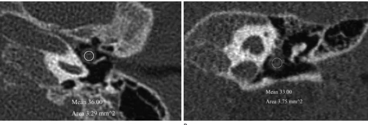
Fig. 1. Congenital cholesteatoma of the left middle ear cavity in a 3-year-old boy. Axial (A) and reformatted coronal (B) temporal bone CT scans demonstrating a well-defined, round soft-tissue mass in the mesotympanum of the left middle ear cavity. The measured Hounsfield unit (HU) is 36 in axial image and 33 in coronal image. The lower HU of the two measured value were selected in this case. This mass considered as congeni- tal cholesteatoma.
B A
sence of bony erosions in the ear ossicle, and the status of the tympanic membrane in the congenital cholesteatoma group.
Statistical Analysis
We performed Student’s t-test using SPSS 20 (SPSS Inc., Chi- cago, IL, USA) for statistical analysis. A p-value < 0.05 was con- sidered significant.
two values measured on axial and reformatted coronal images were selected in each case. The average HU value was calculated from the two lower HU values by the two radiologists. This av- erage HU was defined as the representative value and was used for analysis (Table 1).
In addition, we evaluated the location of the mass, the rela- tionship between the mass and ear ossicle, the presence or ab-
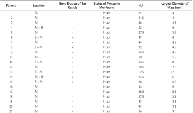
Fig. 2. Otitis media of the left middle ear cavity in a 5-year-old boy. Axial (A) and reformatted coronal (B) temporal bone CT scans demonstrat- ing a well defined soft-tissue mass in epitympanum of the left middle ear cavity. It is difficult to distinguish otitis media from congenital choles- teatoma by image finding alone. The measured Hounsfield unit (HU) is 79 in axial image and 90 in coronal image. The lower HU of the two mea- sured value were selected in this case. This mass considered as otitis media.
B A
Table 1. The Characteristics of Congenital Cholesteatoma
Patient Location Bony Erosion of Ear
Ossicle Status of Tympanic
Membrane HU Largest Diameter of
Mass (mm)
1 M - Intact 34 3
2 M - Intact 31.5 4
3 M - Intact 38 4.5
4 M + H - Intact 42 6
5 M - Intact 27.5 3.5
6 E + M + Intact 43 6
7 M - Intact 44 4.5
8 E + M + Intact 32 4.5
9 M - Intact 34.5 4.5
10 M - Intact 30 5.5
11 E + M - Intact 34.5 8
12 M - Intact 30.5 2.5
13 E + M + Intact 52.5 12
14 M + H + Intact 34.5 6
15 E + M + Intact 36 5.6
16 M - Intact 43 6
17 M - Intact 38.5 5.8
18 M - Intact 37.5 3.3
19 M - Intact 43 3.2
20 M - Intact 44 3.2
21 M - Intact 34 3
Note.-E = epitympanum, H = hypotympanum, HU = Hounsfield unit, M = mesotympanum
ic membrane) are summarized in Table 1. The mean largest diam- eter was 4.98 ± 2.13 mm (range, 2.5–12 mm). The most common location was in the mesotympanum (14/21, 66.5%), followed by the epitympanum, and mesotympanum (5/21, 24%), and then the mesotympanum and hypotympanum (2/21, 9.5%). Five pa- tients (24%) had ear ossicle erosion and 16 patients (76%) did not. All patients had intact tympanic membranes.
DISCUSSION
The HU is a linear transformation of the linear attenuation coefficient measurement, and all biological tissues have a unique HU (9, 10). Although cholesteatoma can be divided into con- genital and acquired types, it consists of a combination of kerati- nous material and stratified squamous epithelium and the two types cannot be distinguished histologically (11). Therefore, congenital cholesteatoma and acquired cholesteatoma will theo- retically have a similar HU. In contrast, non-cholesteatoma tis- sue exhibits a different HU because it has different histological components. A recent study by Park et al. (7) reported a HU of 42.68 ± 24.42 in the cholesteatoma group and 86.07 ± 26.50 in the non-cholesteatoma group. The difference between the two groups improved the diagnosis of cholesteatoma.
In the present study, the HU values of the congenital choleste- atoma group ranged from 27.5 to 52.5 and the mean value was 37.36 ± 6.11. All cases of congenital cholesteatoma had HU val- ues < 64.4, which was consistent with the result of Park et al. (7), who reported a cut-off value of 64.4 HU between cholesteatoma and non-cholesteatoma groups. However, some cases of OM group in our study had HU values < 64.4. Therefore, their cut- off value (64.4 HU) could not be applied in our study. In the present study, no overlap in the value was observed between the cholesteatoma and OM groups. The HU differences between the two groups were significantly different, and the cut-off value was 55.5 HU. This result is probably due to the small sizes of the congenital cholesteatomas in our study. A larger cholesteatoma can lead to obstruction of the Eustachian tube and causes in- creased middle ear effusion and inflammatory changes. The largest diameter in the present study was small (4.98 ± 2.13 mm), and it did not present with these complications. Unlike congenital cholesteatoma, acquired cholesteatoma results from chronic infection with formation of granulation tissue, which
RESULTS
Patients
Twenty-one patients were enrolled in the congenital cholestea- toma group: 16 boys and 5 girls (mean age, 3.9 years; range, 2–7 years). Seventeen patients were incidentally discovered to have a white retrotympanic mass with an intact tympanic membrane during physical examination, including otoscopy, due to symp- toms of the common cold, rhinitis, or enteritis. The other four patients had symptoms of OM such as otalgia or ear fullness and had a history of OM.
Twenty-two patients were enrolled in the OM group: 13 boys and 9 girls (mean age, 6.2 years; range, 1–18 years). Fourteen patients had symptoms of OM, five patients visited for a hearing test, two patients had symptoms of the common cold, and two patients were suspected to have cholesteatoma.
CT Evaluation
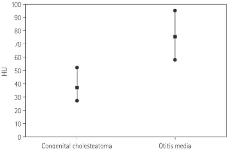
The mean HU value was 37.36 ± 6.11 (range, 27.5–52.5) in the congenital cholesteatoma group and 76.09 ± 8.74 (range, 58.5–
96) in the OM group (p < 0.001) (Fig. 3). The HU cutoff value between the congenital cholesteatoma group and the OM group was calculated using the median value from the maximum HU value in the congenital cholesteatoma group and the minimum value in the OM group. The cut-off value was 55.5 between the two groups. A measured HU value < 55.5 can be considered con- genital cholesteatoma, whereas an HU > 55.5 is considered OM.
The characteristics of congenital cholesteatoma (the largest diameter, location, ear ossicle erosion, and status of the tympan-
Fig. 3. The Hounsfield unit (HU) of congenital cholesteatoma group and otitis media group.
0 10 20 30 40 50 60 70 80 90 100
HU
Congenital cholesteatoma Otitis media
indicator in the diagnosis of congenital cholesteatoma.
REFERENCES
1. McDonald TJ, Cody DT, Ryan RE Jr. Congenital cholestea- toma of the ear. Ann Otol Rhinol Laryngol 1984;93(6 Pt 1):637-640
2. Trojanowska A, Trojanowski P, Olszanski W, Klatka J, Drop A. Differentiation between cholesteatoma and inflamma- tory process of the middle ear, based on contrast-en- hanced computed tomography imaging. J Laryngol Otol 2007;121:444-448
3. De Foer B, Vercruysse JP, Bernaerts A, Meersschaert J, Ke- nis C, Pouillon M, et al. Middle ear cholesteatoma: non- echo-planar diffusion-weighted MR imaging versus de- layed gadolinium-enhanced T1-weighted MR imaging-- value in detection. Radiology 2010;255:866-872
4. Baráth K, Huber AM, Stämpfli P, Varga Z, Kollias S. Neuro- radiology of cholesteatomas. AJNR Am J Neuroradiol 2011;
32:221-229
5. Smith PG, Leonetti JP, Kletzker GR. Differential clinical and radiographic features of cholesterol granulomas and cholesteatomas of the petrous apex. Ann Otol Rhinol Lar- yngol 1988;97(6 Pt 1):599-604
6. Franklin DJ, Jenkins HA, Horowitz BL, Coker NJ. Manage- ment of petrous apex lesions. Arch Otolaryngol Head Neck Surg 1989;115:1121-1125
7. Park MH, Rah YC, Kim YH, Kim JH. Usefulness of computed tomography Hounsfield unit density in preoperative de- tection of cholesteatoma in mastoid ad antrum. Am J Otolaryngol 2011;32:194-197
8. Levenson MJ, Michaels L, Parisier SC. Congenital choleste- atomas of the middle ear in children: origin and manage- ment. Otolaryngol Clin North Am 1989;22:941-954 9. Phelps ME, Hoffman EJ, Ter-Pogossian MM. Attenuation
coefficients of various body tissues, fluids, and lesions at photon energies of 18 to 136 keV. Radiology 1975;117(3 Pt 1):573-583
10. Brooks RA. A quantitative theory of the Hounsfield unit and its application to dual energy scanning. J Comput As- sist Tomogr 1977;1:487-493
11. Caponetti G, Thompson LD, Pantanowitz L. Cholesteatoma.
contributes to its higher HU (12).
Another possible explanation for the lower HU in the present study is that little ear ossicle erosion (24%) was observed, and all cases were confined to the middle ear cavity. The mean HU of congenital cholesteatoma with ear ossicle erosion (39.6 ± 8.29) is higher than without ear ossicle erosion (36.65 ± 5.41) in our study. Bony erosion is rare in congenital cholesteatoma rather than acquired cholesteatoma (4). Although bone destruction is a part of acquired cholesteatoma, involvement of the adjacent bony structure leads to a higher HU because acquired cholestea- toma includes the remaining bone structure after bone destruc- tion. Therefore, the lesser involvement of the adjacent bony structure contributes to the lower HU in the patients with con- genital cholesteatoma. Furthermore, the mean largest diameter of congenital cholesteatoma with ear ossicle erosion (6.82 ± 2.96 mm) was larger than congenital cholesteatoma without ear ossi- cle erosion (4.4 ± 1.50 mm). These results support the hypothe- sis that smaller size and no bony erosion may affect lower HU in congenital cholesteatoma.
In the present study, congenital cholesteatoma was 3.2 times more common in boys, and the mean age was 3.9 years. Previ- ous studies (8, 13, 14) have reported that congenital cholesteato- ma is three times more common in boys than girls, and that the mean age of detection is 4.5 years. Our results are similar to those values.
The cholesteatomas of 17 patients (81%) were incidentally discovered, whereas four patients had symptoms of OM and a history of OM. This agreed with previous reports (8, 13) that most cases of congenital cholesteatoma are asymptomatic and incidentally discovered. In addition, all cases were found in the mesotympanum, in agreement with a previous study (14) that claimed that congenital cholesteatoma is often located in the an- terior superior quadrant of the middle ear, close to the malleus manubrium.
Congenital cholesteatoma is usually considered when it is in- cidentally discovered as a white retrotympanic mass on otosco- py and a sharply marginated round soft tissue mass in the middle ear cavity on temporal bone CT. However, some cases are chal- lenging to diagnose because temporal bone CT has low specifici- ty for cholesteatoma. In the present study, the cut-off value was 55.5 HU between the cholesteatoma group and OM group. In conclusion, HU measurement may be useful as an additional
1):606-613
14. Nelson M, Roger G, Koltai PJ, Garabedian EN, Triglia JM, Roman S, et al. Congenital cholesteatoma: classification, management, and outcome. Arch Otolaryngol Head Neck Surg 2002;128:810-814
Ear Nose Throat J 2009;88:1196-1198
12. Fisch U. Tympanoplasty, Mastoidectomy, and Stapes Sur- gery. New York: Thieme, 1994:146
13. McGill TJ, Merchant S, Healy GB, Friedman EM. Congenital cholesteatoma of the middle ear in children: a clinical and histopathological report. Laryngoscope 1991;101(6 Pt
Congenital Cholesteatoma의 진단에 있어 CT Hounsfield Unit 측정의 유용성1
안상혁
1· 김용우
1· 백승국
1· 황재연
1· 이일우
2목적: Congenital cholesteatoma의 진단에서 CT Hounsfield unit (이하 HU) 측정의 유용성에 대해 평가하고자 하였다.
대상과 방법: Middle ear cavity의 lesion으로 수술을 시행한 43명의 환아가 연구에 포함되었다. 조직학적으로 congenital cholesteatoma로 진단된 환아가 21명이었고, 수술소견으로 otitis media (이하 OM)로 진단된 환아가 22명이었다. 환아의 CT를 후향적으로 분석하였고, middle ear의 연부조직 종괴에 대해 HU를 측정하였다. 추가적으로 cholesteatoma group 에 대해서 연부조직 종괴의 가장 큰 장경과 위치, 귓속뼈의 골미란 유무, 그리고 고막의 상태를 분석하였다.
결과: Congenital cholesteatoma에 대해 측정된 HU의 평균은 37.36 ± 6.11(range, 27.5~52.5)이었고, OM group에서 측정된 HU의 평균은 76.09 ± 8.74(range, 58.5~96)였다. 통계학적으로 두 그룹의 HU는 의미 있는 차이가 있었고(p
< 0.001), 두 그룹을 구분하는 cut-off value는 55.5였다. Congenital cholesteatoma는 고실에 위치한 경우가 가장 많았 고, 5명(24%)의 환아에서 귓속뼈의 골미란이 있었다. 그리고 모든 환아에서 고막은 정상적으로 보였다.
결론: CT HU 측정이 congenital cholesteatoma의 진단에 유용한 추가적인 지표가 될 수 있을 것으로 생각된다.
부산대학교 의과대학 양산부산대학교병원 의학연구소 1영상의학과, 2이비인후과