대 한 방사 선 의 학회 지 1996: 35(3) : 347- 350
섬낭막에서 기원한 일차성 악성 중피종 :1 예 보고1
박희흥 · 최영흐1 . 김태훈 · 이연희 · 김영권 2 . 한동선 2 • 조정흐p . 유필문
심낭막의 일차성 악성 중피종은 매우 드물고 치명적인 질환으로 진단이 어려운 경우가 많으며 대부 분은 사후 부검을 통해서 진단이 내려지고 있다. 저자들은 심낭막의 일차성 악성 중피종 1예를 문헌고 찰과 함께 보고하고자 한다.
컴퓨터단층촬영과 자기공염 영상 소견은 심낭막의 불규칙한 미만성 비대와 심낭막 내부에 연조직 음염이 있고 심근층으로의 결절성 융기 및 침윤성 성장을 하고 심장과 대혈관을 완전히 둘러싸고 있었 다.
서 료응 ‘-
심냥막의 일차성 악성중피종은 매우 치명적인 종양으로 서 효과적인 치료방법은 없으며 가능한 환자의 남은 여생 을 편안하게 하는것이 치료의 목표이며 수술, 방사션 치료,
세포 증식 억제제등이 시도되고 있다. 사망 원인은 심막압 전, 대정맥 폐쇄, 심부전으로 알려져 있고, 급사는 관상동 맥 침범과관계가있는것으로보고되고있다. 저자들은심 막절제술과 부검으로 확진된 심냥막의 일차성 악성 중피종 l 예를 경험하였기에, 컴퓨터단층촬영(이하 CT로 약함)소 견및 자기공명 영상(이하 MRI로 약함)소견을 함께 보고 하고자한다.
증례보고
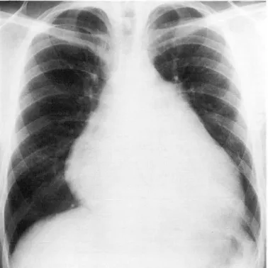
22세 남자환자로 내원하기 하루 전부터 시작된 좌측 측 복부 통증으로 응급실을 통해서 내원하였고, 좌측 늑골척 추각 압통 (CV A tenderness) 과 빈뇨가 있었고, 소변에서 케톤과 나이트라이트가 발견되었다. 과거력상 4개월전에 심냥 삼출액이 있어 섬막천자술을 시행하였고 만성 육아성 염증세포가 발견되어 결핵성 심낭염 의증하에 항결핵약으 로 투약하면서 경과 관찰중이었다. 단순흉부촬영 (Fig. 1) 에서 심비대가 있고 폐혈관 음영은 정상이어서 심낭 삼출 액의 가능성을 생각하였다. 심전도상 동빈맥과 비특이성 ST-T변화를 보였고, 이원성 심초음파술 (2-
D
Echocardi1단국대학교 의과대학 방사선과학교실
2단국대학교 의과대학 내과학교실
3단국대학교 의과대학 병리학교실
이 논문은 1996년 3월 2일 접수하여 1996년 7월 8일에 채택되었응
ography) 에서 소량의 심낭 삼출액이 있었고 좌심실의 크 기와 수축력은 정상이었다. 심막천자술을 시행하여 약
300
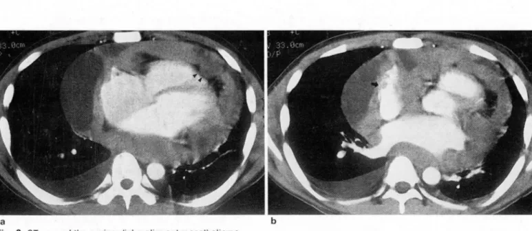
cc의 삼출액을 배액하였는데 악성 세포는 발견되지 않았 고 많은 성숙 임파구만 발견되었다. 단순흉부촬영 2주후에 시행한 CT소견상 심냥막이 불규칙하게 미만성으로 두꺼 워져 있었고 심냥막 내부엔 낭성 병소를 가진 연조직 음영 이 심근층을 둘러싸고 있으며 침윤성 성장을 보였다 (Fig.
2).
다발성으로 좌측 신장 경색이 있었다. 단순흉부촬영4
주후에 시행한 MRI소견은 심냥막이 미만성으로 두꺼워
Fig. 1. Chest radiograph shows cardiomegaly with sagging appearance and small amount 01 left pleural ellusion
H
”
대 한 방사 선 의 학회 지 1996; 35(3) : 347-350
져 있으며 T1 강조 축상영상에서 심근층과 등신호 강도를 보이고 T2강조 축상영상에서 심근층보다 약간의 고신호 강도를 보이는 연조직 음영이 있고 섬외막 지방층이 종괴 에 의해 부분적으로 소실되어 있으며 T1 강조 관상영상 (Fig.3) 에서 종괴가 대동맥궁 위치까지 심장과 두 대혈관 을한전히 툴러싸며 침윤성 성장을하고있였다.
심막절제술 (Pericardiectomy) 를 통해 시행한 생겸상 침윤성 암으로 진단되였으며 악성 중피종과 전이성 암종 의 감별이 어려웠다. 환자는 내원 21일후에 잡지가 호흡곤 란과 청색증이 생켰고 호흡마비와 심부전으로 사망하였
’
다. 사망 후에 시행한 부검상 악성 중피종우로 확진되였다.
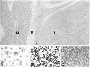
부분적으로 절제된 심냥막의 육안소견상 종양은 섬낭강을 두껍게 둘러싸며 침윤하고 있었으며 태l 교적 단단하고 생 선살 양상의 점액성을 띠는 소견을 보였고 육안으로도 섬 외막 지방층으로의 종양의 침윤이 관찰되었다 (Fig.4). 현 미경 소견상 종양세포는 주로 입방세포 (cuboidal cell) 틀 로 이루어져 있으며 대부분 미만성의 배열을 보였으며 부 분적으로 유두양 소견을 보이는 특정적인 상피세포형 (epi - thelial type) 중피종의 소견을 보였다 (Fig.5a). 면역조직 학적 염색의 결과 이들 세포는 cytokeratin antibody 에 주
a
Fig. 2. CT scan of.the pericardial malignant mesothelioma
b
a. Pericardium is diffusely irregularly thickend with cystic portion. Tumor growth showed nodular bulging into the mycardium (arrowheads)
b. CT scan through slightly higher level shows tumoral infiltration into the right atrium(arrow) and obliteration of the epicardial fat. Left atrium and the pulmonary vein are compressed by the tumor
Fig. 4. Grossly, the tumor (T) is a diffuse fish-flesh lesion that spreads widely in the pericardial space. Epicardial fat (E) and Fig. 3. Coronal T1 weighted image shows diffuse pericardial parietal pericardium (p) delinate the infiltrating tumor. M;
thickening encasing the hearÎ and two great vessels above myocardium aortic arch level
m
생
Jι‘
Fig. 5. The tumor (T) inliltrates into the epicardial lat (E) and locally into the myocardium (M). Neoplastic cells consist 01 cuboidal cells lorming partly papillary structure (epithelial type) (H&E, X 40). Inset: A, multinucleated giant cells and pleomorphic cells with myxoid stroma (H&E, X 400); B, strong positive staining lor low-molecular weight cytokeratin protein (Immunohistochemical staining, X 400); C, positive staining lor acid mucopolysaccharide (Alcian Blue staining, X 400)
로 헥 주변으로 강한 양성 의 소견을 보였으며 (Fig. 5b)
Acid mucin을 위한 특수 염색에서는 세포의 세포막 및 세
포사이의 기질에 양성의 소견을 보임으로써 선암종과 구별 되는 중피종 세포의 특정적인 소견을 더해주었다 (Fig. 5c) 또한 전지현미경 소견에서도 다른 선암종과 구별되는 특정 적인 긴 미세융모 (microvilli) 및 많은 세포질내의 당김세 사 (tonofilaments) 가 관찰되 였다 (Fig.6).
고 찰
심장종양은 매우 드문 질환으로서 그 발생빈도는
O .
0017-0.28%로 보고되고 있으며 악성 중피종은 횡문근 암 종과 맥관 육종 다음으로 흔하며 일차성 심장종양중
2 -4%
정도로 보고되고 있다(1). 악성 중피종은 흉막, 심 냥막, 횡근막, 복막, 정소집막 (tunica vaginalis testis) 을 포함하는모든장막조직에서 광범위하게 발생할수있는것 으로 알려져 있고 특히 심냥막의 악성 중피종은 매우 드문 것으로 알려져 있다 (2). 악성 중피종은 생전에 진단이 어 렵고환자의 20%에서만성공적으로진단이 되었다고보고 된 바 있는데 임상증상이 없거나 비특이적이고, 심전도소 견도 비특이적인 경우가 많으며, 대개 섬낭 삼출액, 섬냥막 압전,교착성 심냥염, 울혈성 심부전의 형태로나타나기 때 문이다(1 ).악성 중피종은 석면 노출과 밀접한 관계가 있는 것으로 알려져 있는데 원인은 아직 알려진 바 없으며 석변에 심하 게 노출된 사람은 일생동안 7-10%의 높은 발생빈도를 보 이는 것으로 보고되고 있고 (2) , 대개 육종형( sarcomatous
박희홍 외 : 심 낭막에서 기원한 일차성 악성 중피종
Fig. 6. On electron microscopy, the presence 01 long micr。
villi and abundant tonofilaments are observed, but microvilli rootlets and lamella bodies are absent (EM, X 10000)
type) 보단 상피세포형이 많으며 암세포는 풍부한 글리코 겐과 하이알루로닉 산을 가지고 있으며 대개 심근층을 잘 침범하며 30-50%에서 전이가 일어나는데 주로 임파절이 나 폐, 간흑 신장으로의 전이가 보고되고 있다(1, 3). 본 증 례에서는 석면에 노출된 과거력은 없었고 상피세포형이며 신장경색은 아마도 신장으로의 종양 색전에 의한 것으로 추정하였다.
Thomason등 (4) 은 총 28예를 분석하였는데 남자가 두배 많고다양한나이 분포를가지며 (7세 .평균 47서]) 석면 노 출의 빈도는 14%이고 흔히 사용되는 영상 진단술은 심초 음파가
12%
, CT는 44%로 낮은 민감도를 보였고 병리학 적으로 72%에서 미만성 성장을하는형태로보고하였다.악성 중피 종의 진단방법으로는 심초음파,
CT
, MRI, 조 직학적 검사가흔히 이용되나, 67Ga 스캔이 진단에 도움을 준다는 보고가 있다 (5).임상 증상이나 이학적 검사상 심장종양이 의심된다면 우 선적으로 심초음파를 통해서 심냥막비후나 심장내 종괴를 확인할 수 있고 (6) ,
CT
및 MRI를 시행하여 심냥막의 불 규칙한 미만성 비후나 연조직 음영을 발견함드로써 진단의 정 확도가 높아질 수 있다(7). 특히 MRI는 종양의 크기 와 범위를알수있고섬벽과주위 조직에로의 침범을규명하는 데 매우 유용한 것으로 보고되고 있다 (8). 본 증례의CT
및 MRI소견에서 기 존에 알려진것과 다른 특이한 소견은
없였으나 심낭막의 결절성 비후와 심외믹 지방층 소실및 침윤성 성장이 섬냥막의 악성종양을 시사하는 소견으로 생 각되어진다.
악성 중피총은 선암과의 감별이 간흑 어려운 것으로 얼
349 -
대 한 방사 선 의 학회 지 1996; 35(3) : 347-350
려져 있는데 면역조직화학검사가 감별에 유용하며 선암과 달리 암배아성 항원 (CEA) 음성, 케라턴 단백 양성이 나타 나고프로콜라겐 l형에 대한면역염색。l 감별에 유용한것 으로 알려져 있다 (1 , 9). 그러나 이러한 면역조직화학적 방 법에도 불구하고 생검에서는 진단이 어려운 경우가 많으며 대개의 진단은 부검에 의해 이루어지고 있다.
고
참
헌 ==
1. Fazekas T, Ungi 1, Tiszlavicz L. Primary malignant meso.
thelioma of the pericardium. Am Heart J 1992; 124: 227.231 2. Watanabe M‘ Kimura N, Kato M, Iwami 0, Takahashi M,
Nagura F. An autopsy case of malignant mesothelioma associated with asbestosis. Pathol Int 1994; 44: 785.792 3. Loire R, Tabib A. Malignant mesothelioma of the pericar.
dium. An anatomo.clinical study of 10 cases. Arch des Malad du Coeur et des Vaiss 1994; 87: 255.62
4. Thomason R, Schlegel W, Lucca M, Cummings S, Lee S. Pri.
mary malignant mesothelioma of the pericardium. Case re.
poη and literature review. Tex Heart Inst J 1994;21 170-174
5. Taguchi T, Fujiwara Y, Ichiki H, Kohno N, Hiwada K. A case of malignant pericardial mesothelioma detected by gallium-67 scintigraphy. Kaku Igaku 1991 ; 28: 281-4
6. Chen HZ, Jiang L, Rong WH. Tumors of the heart. Chin Med J 1 992 ; 1 05 : 1 53-1 58
7. Permutti B‘ Cardin G, Pirrelli M, et al. Primary mesothelioma of the pericardium: diagnostic methods. A case report Cardiologia 1993; 38: 59-63
8 임 명관, 박재형, 정진욱, 김지혜, 최연현, 한만청 원발성 심장 및 심 막 종앙의 자기공명영상 대한방사선의학회지 1995; 32 : 249-254 9. Topov J‘ Georgiev T, Alexiev B. Oiffuse malignant mesotheli-
。 maof the pericardium. Zentralbl fur Pathol 1991 ; 137: 164-6
Journal of the Korean Radiological Society 1996: 35(3) : 347-350
Primary Malignant Mesothelioma of the Pericardium:
A Case Report'
Hee Hong Park, M.D., Young Hi Choi, M.D., Tae Hoon Kim, M.D., Yeon Hee Lee, M.D., Young Kwon Kim, M.D.2
, Dong Sun Han, M.D.2 , Jeong-Hee Cho, M.D.3, Pil Mun Yu, M.D.
1 Department of Diagnostic Radiology, Dankook University College of Medicine
2Department of Internal Medicine, Dankook University College of Medicine 3Department of Pathology, Dankook University College of Medicine
Primary malignant mesothelioma of the pericardium is a very rare and highly lethal neoplasm. Diagnosis is a difficult problem and most of the cases reported in the literature were diagnosed at postmortem. We report a case of primary malignant mesothelioma of the pericardium in a 22 year-old man
CT and MR imaging both showed diffuse irregular pericardial thickening, soft tissue density with cystic lesion, nodular bulging into the myocardium, permeative growth of the tumor, and encasement of the heart and tw。
great vessels
Index Words: Pericardium, MR Pericardium, CT Pericardium, neoplasms
Address reprint requests to : Hee Hong Park, M.D., Department of Diagnostic Radiology, Dankook University Hospital,
~ 29 Anseo-dong, Chonan, Choongnam, 330-715 Korea. Tel. 82-417-550-6921 Fax. 82-417-552-9674