2020 Korean Society for Surgery of the Hand, Ko- rean Society for Microsurgery, and Korean Society for Surgery of the Peripheral Nerve. All Rights re- served.
This is an open-access article distributed under the terms of the Creative Commons Attribution Non-Commercial license (http://creativecommons.
org/licenses/by-nc/4.0/), which permits unrestricted non-commercial use, distribution, and reproduction in any medium, provided the original work is prop- erly cited.
INTRODUCTION
Poisonous snake has specialized teeth which release venom from glands linked to the fangs. Skin necrosis at the bite site, to systemic symptoms including acute kidney injury, acute respiratory failure, acute myocardial infarction, disseminated intravascular, coma, coagulation, and death [1]. In a 2008 World Health Organi- zation survey, about 421,000 people are bitten by snakes annually worldwide, of which 20,000 are killed. In South Korea, 192 to 621 people are bitten by snakes annually, and an average of 5 people are killed per year [2]. Especially in South Korea, distal parts of upper extremities are the most common sites of snakebite injuries [3]. Various severe complications in hands have been reported, including compartment syndrome, occasionally resulting in amputation. Given the impor- tance of the functions of extremities, even localized symptoms of snakebites on extremities should be treated with the utmost care.
In the current study, we present a case of snakebite on the left ring finger, treat- ed with surgical intervention and topical oxygen therapy (TOT) which has not
증례보고
최환준, 이다운, 류형래, 김준혁, 이준호
순천향대학교 부속 천안병원 성형외과
Case of Treatment of Snakebite on Finger with Topical Oxygen Therapy
Hwan Jun Choi, Da Woon Lee, Hyeong Rae Ryu, Jun Hyuk Kim, Jun Ho Lee
Department of Plastic and Reconstructive Surgery, Soonchunhyang University Cheonan Hospital, Cheonan, Korea
Snakebites, though uncommon, are a potentially serious cause of disability or death.
Local symptoms may include pain, edema, or ecchymosis that may progress to skin necrosis or compartment syndrome. This study explores the case of a 4-year-old male patient bitten by a snake on the distal volar aspect of his left ring finger. On physical examination, there were moderate swelling, hemobullae formation, and the skin ne- crosis was progressing on middle phalanx of ring finger. Fasciotomy and topical oxy- gen therapy was performed. The topical oxygen therapy (TOT) was started once a day for 90 minutes with 4 L/minute of oxygen flow. TOT is a method of delivering humidi- fied oxygen directly to the wound bed to support the healing of chronic and hypoxic wounds. There is no report on TOT for snakebite injury. In this report, we would like to report on the clinical experience of early surgery and adjuvant TOT with literary con- sideration.
Keywords: Snakebites, Topical, Oxygen therapy, Adjuvant pISSN 2586-3290 · eISSN 2586-3533
Arch Hand Microsurg 2020;25(4):292-296 https://doi.org/10.12790/ahm.20.0061
Received: October 12, 2020 Revised: November 9, 2020 Accepted: November 10, 2020 Corresponding author:
Hwan Jun Choi
Department of Plastic and
Reconstructive surgery, Soonchunhyang University Cheonan Hospital, 31 Sooncheonhyang 6-gil, Dongnam-gu, Cheonan 31151, Korea
Tel: +82-41-570-2195 Fax: +82-41-574-6133 E-mail: [email protected] ORCID:
https://orcid.org/0000-0002-0752-0389
Case Report
been reported in previous studies. We believe this study will contribute to improved treatment of snakebite-induced soft tis- sue necrosis. The study was performed in accordance with the principles of the Declaration of Helsinki.
CASE REPORT
A 4-year-old boy who was healthy and never had undergone any disease was admitted with snakebite of his ring finger. He was bitten by a striped brown snake while going to the valley.
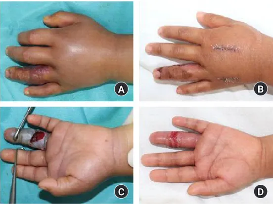
After the injury, he was treated his finger by simple dressing with the laceration, empirical antibiotics, and antivenom at lo- cal clinic. After that, he transferred to our department for man- agement on the same day. On physical examination, there were moderate tenderness, swelling, bullae formation, and skin ne- crosis that was progressing on the middle phalanx of ring fin- ger (Fig. 1). But, there was neither exquisite tenderness along flexor tendon sheath nor marked pain on the proximal part of ring finger. Therefore, the possibility of tenosynovitis (TS) was low. On admission, he had a temperature of 37.3°C. Blood cul- tures, a full blood count, and a wound microbiology swab were taken and he was promptly commenced on intravenous cefa- clor, analgesia, hand elevation, immobilization, and general bed rest. Radiologic inspection was carried out to confirm the pres-
ence of minute foreign body. There was no radiopaque densi- ties seen as foreign body. The full blood count showed minor leukocytosis (14.4×10⁹/L). There was no growth in wound cul- tures.
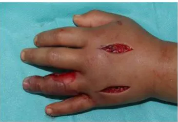
Although the treatment was maintained for 2 days with sim- ple dressing and antivenom, his finger was exacerbated and seemed to be more swollen. So 3 days after the initial treat- ment, fasciotomy was performed on the hand dorsum and the ring finger for the lateral aspect, judging that the compartment syndrome could proceed due to the worsening of the swelling.
Fasciotomy allowed oxygen to be supplied to deeper tissue.
Also, negative pressure wound therapy was performed on the hand dorsum (Fig. 2). On the same day, TOT was started once a day for 90 minutes with 100% oxygen before daily dressing.
After sealing the hand with an aseptic fluid pack, an O2 catheter was connected (Fig. 3). Antibiotic ointment was superficially applied on wound margin to prevent drying up. On TOT day 6%, 10%, 50%, and 80% epithelization was progressed respec- tively. TOT was carried out for a total of 12 days. He responded well to the treatment and was discharged after 19 days of hospi- talization.
B
D A
C
Fig. 1. (A, B) Preoperative finding. Moderate swelling and skin necrosis were observed. (C, D) Findings of postoperative day 10. The swelling was subsided and epithelization was progressed.
DISCUSSION
A total of 16 species of snakes are known to exist in Korea, of which only four are poisonous; three species of the genus Gloy- dius (G. brevicaudus, G. saxatilis, G. ussuriensis) and one spe- cies of the genus Rhabdophis (R. tigrinus) [4]. Studies so far have reported G. ussuriensis to be the predominant cause of snakebite due to its large population and easily accessible habi- tation area at the base of mountain or in farming area. There are no accurate statistics on the frequency of venomous snake- bite injuries in Korea, although an average of 409.6 patients are reported to visit the hospital annually, which may increase when including patients with minor injuries who do not visit the hospital [5]. Patients with snakebite injuries develop a wide variety of clinical courses, from mild symptoms such as edema
and pain at the injury site to serious complications including acute kidney injury, acute respiratory failure, acute myocardial hemoptysis, infarction, coma, disseminated intravascular coag- ulation, and death [1]. In our case, the patient had no systemic complication but had localized tenderness, swelling, and skin necrosis.
The hands especially the distal areas such as the fingers and palm, were the predominant sites of injury [3]. In this case, the patient was injured his left ring finger. Because hands, especial- ly fingers, are one of the most important functional areas, so if not treated properly, it can cause serious side effects. In partic- ular, the patient is not yet fully grown, which can lead to more fatal consequences [6].
In this case, we ruled out TS. But because snakebite injury is commonly accompanied by TS, physicians always should keep in mind the TS on patients with snakebite injury. TS initially starts with exudates within the tendon sheath leads to increased pressure, leading to ischemia. There are several signs which called Kanavel signs. There is tenderness along the sheath and finger sits in a resting flexed posture in patients with TS. Also, pain is more severe at the proximal part and fusiform swelling of the finger can be found. Treatment of TS involves broad-spectrum antibiotics and rapid surgery to debride tissues and irrigation [7].
Treatment of snakebite injury can be divided into surgical and nonsurgical treatment. From a surgical perspective, the first dilemma the clinician faces in managing a snakebite wound is determining whether to perform debridement on the wound site. In principle, trauma patients should undergo de- bridement as soon as possible, thereby removing the necrotized tissue in order to prevent further infection. In the past, early re- moval of snake venom by surgical methods was recommended as immediate management [6].
However, the majority of such treatments resulted in soft tis- sue damage, leading to failed skin graft or flap, amputation, and osteomyelitis, ultimately leading to a poor prognosis. Immedi- ate debridement is no longer the treatment of choice in manag- ing snakebites; rather, the current standard of care is adminis- tration of antivenom followed by delayed debridement. A re- cent study using an electron microscope observed viable tissues buried inside necrotized muscles of the snakebite wound, pro- viding additional evidence for preemptive antivenom use fol- lowed by delayed debridement [6].
Oxygen therapy can be divided into hyperbaric oxygen ther- apy (HBOT) and TOT. As HBOT is systemic and raises the pressure of oxygen (pO2), there is a risk of complications such as seizures, damage to the tympanic membrane of the ear, and Fig. 2. Intraoperative finding. Fasciotomy and negative pressure
wound therapy was performed.
Fig. 3. The image of topical oxygen therapy. Wound was sealed with aseptic fluid bag which can be connected with an oxygen cannula. The fluid bag expanded in balance with the external pressure of 1 atmosphere.
damage to the retinal nerve. If patients have diabetes, their glu- cose levels could also be affected by an increased pO2. Also, HBOT can only be performed in hospitals that have chamber and it is not cost-effective [8].
A study performed by Fries et al. [8] demonstrated that TOT promotes vascular endothelial growth factor expression in wound and neovascularization via pig models. Four female pathogen-free domestic pigs each had five wounds located on their backs, for a total of 20 wounds. Each wound consisted of a 2.5 ×2.5-cm excised full-thickness skin. Half of the wounds were treated with TOT using 100% oxygen at 1 atmosphere of pressure for 3 hours per day for the first 7 days of wounding;
the other half of the wounds were treated with room air, dressed with a transparent dressing, and allowed to heal by sec- ondary intention.
Studies have shown that TOT elevates tissue oxygen levels to a depth of 2 mm within the wound bed, stimulates neovascu- larization, supports synthesis and maturity of collagen deposi- tion, leading to increased tensile strength and decreased recur- rence of the wound [5]. Increased oxygen levels at the wound site have shown to lead to the timely closure of wounds. In this case, the patients had epidermal to dermal layer necrosis. And fasciotomy was performed, so oxygen could be supplied to the deep layer of wound sufficiently and may stimulate neovascu- larization and maturation of collagen deposition.
TOT is a noninvasive, portable therapy that uses a reusable acrylic chamber, vinyl extremity boot, or vinyl multipurpose bag to deliver oxygen directly to the wound bed. This method of delivery achieves tissue penetration and increased oxygen levels in the open wound without risk of systemic oxygen toxic- ity. TOT has merit that can be applied anytime, anywhere 24 hours a day in a cost-effective manner, and there is no risk such as systemic oxygen poisoning.
So far, there have been no reports of TOT for the snakebite injury. This case report is meaningful in that it showed the pos- sibility that the TOT may be helpful in snakebite injury. When patients with snakebite visit, because TOT is cost-effective, noninvasive, and there are few complications, physicians can
consider trying it as adjuvant therapy.
CONFLICTS OF INTEREST
The authors have nothing to disclose.
ACKNOWLEDGEMENTS
This work was supported by the National Research Founda- tion of Korea (NRF) grant funded by the Korea government (MSIT) (2020R1A2C1100891), and was supported by Soon- chunhyang University research fund.
REFERENCES
1. Russell FE. Snake venom poisoning in the United States.
Annu Rev Med. 1980;31:247-59.
2. Kasturiratne A, Wickremasinghe AR, de Silva N, et al. The global burden of snakebite: a literature analysis and modelling based on regional estimates of envenoming and deaths. PLoS Med. 2008;5:e218.
3. Shin CS, Bae JS, Sohn KS. Clinical analysis on venomous snake bite in Korea. J Korean Surg Soc. 1984;27:245-54.
4. Lee BJ, Hong SI, Kim HS, et al. Hematological features of co- agulopathy and the efficacy of antivenin therapy for a Korean snakebite. J Korean Surg Soc. 2007;72:18-26.
5. Orsted HL, Poulson R, Advisory Group, et al. Evidence-based practice standards for the use of topical pressurised oxygen therapy. Int Wound J. 2012;9:271-84.
6. Rha JH, Kwon SM, Oh JR, Han BK, Lee KH, Kim JH. Snake- bite in Korea: a guideline to primary surgical management.
Yonsei Med J. 2015;56:1443-48.
7. Pang HN, Teoh LC, Yam AK, Lee JY, Puhaindran ME, Tan AB. Factors affecting the prognosis of pyogenic flexor tenosy- novitis. J Bone Joint Surg Am. 2007;89:1742-8.
8. Fries R, Wallace W, Roy S, et al. Dermal excsional wound healing in pigs following treatment with topically applied pure oxygen. Mutat Res. 2005;579:172-81.
뱀 교상 환자에서 국소 산소치료에 대한 증례보고
최환준, 이다운, 류형래, 김준혁, 이준호
순천향대학교 부속 천안병원 성형외과
뱀 교상은 흔하지 않지만 국소 통증, 종창, 홍반, 구획증후군, 조직괴사 등의 국소 부위 증상을 보이며, 전신적인 증상으로 출혈, 쇼크, 혼수, 심한 경우 사망에 이를 수도 있다. 이번 연구는 4살 남아에서 좌측 수부 제4수지 중위지부 뱀 교상 후 중등도의 부종과 수포, 피부 괴사가 진행된 사례를 소개한다. 입원 3일째 근막절개술 및 국소 산소요법을 시행하였다. 국소 산소요법은 하루 한 번, 분당 4 L 속도의 100%
산소농도로 90분간 진행하였다. 환자는 수술 후 추가적인 조직괴사 없이 상피화되어 퇴원하였다. 국소 산소요법은 창상에 습윤한 고압의 산소환경을 제공하여 만성 창상 또는 저산소성 창상의 치유에 도움을 줄 수 있다. 현재 국내 대표적인 독성동물인 뱀 교상에 대해 보조적으로 국소 산소요법을 실시한 보고는 없다. 이에 이번 증례보고에서는 조기에 수술과 보조적 국소 산소요법을 병행하여 치료한 임상적 경험을 문헌적 고찰과 함께 보고한다.
색인단어: 뱀 교상, 국소 산소요법, 보조요법
접수일 2020년 10월 12일 수정일 2020년 11월 9일 게재확정일 2020년 11월 10일 교신저자 최환준
31151, 천안시 동남구 순천향6길 31, 순천향대학교 부속 천안병원 성형외과 TEL 041-570-2195 FAX 041-574-6133 E-mail [email protected] ORCID https://orcid.org/0000-0002-0752-0389