ISSN 0378-6471 (Print)⋅ISSN 2092-9374 (Online)
https://doi.org/10.3341/jkos.2018.59.4.347
Original Article
특발망막앞막에서 수술 전 정면빛간섭단층촬영 영상과 수술 후 시력 호전과의 관계
The Correlation between Preoperative En Face Optical Coherence Tomography and Postoperative Visual Prognoses in Idiopathic Epiretinal Membranes
안성현1⋅이태은1,2,3⋅유인천1,2,3⋅안 민1,2,3⋅조남천1,2,3⋅정진구1,2,3
Sung Hyun Ahn, MD1, Tae Eun Lee, MD, PhD1,2,3, In Cheon You, MD, PhD1,2,3, Min Ahn, MD, PhD1,2,3, Nam Chun Cho, MD, PhD1,2,3, Jin Gu Jeong, MD1,2,3
전북대학교 의학전문대학원 안과학교실1, 전북대학교 임상의학연구소2, 전북대학교병원 의생명연구원3 Department of Ophthalmology, Chonbuk National University Medical School1, Jeonju, Korea
Research Institute of Clinical Medicine, Chonbuk National University2, Jeonju, Korea Biomedical Research Institute, Chonbuk National University Hospital3, Jeonju, Korea
Purpose: To investigate the correlation between en face optical coherence tomography and improvements in the postoperative prognoses of idiopathic epiretinal membranes.
Methods: The medical records of 59 epiretinal membrane patients who had epiretinal membrane peeling between January 2005 and January 2016, and were followed up for > 12 months, were retrospectively reviewed. The preoperative en face images were divided into four sections involving three circular areas (6,000 µm diameter circle, 3,000 μm diameter circle, and 1,000 μm diam- eter circle) and one square (6,000 × 6,000 μm). The surface area where no epiretinal adhesion was present was quantified by measuring the number of black pixels using image-editing software (Adobe Photoshop CS6, Adobe Systems, San Jose, CA, USA). Then the correlations among the value of black pixels, preoperative and postoperative visual acuities, and central retinal thickness were analyzed.
Results: The best-corrected visual acuity (BCVA) was significantly increased after epiretinal membrane peeling (p < 0.001), and the central retinal thickness was significantly decreased (p < 0.001). As the number of black pixels in the circles and the square in the en-face images increased, the postoperative BCVA significantly increased (r = 0.645, p < 0.001; r = 0.590, p < 0.001, respectively).
Conclusions: As the nonadhesive surfaces of the epiretinal membrane and the retina in preoperative en face images became wider, the increments of the BCVA after surgery were greater. Therefore, en face optical coherence tomography can be used to predict prognosis after epiretinal membrane peeling.
J Korean Ophthalmol Soc 2018;59(4):347-354
Keywords: En face image, Epiretinal membrane, Optical coherence tomography
■Received: 2017. 9. 21. ■ Revised: 2018. 1. 17.
■Accepted: 2018. 3. 29.
■Address reprint requests to Jin Gu Jeong, MD
Department of Ophthalmology, Chonbuk National University Hospital, #20 Geonji-ro, Deokjin-gu, Jeonju 54907, Korea Tel: 82-63-259-3473, Fax: 82-63-250-1960
E-mail: [email protected]
* The study was presented as an e-poster at the 117th Annual Meeting of the Korean Ophthalmological Society 2017.
*Conflicts of Interest: The authors have no conflicts to disclose.
ⓒ2018 The Korean Ophthalmological Society
This is an Open Access article distributed under the terms of the Creative Commons Attribution Non-Commercial License (http://creativecommons.org/licenses/by-nc/3.0/) which permits unrestricted non-commercial use, distribution, and reproduction in any medium, provided the original work is properly cited.
망막앞막은 Iwanoff에 의해 1865년 처음 기술되었으며,1 내경계막 표면에 여러 종류의 세포들이 증식하면서 섬유 세포막을 형성하여 발생하는 것으로 알려졌다.2 이러한 섬 유세포막의 견인력은 황반의 구조적 손상 및 황반부종을 일으킬 수 있으며, 이로 인해 시력저하나 변시증 등의 증 상이 나타난다.3-5 1978년 Machemer가 처음으로 유리체절 제술을 통한 망막앞막의 제거를 보고하였고, 현재 시력저
하 및 변형시 등의 증상이 동반된 경우 수술적 제거가 치 료로 선호되고 있다.6-8
빛간섭단층촬영(optical coherence tomography, OCT)은 망막의 두께 및 부피를 정량적으로 측정할 수 있기 때문 에 망막앞막과 같은 중심오목 질환의 진단 및 경과 평가에 유용하다. 최근에는 스펙트럼영역 빛간섭단층촬영(spectral domain OCT, SD-OCT)의 발달로 더욱 빠른 속도로 고해 상도 검사가 가능하게 되었고, 이로 인해 망막의 미세구조 와 시력에 대한 연구가 활발해졌다.9-11
망막앞막 수술의 예후인자로서 수술 전 시력, 증상이 나 타난 기간, 유리체망막견인 유무, 변시증 정도, 망막앞막의 두께, 수술 전 황반부종 유무 등이 제시되고 있다.12-15 또한 망막앞막의 OCT를 통한 분석이 망막앞막 수술의 예후를 예측하는 데 도움이 된다고 알려져 있다.12,16-18 Kim et al15 및 Kim et al19은 후향적 및 전향적 관찰증례연구에서 SD-OCT를 통한 망막앞막의 유착 정도가 수술 전 수술의 난이도를 평가하는 데 유용하며, 망막앞막 제거의 시작부 위를 결정하는 데 도움을 준다고 하였다. 또한 Seidel et al18 및 Kim et al19은 망막앞막이 침윤된 범위가 넓을수록 망막 조직의 손상이 커서, 망막앞막과 망막의 유착정도를 파악하는 것이 수술 후 예후를 예측하는 데 도움이 된다고 하였다.
이에 저자들은 망막과 유착이 없는 망막앞막의 상승 부 위를 파악하는 것이 수술 후 예후를 예측하는 데 유용할 것으로 생각하였다. 따라서 저자들은 망막과 유착이 없는 망막앞막의 범위에 대해 OCT En Face 영상을 통해 정량 화를 시도하였으며, 시력예후 및 망막중심두께의 변화와 의 상관관계를 알아보고자 하였다.
대상과 방법
2005년 1월부터 2016년 1월까지 전북대학교병원 안과 를 내원하여 특발망막앞막으로 진단된 후 망막앞막 제거 술을 받은 환자 중 12개월 이상 경과 관찰한 환자 59명 59 안의 의무기록을 후향적으로 분석하였다. 20/50 이하의 시력감소가 보이는 경우, 20/50 이상의 시력이라도 변시 증 등의 증상이 심한 경우에 수술적 처치를 시행하였다.
수술은 유리체절제술을 시행하였고, 경도의 백내장이 있 는 경우에는 백내장수술을 동시에 시행하였다. 특발망막 앞막은 망막앞막이 후유리체박리 이외에 다른 안과적 질 환 없이 황반에 국한된 경우로 하였다. 포도막염, 외상, 망 막혈관질환, 망막박리수술 등에 의한 이차망막전막은 대 상군에서 제외하였다. 당뇨망막병증, 나이관련황반변성, 녹내장 등 시력에 영향을 미칠 수 있는 안과적 질환이 있
는 환자도 제외하였다.
Lens Opacities Classification System (LOCS) Ⅲ 분류에 서 NO3, C3, P3 이상의 백내장을 동반한 경우는 수술 전 빛간섭단층촬영 신호강도에 영향을 미칠 수 있고, 또한 이 러한 환자에서 망막앞막 제거술과 백내장수술을 같이 시 행한 경우 시력 호전 정도가 커서 연구 결과에 영향을 미 칠 수 있기 때문에 대상군에서 제외하였다. 또한 유리체절 제술만 시행하였으나 경과 관찰 중 백내장이 발생한 경우 도 대상군에서 제외하였다.
환자군의 나이, 성별, 술 전 및 술 후 12개월째 최대교 정시력을 조사하였다. 술 전 CIRRUSTM HD-OCT4000 (Carl Zeiss, Oberkochen, Germany)을 이용한 빛간섭단층 촬영을 시행하였고, 이를 통해 술 전 및 술 후 12개월째 중심망막두께(central retinal thickness, CRT)를 조사하였 다. 또한 macular cube 512 × 128 scan을 “advanced visu- alization” 모드를 통해 internal limiting membrane (ILM) slab을 기준으로 En Face OCT 영상을 얻었다. OCT 내 소 프트웨어를 통해 얻어진 이 영상에서 망막앞막의 상승부 위가 line scan ophthalmoscope (LSO) image 위에 자동적 으로 오버레이 되기 때문에 이 부위는 black pixel로 나타 난다. 이렇게 얻어진 영상들을 early treatment diabetic ret- inopathy study (ETDRS) subfields에 준하여, 3개의 원 (6,000 μm diameter circle, 3,000 μm diameter circle, 1,000 μm diameter circle)과 1개의 사각형(6,000 × 6,000 μm square), 총 4가지로 영상의 크기를 편집하였다. 그리 고 각각의 이미지를 그래픽 편집 소프트웨어(Adobe Photoshop CS6, Adobe Systems, San Jose, CA, USA)에서 불러온 뒤, “창” 메뉴의 “막대 그래프” 항목에서 256색 중 검은색에 해당하는 레벨0 (레벨0–레벨255)의 픽셀 값을
“black pixel 값”으로 정하였다. 따라서 영상에서 검은색이 많을수록 black pixel 값이 높게 나타나며, 이 부위를 망막 앞막의 유착이 없는 부위라고 생각할 수 있다(Fig. 1-3). 이 를 바탕으로 black pixel 값, 수술 전후 최대교정시력 및 수술 전후 중심망막두께와의 상관관계에 대하여 분석하 였다.
모든 환자에서 전신마취 또는 2.0% Lidocaine과 0.5%
Bupivacaine을 이용한 구후마취 후 수술을 진행하였다. 수 술은 두 명의 망막전문의에 의해 시행되었으며, 23게이지 평면부 유리체절제술을 시행하였다. 필요에 따라 수정체 유화술 및 인공수정체삽입술을 함께 시행하였다. 망막앞 막은 안내 집게 및 흡입바늘을 사용하여 제거하였다. 필요 에 따라 내경계막 제거를 함께 시행하였으며, 1.0 mg/mL 의 인도사이아닌그린 염색약을 사용하여, 30초간 유입용 액을 정지시켜 충분히 염색시킨 후 내경계막 및 망막앞막
A B C
D
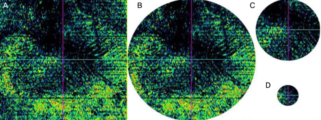
Figure 1. Four types of En Face optical coherence tomography (OCT) images. 6,000 × 6,000 μm square (A), 6,000 μm diameter
circle (B), 3,000 μm diameter circle (C), and 1,000 μm diameter circle (D). The original En Face OCT image (A) was edited in three circles according to the early treatment diabetic retinopathy study subfields. Then we obtained the black pixel values of each of the images.A B
Figure 2. Large area of elevated epiretinal membrane (ERM) peeling. Line scanning ophthalmoscope image (A) and En Face image (B).
Black pixelated areas calculated automatically reveal elevated ERM in (B). It shows that the retina and the ERM have a wide area of contact in (A). And the larger the area of contact, the lower the black pixel value may be considered as shown as the narrow area of black color in (B).
A B
Figure 3. Small area of elevated epiretinal membrane (ERM) peeling. Line scanning ophthalmoscope image (A), and En Face image (B).
It shows that the retina and the ERM have a narrow are of contact in (A). And the narrower the area of contact, the greater the black pixel value may be considered as shown as the greater area of black color in (B).
Table 1. Demographic characteristics and clinical features of the patients
Characteristics Value
Number of patients 59
Sex (male:female) 27:32
Age at surgery (years) 64.8 ± 8.9
Preoperative lens state (phakic:pseudophakic) 36:23
Types of operation (phacovitrectomy:vitrectomy) 15:44
BCVA (logMAR) (preoperative:postoperative) 0.51 ± 0.29:0.24 ± 0.23*
BCVA change (logMAR) 0.27 ± 0.24
Values are presented as mean ± SD unless otherwise indicated.
SD = standard deviation; BCVA = best corrected visual acuity
*p-value < 0.001, Wilcoxon signed rank test.
Table 2. Central retinal thickness after and before epiretinal membrane peeling (ERMP), and values of the black pixelated area on
En Face imagesVariables Value
CRT (μm) (preoperative:postoperative) 468.97 ± 101.44:354.75 ± 63.20*
CRT chage (μm) 113.53 ± 86.11
6,000 × 6,000 μm square (pixels) 42,893.92 ± 6,826.66
6,000 μm diameter circle (pixles) 33,799.24 ± 5,652.37
3,000 μm diameter circle (pixles) 8,094.85 ± 1,958.61
1,000 μm diameter circle (pixles) 861.90 ± 391.41
Values are presented as mean ± SD unless otherwise indicated.
CRT = central retinal thickness; SD = standard deviation.
*p-value < 0.001, Wilcoxon signed rank test.
Table 3. Correlation between the visual prognosis and values of the black pixelated area on En Face images
Variables Preoperative BCVA Postoperative BCVA
(12 months)
BCVA change (12 months)
r* p-value r* p-value r* p-value
6,000 × 6,000 μm square pixels 0.300 0.021† -0.312 0.016† 0.645 <0.001†
6,000 μm diameter circle pixels 0.247 0.059 -0.321 0.013† 0.590 <0.001†
3,000 μm diameter circle pixels 0.199 0.131 -0.125 0.344 0.352 0.006†
1,000 μm diameter circle pixels 0.162 0.220 0.035 0.792 0.163 0.218
BCVA = best corrected visual acuity.
*Spearman correlation test; †Statistically significant.
을 제거하였다.
본 연구는 전북대학교병원 생명의학연구 윤리심의위원 회(institutional review board, IRB)의 승인을 받았으며(승인 번호: 2017-06-014), 헬싱키선언을 준수하였다. 통계는 SPSS statics software package version 20.0 (IBM, Armonk, NY, USA)을 사용하였고, 단변량 분석은 Wilcoxon signed rank test로 검정하였고, black pixel 값, 수술 전후 최대교정시 력 및 수술 전후 중심망막두께 등의 인자들 간의 분석은 선형 회귀모형을 사용하였다. 또한 p값이 0.05 미만인 경 우를 통계학적으로 의의가 있는 것으로 판단하였다.
결 과
대상 환자 59명 59안 중 남자는 27명, 여자는 32명이었 으며, 수술 시 나이는 41세에서 80세로 평균 64.8 ± 8.9세
였다. 59안 중 23안은 인공수정체안이었으며, 36안은 수 정체안이었다. 수정체안 36안 중 15안은 망막앞막 제거술 과 백내장수술을 동시에 시행하였다. 술 전 평균 최대교정 시력은 0.51 ± 0.29 logMAR였고, 수술 후 12개월째 평균 최대교정시력은 0.24 ± 0.23 logMAR로 유의한 시력변화 가 관찰되었다(p<0.001) (Table 1).
수술 전 평균 중심망막두께는 468.97 ± 101.44 μm, 수 술 후 평균 중심망막두께는 354.75 ± 63.20 μm로 유의하 게 감소하였고(p<0.001), 수술 전후 중심망막두께 차이는 평균 113.53 ± 86.11이었다(Table 2). 또한 수술 전 En Face 영상의 평균 black pixel 값은 6,000 × 6,000 μm square (6,000 squre), 6,000 μm diameter circle (6,000 cir- cle), 3,000 μm diameter circle (3,000 circle), 1,000 μm di- ameter circle (1,000 circle)에서 각각 42,893.92 ± 6,826.66, 33,799.24 ± 5,652.37, 8,094.85 ± 1,958.61, 861.90 ±
A B
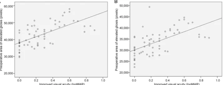
Figure 4. Scattergram showing the significant positive correlation between the change in best corrected visual acuity (BCVA) and
preoperative epiretinal membrane area (pixels) with no retinal contact. (A) BCVA and 6,000 × 6,000 μm square (r = 0.645, p <0.001), and (B) BCVA and 6,000 μm diameter circle (r = 0.590, p < 0.001).
Table 4. Correlation between the values of the black pixelated area on En Face images and the values of central retinal thickness
(CRT) changeVariables CRT change (12 months)
r* p-value
6,000 × 6,000 μm square pixels 0.055 0.680
6,000 μm diameter circle pixels 0.042 0.758
3,000 μm diameter circle pixels 0.081 0.540
1,000 μm diameter circle pixels 0.040 0.764
*Spearman correlation test.
Table 5. Correlation between the visual prognosis and the values of the central retinal thickness (CRT)
VariablesPostoperative BCVA (12 months)
BCVA change (12 months)
r* p-value r* p-value
Preoperative CRT 0.360 0.005† -0.026 0.843
CRT change (12 months) 0.286 0.028† 0.179 0.174
Preoperative BCVA 0.580 <0.001† 0.663 <0.001†
BCVA = best corrected visual acuity.
*Spearman correlation test; †Statistically significant.
Table 6. Correlation between the values of the central retinal thickness (CRT)
VariablesPostoperative CRT (12 months)
CRT change (12 months)
r* p-value r* p-value
Preoperative CRT 0.536 <0.001† 0.786 <0.001†
*Spearman correlation test; †Statistically significant.
391.41이었다(Table 2).
시력호전 정도와 6,000 square 및 6,000 circle의 En Face 영상 black pixel 값은 각각 높은 상관관계를 보였다 (r=0.645, p<0.001) (r=0.590, p<0.001) (Table 3, Fig. 4).
또한 3,000 circle의 black pixel 값도 시력호전 정도와 유 의한 상관관계를 보였으나(r=0.352, p=0.006), 1,000 circle 의 black pixel 값과 시력호전 정도는 통계학적으로 유의
하지 않았다(p=0.218) (Table 3). 반면, En Face 영상 black pixels 값과 수술 전후 중심망막두께 변화량은 통계 적으로 유의하지 않았다(Table 4).
시력과 중심망막두께와의 상관관계를 분석한 결과, 수술 전 중심망막두께 및 수술 전후 중심망막두께 변화량은 수술 후 시력과 유의한 상관관계를 보였으나(r=0.360, p=0.005) (r=0.286, p=0.028), 시력호전 정도와는 통계학적으로 유
의하지 않았다(Table 5). 또한 수술 전 중심망막두께는 수 술 후 중심망막두께 및 수술 전후 중심망막두께 변화량과 높은 상관 관계를 보였다(r=0.536, p<0.001) (r=0.786, p<0.001) (Table 6). 수술 전 시력과 수술 후 시력은 유의 한 상관관계를 가졌고(r=0.580, p<0.001), 수술 전 시력과 시력호전 정도의 상관관계도 유의한 결과를 보였다 (r=0.663, p<0.001) (Table 5).
고 찰
망막앞막 제거술은 난이도 및 위험도가 높은 수술인데, 이는 안내 집게를 이용하여 망막앞막을 잡을 때 망막앞막 아래에 있는 망막을 잡으면서 신경절세포층에 비가역적 인 손상을 초래할 수 있기 때문이다. 따라서 망막과의 유 착이 없는 망막앞막 부위의 범위가 넓을수록 집게를 이용 해서 잡기가 쉬우며, 안전하게 망막앞막을 제거하는 것을 용이하게 해준다.
Wilkins et al20은 OCT를 통해 망막앞막과 망막표면 사 이의 분리가 없는 전반적 유착(global adherence)과 망막 표면과 일부 유착이 있으나 망막표면과 명확한 분리가 관 찰되는 국소적 유착(focal adherence)으로 망막앞막을 구 분하였고, 전반적 유착 형태가 국소적 유착을 보이는 망막 앞막보다 수술이 더 어렵고 합병증 발병이 높을 것이라고 주장하였다. 또한 Kim et al19은 SD-OCT를 이용하여 망막 앞막의 유착 형태를 국소적 유착(focal), 전반적 유착 (broad), 완전 유착(complete) 3가지로 분류하여 망막제거 술의 난이도와의 상관관계를 분석하였고, 완전 유착의 경 우 국소적 유착보다 수술의 난이도가 8.6배 증가한다고 주장하였다. 따라서 망막앞막과 망막표면 사이의 유착이 심할수록 수술 난이도가 증가하여 시력 예후가 좋지 않을 것으로 생각해 볼 수 있다.
또한 Seidel et al18 및 Kim et al19은 망막앞막의 침윤 (infiltration) 정도, 제거술 후 남아있는 내경계막, 그리고 시력예후와의 관계에 대하여 연구하였는데, 망막앞막 그 자체로도 망막 조직에 손상을 주기 때문에, 망막앞막이 침 윤된 범위가 넓을수록 망막 조직이 더 많이 손상을 받는 다고 하였다.
본 연구에서는 HD-OCT를 이용하여 망막앞막을 분석 하였는데, 망막앞막과 망막의 접촉이 클수록 En Face 영 상에서 검은색으로 나타나는 부위의 면적이 작고(Fig. 2), 접촉이 작을수록 검은색으로 나타나는 부위의 면적이 큰 것을 알 수 있었다(Fig. 3). 따라서 그래픽 편집 소프트웨 어를 이용하여 이러한 En Face 영상의 black pixel 값을 구하여 망막과의 접촉이 없는 부위를 수치화하였다. 한편
Pavlidis et al21도 En Face OCT 영상의 black pixel 값을 이용하여, 유리체절제술 후 망막앞막의 예후인자로서 En Face OCT의 유용성에 대하여 연구하였는데, Pavlidis et al21의 연구에서는 6,000 square En Face OCT 영상의 black pixel 값만을 분석한 반면, 본 연구에서는 ETDRS subfields에 준하여 En Face OCT 영상을 더 세분화하였다 는 점에서 차이가 있다.
한편, 본 연구에서는 3,000 circle의 black pixels 값보다 6,000 square 및 6,000 circle의 black pixel 값이 시력호전 정도와 더 높은 상관관계를 보였고, 저자들은 추가적으로 6,000 square에서 3,000 circle을 제외한 영역의 black pix- el 값(6,000 square black pixel 값–3,000 circle black pixel 값)과 6,000 circle의 black pixel 값에서 3,000 circle을 제 외한 영역의 black pixel 값(6,000 circle black pixel 값–
3,000 circle black pixel 값)을 구하였고 시력호전 정도와의 상관관계를 분석하였는데, 각각 3,000 circle보다 더 높은 상관관계를 보였다(r=0.631, p<0.001) (r=0.580, p<0.001).
망막앞막 수술의 경우 막을 얼마나 제거해야 하는지에 대 한 지침이 없으나, 대부분의 술자들은 망막혈관궁 내에는 견인이 없어야 한다고 믿으며, 망막손상 없이 모든 방향으 로 많이 제거해야 한다고 생각한다. 그러므로 본 연구의 결과는 중심부분뿐만 아니라, 중심 이외 부분의 망막앞막 유착도 시력예후에 중요하며, 따라서 수술 시 가능한 한 넓은 범위의 망막앞막을 제거해야 한다는 주장을 뒷받침 한다고 생각하였다. 추후 망막앞막의 망막유착 범위, 망막 앞막 제거 범위 및 시력예후와의 관계에 대한 추가적인 연구가 필요할 것으로 생각된다.
본 연구에서는 그래픽 편집 소프트웨어(Adobe Photoshop CS6, Adobe Systems)에서 레벨 0에 해당되는 검은색만 black pixel로 정의하여 “black pixel 값”을 구하였는데, En Face 이미지에서 어둡게 나타나는 부분을 다른 어두운 색 의 레벨로 확장하여 pixel 값을 구한다면 결과에 변화가 있을 수 있기 때문에 이는 본 연구의 한계점으로 생각된 다. 수술 후 시력에 영향을 주는 예후 인자로 지금까지 제 시된 것들은 저자마다 다양하다.
수술 전 중심망막두께
중심망막두께와 관련하여 Kim et al15와 Kwon et al22의 연구에서 술 전 망막중심두께가 최종시력과 상관관계를 보였는데, 본 연구에서도 술 전 망막중심두께와 12개월째 최 대교정시력이 유의한 상관관계를 보였다(r=0.360, p=0.005).
또한 Suh et al23의 연구에서는 술 전 중심망막두께는 술 후 중심망막두께, 술 후 시력, 시력호전 정도와 상관관계 가 있다고 하였고, 시력호전 정도를 제외하고는(p=0.843),
본 연구에서도 술 전 중심망막두께가 술 후 중심망막두께 (r=0.536, p<0.005) 및 술 후 시력과 유의한 상관관계를 보였다(r=0.360, p=0.005) (Table 5, 6).
수술 전후 중심망막두께 변화
반면, Suh et al23의 연구에서 중심망막두께 변화 정도는 술 후 시력 및 시력호전 정도와 상관관계가 없다고 보고 하였는데, 본 연구에서는 술 후 시력과는 유의한 상관관계 를 보였으나(r=0.286, p=0.028), 시력호전 정도와는 통계 학적으로 유의하지 않았다(p=0.174) (Table 5). 따라서 중 심망막두께가 시력예후에 미치는 영향에 대해서는 추후 다양한 연구가 필요할 것으로 보인다. 또한 본 연구에서는 En Face 영상의 black pixel 값과 수술 전후 중심망막두께 변화량은 통계학적으로 유의하지 않았는데, 망막앞막의 침윤 정도 외에 중심망막두께에 영향을 주는 다른 요인이 있는지에 대한 연구가 필요할 것으로 보인다.
수술 전 시력
많은 연구들에서 수술 전 시력은 수술 후 최종 시력에 영향을 주는 예후인자로서 알려져있다.15,22,24 본 연구에서 도 수술 전 시력은 수술 후 12개월째 시력과 유의한 상관 관계를 가졌고(r=0.580, p<0.001), 시력호전 정도와도 높 은 상관관계를 보였다(r=0.663, p<0.001) (Table 5). 따라 서 수술 전 시력은 망막앞막에서 중요한 예후인자로 생각 된다.
망막앞막에서 유리체절제술 후 수정체핵경화의 빠른 진행이 보고되었으며, 수술적 치료에 따른 가장 많은 합병 증으로 알려져 있다.7,24-26 Ando et al27은 망막앞막 환자에 서 유리체절제술만 시행한 환자와 백내장수술을 동시에 받은 환자를 비교하였는데, 2년 경과 관찰 후 유리체절제 술만 받은 환자군의 65%가 백내장 진행으로 인해 시력이 저하되었다고 보고하였다. 본 연구에서는 유리체절제술만 시행한 환자와, 백내장수술을 동시에 시행한 환자를 따로 분류하지 않았으며, 수술 전 중등도 이상의 백내장이 있는 경우는 대상군에서 제외하였다. 따라서 백내장수술 여부 및 수술 전 백내장 정도에 대한 분석이 없다는 점은 본 연 구의 제한점으로 생각한다. 이외에도 후향적 연구이며 대 상 환자 수가 많지 않았다는 점, 상대적으로 경과 관찰 기 간이 짧아 재발에 대한 분석을 시행하지 못했다는 제한점 이 있다. 따라서 향후, 백내장에 의한 영향까지 고려하여 더 많은 환자를 대상으로 장기간의 전향적 연구가 필요하다.
결론적으로, 유리체절제술을 통한 망막앞막 제거술을 시행하는 경우 망막과의 유착이 없는 부위가 넓을수록 더 욱 좋은 예후를 기대할 수 있으며, 수술 전 정면빛간섭단
층촬영 영상을 통해 이러한 부위를 파악하여 수술을 용이 하게 할 수 있다. 따라서 망막앞막의 수술 후 시력 예후 예측 인자로서 정면빛간섭단층촬영 영상이 유용할 것으 로 생각된다.
REFERENCES
1) Koutsandrea CN, Apostolopoulos MN, Alonistiotis DA, et al.
Indocyanine green-assisted epiretinal membrane peeling evaluated by optical coherence tomography and multifocal electroretino- graphy. Clin Ophthalmol 2007;1:535-44.
2) Smiddy WE, Maguire AM, Green WR, et al. Idiopathic epiretinal membranes. Ultrastructural characteristics and clinicopathologic correlation. Ophthalmology 1989;96:811-20.
3) Michels RG. Vitreous surgery for macular pucker. Am J Ophthalmol 1981;92:628-39.
4) de Bustros S, Rice TA, Michels RG, et al. Vitrectomy for macular pucker: use after treatment of retinal tears or retinal detachment.
Arch Ophthalmol 1988;106:758-60.
5) Ando F, Ohba N, Touura K, Hirose H. Anatomical and visual out- comes after episcleral macular buckling compared with those after pars plana vitrectomy for retinal detachment caused by macular hole in highly myopic eyes. Retina 2007;27:37-44.
6) McDonald HR, Verre WP, Aaberg TM. Surgical management of idiopathic epiretinal membranes. Ophthalmology 1986;93:978-83.
7) de Bustros S, Thompson JT, Michels RG, et al. Nuclear sclerosis after vitrectomy for idiopathic epiretinal membranes. Am J Ophthalmol 1988;105:160-4.
8) Margherio RR, Cox MS Jr, Trese MT, et al. Removal of epimacular membranes. Ophthalmology 1985;92:1075-83.
9) van Velthoven ME, Faber DJ, Verbraak FD, et al. Recent develop- ments in optical coherence tomography for imaging the retina.
Prog Retin Eye Res 2007;26:57-77.
10) Ko TH, Fujimoto JG, Schuman JS, et al. Comparison of ultrahigh- and standard-resolution optical coherence tomography for imaging macular pathology. Ophthalmology 2005;112:1922.e1-15.
11) Schmidt-Erfurth U, Leitgeb RA, Michels S, et al. Three-dimen- sional ultrahigh-resolution optical coherence tomography of mac- ular diseases. Invest Ophthalmol Vis Sci 2005;46:3393-402.
12) Odrobina D, Michalewska Z, Michalewski J, et al. Long-term eval- uation of vitreomacular traction disorder in spectral-domain opti- cal coherence tomography. Retina 2011;31:324-31.
13) Kinoshita T, Kovacs KD, Wagley S, Arroyo JG. Morphologic dif- ferences in epiretinal membranes on ocular coherence tomography as a predictive factor for surgical outcome. Retina 2011;31:1692-8.
14) Falkner-Radler CI, Glittenberg C, Hagen S, et al. Spectral-domain optical coherence tomography for monitoring epiretinal membrane surgery. Ophthalmology 2010;117:798-805.
15) Kim J, Rhee KM, Woo SJ, et al. Long-term temporal changes of macular thickness and visual outcome after vitrectomy for idio- pathic epiretinal membrane. Am J Ophthalmol 2010;150:701-9.e1.
16) Shiono A, Kogo J, Klose G, et al. Photoreceptor outer segment length: a prognostic factor for idiopathic epiretinal membrane surgery. Ophthalmology 2013;120:788-94.
17) Theodossiadis PG, Theodossiadis GP, Charonis A, et al. The pho-
= 국문초록 =
특발망막앞막에서 수술 전 정면빛간섭단층촬영 영상과 수술 후 시력 호전과의 관계
목적: 특발망막앞막 환자에서 수술 전 정면빛간섭단층촬영(En Face optical coherence tomography) 영상과 술 후 예후와의 관련성 에 대하여 알아보고자 한다.
대상과 방법: 2005년 1월부터 2016년 1월까지 특발망막앞막으로 진단된 후 망막앞막 제거술을 받은 환자 중 12개월 이상 경과 관찰한 환자 59명 59안의 의무기록을 후향적으로 분석하였다. 술 전 En Face 영상을 early treatment diabetic retinopathy study (ETDRS) subfields에 준하여, 3개의 원(6,000 μm diameter circle, 3,000 μm diameter circle, 1,000 μm diameter circle)과 1개의 사각형 (6,000 × 6,000 μm square), 총 4가지로 나누었고, 그래픽 편집 소프트웨어(Adobe Photoshop CS6, Adobe Systems, San Jose, CA, USA)를 통해 영상의 black pixel 값을 측정하여, 망막앞막의 유착이 없는 부위를 수치화하였다. 이후 Black pixel 값, 수술 전후 시력 및 수술 전후 중심망막두께와의 상관관계에 대하여 분석하였다.
결과: 망막앞막 제거술 후 최대교정시력은 유의하게 증가하였고(p<0.001), 중심망막두께는 유의하게 감소하였다(p<0.001). 6,000
× 6,000 μm square 및 6,000 μm diameter circle의 En Face 영상 black pixel 값이 증가할수록 수술 전후 최대교정시력 호전 정도가 증가하였으며, 이는 통계학적으로 유의하였다(r=0.645, p<0.001) (r=0.590, p<0.001).
결론: 망막앞막 환자의 수술 전 En Face 영상에서 망막앞막과 망막의 유착이 없는 부위의 면적이 클수록 수술 후 최대교정시력의 증가량이 많았다. 따라서 망막앞막의 수술 후 시력 예후 예측 인자로서 정면빛간섭단층촬영 영상이 유용할 것으로 생각된다.
<대한안과학회지 2018;59(4):347-354>
toreceptor layer as a prognostic factor for visual acuity in the sec- ondary epiretinal membrane after retinal detachment surgery:
imaging analysis by spectral-domain optical coherence tomography.
Am J Ophthalmol 2011;151:973-80.
18) Seidel G, Weger M, Stadlmüller LG, et al. Association of pre- operative optical coherence tomography markers with residual in- ner limiting membrane in epiretinal membrane peeling. PLoS One 2013;8:e66217.
19) Kim JS, Chhablani J, Chan CK, et al. Retinal adherence and fibril- lary surface changes correlate with surgical difficulty of epiretinal membrane removal. Am J Ophthalmol 2012;153:692-7, 697.e1-2.
20) Wilkins JR, Puliafito CA, Hee MR, et al. Characterization of epiretinal membrane using optical coherence tomography. Ophthalmology 1996;103:2142-51.
21) Pavlidis M, Georgalas I, Körber N. Determination of a new param- eter, elevated epiretinal membrane, by En Face OCT as a prog- nostic factor for pars plana vitrectomy and safer epiretinal mem- brane peeling. J Ophthalmol 2015;2015:838646.
22) Kwon SI, Ko SJ, Park IW. The clinical course of the idiopathic epi-
retinal membrane after surgery. Korean J Ophthalmol 2009;23:
249-52.
23) Suh MH, Seo JM, Park KH, Yu HG. Associations between macular findings by optical coherence tomography and visual outcomes af- ter epiretinal membrane removal. Am J Ophthalmol 2009;147:
473-80.e3.
24) Rice TA, de Bustros S, Michels RG, et al. Prognostic factors in vi- trectomy for epiretinal membranes fo the macula. Ophthalmology 1986;93:602-10.
25) Pournaras CJ, Donati G, Brazitikos PD, et al. Macular epiretinal membranes. Semin Ophthalmol 2000;15:100-7.
26) Donati G, Kapetanios AD, Pournaras CJ. Complications of surgery for epiretinal membranes. Graefes Arch Clin Exp Ophthalmol 1998;236:739-46.
27) Ando A, Nishimura T, Uyama M. Surgical outome on combined procedures of lens extraction, intraocular lens implantation, and vi- trectomy during removal of the epiretinal membrane. Ophthalmic Surg Lasers 1998;29:974-9.