J Korean Ophthalmol Soc 2014;55(5):686-692 pISSN: 0378-6471⋅eISSN: 2092-9374
http://dx.doi.org/10.3341/jkos.2014.55.5.686
Original Article
좋은 시력을 가진 특발성 망막앞막의 수술결과
Surgical Outcomes of Idiopathic Epiretinal Membrane with Good Visual Acuity
김성일1⋅박성후1⋅변익수2,3⋅이지은1,4
Sung Il Kim, MD1, Sung Who Park, MD1, Ik Soo Byon, MD2,3, Ji Eun Lee, MD, PhD1,4
부산대학교병원 안과1, 양산부산대학교병원 안과2, 양산부산대학교병원 의생명융합연구소3, 부산대학교 의학전문대학원 안과학교실4 Department of Ophthalmology, Pusan National University Hospital1, Busan, Korea
Department of Ophthalmology, Pusan National University Yangsan Hospital2, Yangsan, Korea Biomedical Research Institute, Pusan National University Yangsan Hospital3, Yangsan, Korea Department of Ophthalmology, Pusan National University Graduate School of Medicine4, Busan, Korea
Purpose: To evaluate surgical outcomes of idiopathic epiretinal membrane (ERM) with good visual acuity.
Methods: We evaluated patients who were diagnosed with idiopathic ERM with best corrected visual acuity (BCVA) greater than 20/40 and who were followed-up for 12 months or longer after vitrectomy and membrane removal. BCVA, metamorphopsia, cen- tral subfield macular thickness (CSMT), foveal contour, and status of photoreceptor inner/outer segment (IS/OS) junction were retrospectively assessed based on the medical records and optical coherence tomography (OCT) images.
Results: Twenty-four eyes were included in the present study. The mean BCVA (log MAR) did not significantly improve from baseline to 12 months after surgery (0.26 ± 0.06 and 0.25 ± 0.19, respectively). Six eyes showed improved vision of two or more lines, and six eyes had decreased vision of two or more lines. Metamorphopsia remained in all four eyes with preoperative symp- toms until 12 months postoperatively. CSMT decreased significantly from 418 ± 86 μm at baseline to 343 ± 45 μm at 12 months (p < 0.01). Among 17 eyes without foveal depression at baseline, 11 eyes recovered a foveal depression at an average of 6.6 months after surgery. IS/OS status at baseline was intact in 19 eyes, attenuated in three eyes, and disrupted in two eyes and did not change significantly at 12 months.
Conclusions: Surgical treatment for idiopathic ERM with good visual acuity resulted in anatomical but not functional improvement. Choosing surgery for idiopathic ERM with good visual acuity should be considered carefully because decreased visual acuity could result in some patients.
J Korean Ophthalmol Soc 2014;55(5):686-692
Key Words: Good visual acuity, Idiopathic epiretinal membrane, Inner limiting membrane, Removal of membrane, Vitrectomy
■Received: 2013. 6. 27. ■ Revised: 2013. 9. 12.
■Accepted: 2014. 3. 15.
■Address reprint requests to Ik Soo Byon, MD
Department of Ophthalmology, Pusan National University Yangsan Hospital, #20 Geumo-ro, Mulgeum-eup, Yangsan 626-770, Korea
Tel: 82-55-360-2131, Fax: 82-55-360-2161 E-mail: [email protected]
ⓒ2014 The Korean Ophthalmological Society
This is an Open Access article distributed under the terms of the Creative Commons Attribution Non-Commercial License (http://creativecommons.org/licenses/by-nc/3.0/) which permits unrestricted non-commercial use, distribution, and reproduction in any medium, provided the original work is properly cited.
망막앞막은 1865년 Iwanoff1에 의해 처음 소개된 질환으 로, 망막의 내경계막 표면에서 교세포의 증식으로 인해 무
혈관성 세포막이 발생하는 질환이다. 약 7-11.8%의 유병률 을 보이며 연령이 증가할수록 발생률은 증가한다고 알려졌 다.2-4 대부분은 특발성으로 발생하며, 염증성 질환, 망막혈 관질환, 외상, 망막박리 등에서 이차적으로 발생할 수도 있 다.5-8 망막앞막에 의한 차폐(covering), 막의 수축에 의한 황 반부의 뒤틀림(distorting)과 망막 견인, 혈관 누출에 의한 망막부종, 축삭 이동 차단 등에 의해 시력저하, 변형시, 단 안복시 등을 일으킨다.9
망막앞막의 제거는 1978년 Machemer10가 유리체절제술 과 막제거술을 통해 최초로 성공을 보고한 이후 최근에는
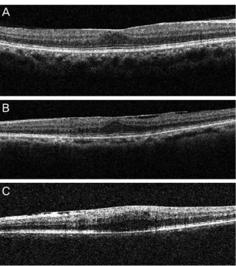
Figure 1. Classification of the inner segment/outer segment
(IS/OS) junction of the photoreceptor layer according to opti- cal coherence tomography scan. (A) Intact type; continuous hyper-reflectivity line of the IS/OS junction. (B) Attenuated type; decreased reflectivity line of the IS/OS junction. (C) Disrupted type; absent or discontinuity of the IS/OS junction.약 90%의 높은 성공률을 보이고 있다.11-13 수술의 적응증은 명확히 정해진 것이 없으나 수술 전 시력이나 환자의 주관 적 증상에 기초해, 일반적으로 20/50 이하의 시력, 심한 변 시증, 단안복시 등을 호소할 때 수술을 고려한다. 하지만, 최근에는 수술 기구와 기술의 발전으로 유리체절제술의 적 응증이 확대되어 더 좋은 시력과 높은 입체시를 얻기 위해 수술적 치료를 일찍 고려할 수도 있다.14
특발성 망막앞막의 수술적 치료의 우수한 결과와 관련인 자들에 대해 많은 보고들이 있어 왔으나 대부분이 진행된 망막앞막에 의해 시력이 낮은 환자들을 대상으로 시행한 연구들이다. 좋은 시력을 가진 특발성 망막앞막에 대한 조 기수술결과를 연구한 경우는 많지 않으며 아직 국내의 보 고는 없다. 본 연구는 특발성 망막앞막의 조기 수술적 치료 에 대한 타당성을 알아보기 위해 좋은 시력을 가진 망막앞 막 환자의 유리체절제술과 막제거술 후 시력변화와 황반부 의 해부학적 변화에 대해 조사하였다.
대상과 방법
2004년 1월부터 2011년 4월까지 부산대학교병원에서 특 발성 망막앞막으로 진단받고 뚜렷한 황반부의 부종(300 μm 이상)이 있거나 변형시를 호소하여 유리체절제술과 막제거 술을 시행한 환자들 중에서 수술 전 최대교정시력이 20/40 이상(logMAR: 0.3 이하)이면서 수술 후 12개월 이상 경과 관찰이 가능하였던 환자들을 후향적으로 분석하였다. 당뇨 망막병증, 망막혈관폐쇄, 망막열공, 망막박리, 포도막염 등 에 의한 이차성 망막앞막은 제외하였으며, 이전의 백내장 수술을 제외한 안내수술이나 안내주사, 안외상의 과거력이 있는 환자도 본 연구 대상에서 제외하였다.
수술 전과 수술 후 1, 3, 6, 12개월에 최대교정시력, 세극 등검사, 안저검사, 빛간섭단층촬영 영상을 분석하였으며, 수술 전과 수술 후 12개월의 변시증 유무를 조사하였다. 수 술 전후의 최대교정시력은 Snellen 시력표를 이용하여 측정 하였으며, 통계적 분석을 위해 logarithm of the minimum angle of resolution (logMAR) 시력으로 변환하였다. 수술 전과 비교해 수술 후 12개월의 시력이 2줄 이상 상승, 2줄 이상 저하, 변화 없었던 세 군으로 나누어 추가 분석을 시 행하였다.
빛간섭단층촬영 영상은 신호강도(signal strength)가 8 이 상인 것을 대상으로 중심막막두께, 중심오목의 형태, 시세 포 내절/외절의 상태를 분석하였다. 중심망막두께는 중심부 1 mm 값인 central subfield macular thickness를 조사하였는 데, 2008년 10월 이전은 시간영역 빛간섭단층촬영장치(time domain optical coherence tomography)인 stratus OCT (Carl
Zeiss, CA, USA)가, 2008년 11월 이후로는 공간영역 빛간 섭단층촬영장치(spectral domain optical coherence tomog- raphy)인 Cirrus-HD (Carl Zeiss, CA, USA)가 사용되었기에 두 장비 사이의 오차를 보정하기 위해 시간영역 빛간섭단 층촬영장치에서 측정된 값은 50 μm를 더하여 보정하였 다.15 중심오목형태는 중심오목의 소실 및 회복 여부를 확 인하였다. 시세포 내절/외절의 상태는 수직, 수평단층촬영 의 두 영상에서 중심 3 mm 지역을 분석하였는데, 균일하고 밝은 반사를 보이는 것을 정상형(intact type), 형태는 양호 하나 밝기가 감소되어 있는 것을 감쇄형(attenuated type), 단절된 부분이 존재하는 것을 단절형(disrupted type)으로 구분하여 조사하였다(Fig. 1).
55세 이상의 환자들 중에서 수정체가 LOCS III 기준으로 2단계 이상이 있는 경우에는 백내장수술을 유리체절제술 및 막제거술과 함께 시행하였으며, 수정체를 보존한 경우 에는 수술 후 시력에 영향을 줄 수 있는 백내장(LOCS III 기준으로 수술 전과 비교하여 2단계 이상 악화)의 발생 여 부를 조사하였다.16
수술방법은 23게이지 삽입관으로 섬모체평면부 세 부분 에 공막창을 만들고 중심부 유리체를 절제한 뒤 트리암시 놀론(탐세톤 주, 한올바이오파바)을 주입하여 뒤유리체박
A
B
C
Table 1. Baseline characteristics
Parameters Values
Laterality (right:left) 11:13
Sex (M:F) 4:20
Age (years) 57.8 ± 9.4
BCVA (log MAR) CSMT (μm)
Lens state (Phakia:Pseudophakia)
0.26 ± 0.06 417.8 ± 86.4
24:0 Combined cataract surgery (%)
ILM peeling (%) ICG assisted (%) TA assisted (%) No adjuvant (%)
16 (66.7) 21 (87.5) 12 (50.0) 7 (29.2) 2 (8.3) Values are presented as mean ± SD.
BCVA = best corrected visual acuity; CSMT = central subfield macular thickness; ILM = internal limiting membrane; ICG = indocyanine green; TA = triamcinolone acetonide.
(log MAR) ( m)µ
0.45 0.4 0.35 0.3 0.25 0.2 0.15 0.1 0.05 0
750
650
550
450
350
250
BCVA CSMT 0.26 0.06±
0.32 0.13*± 0.25 0.18±
0.26 0.22± 0.25 0.19±
417.8 86.4± 380.5 60.6*±
348.7 49.5*± 334.2 45.4*± 342.7 44.9*±
Figure 2. Changes in central subfield macular thickness (CSMT)
and the best corrected visual acuity (BCVA). Values are pre- sented as mean ± SD. CSMT improved significantly 1 month after surgery (*p < 0.01), but BCVA did not.리를 확인하였다. 황반부에 완전한 뒤유리체박리가 없는 경우에는 유리체절제침이나 막쑤시게를 이용하여 뒤유리 체박리를 만들었다. 망막앞막을 제거할 때에는 견인을 최 소화하기 위해 눈속집게를 이용하여 막의 끝을 잡고 망막 과 접선방향으로 중심오목을 중심으로 둥글게 제거하였다.
혈관궁 안쪽에 존재하는 막은 최대한 제거하였다. 내경계 막을 추가로 제거할 때에는 인도사이아닌그린(인도시아닌 그린 주, 동인당제약) 또는 트리암시놀론을 사용하였다. 백 내장 수술을 함께 시행한 경우에는 각막절개를 통해 수정 체초음파유화술과 인공수정체삽입술을 먼저 시행하고 그 뒤에 유리체절제술을 시행하였다.
SPSS 18.0 Kr for window 프로그램을 사용하여 통계학 적 분석을 시행하였다. 수술 전과 수술 후 최대교정시력 및 중심망막두께 변화는 Wilcoxon signed-rank test를, 시세포 내절/외절의 상태와 내경계막 제거 여부는 Fisher’s exact test를 시행하였으며, 시력변화에 따른 세 군간의 최대교정 시력과 중심망막두께 변화는 Kruskal-Wallis H test를 시행 하여 p값이 0.05 미만인 경우를 통계적으로 유의하다고 판 단하였다.
결 과
대상은 총 24명 24안으로 남자가 4명, 여자가 20명이었 다. 평균 나이는 57.8 ± 9.4세(36-75세) 였다. 수술 전 증상 호소 기간은 평균 9개월이었다. 수술 전 경과관찰기간은 평 균 3개월로, 경과관찰 기간 동안 변형시와 경도의 시력 저 하, 빛간섭단층촬영의 객관적인 황반부종이 지속되었다. 대 상안 모두 수정체안이었으며 16안에서 수정체초음파유화 술 및 인공수정체삽입술을 함께 시행하였고, 8안은 수정체 를 보존하였다. 백내장을 동시에 수술한 경우에 백내장 수
술과 관련된 합병증은 없었다. 추가적으로 내경계막을 제 거한 경우는 21안(87.5%)이었다. 인도사이아닌 그린을 이 용한 경우가 12안, 트리암시놀론을 이용한 경우는 7안이었 으며 2안에서는 염색하지 않고 제거하였다. 수술 전 최대교 정시력은 20/25 1안, 20/30 8안, 20/40 15안으로 평균최대교 정시력(logMAR)은 0.26 ± 0.06이었다. 평균중심망막두께 는 417.8 ± 86.4 μm였다(Table 1).
최대교정시력은 수술 후 1개월 0.32 ± 0.13, 3개월, 0.25
± 0.18, 6개월 0.26 ± 0.22, 12개월에 0.25 ± 0.19로 수술 1 개월째에 일시적인 시력저하가 있었으나 수술 3개월째부터 는 수술 전과 비교하여 유의한 차이는 없었다(p=0.032, 0.788, 0.969, 0.654, Fig. 2). 2줄 이상의 시력 호전을 보인 경우와 저하된 경우는 각각 6안씩 있었으며, 시력변화가 없 는 경우는 12안이었다. 수술 전 변시증을 호소했던 4안은 모두 수술 후 12개월까지 증상이 남았다.
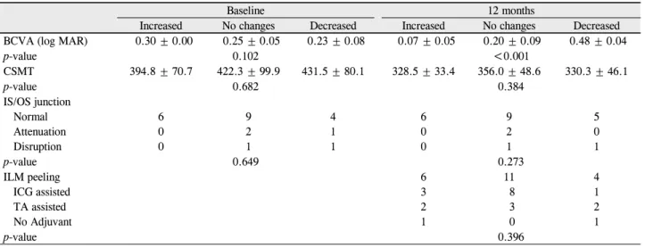
중심망막두께는 수술 후 1개월에 380.5 ± 60.6 μm, 3개 월에 348.7 ± 49.5 μm, 6개월에 334.2 ± 45.4 μm, 12개월에 342.7 ± 44.9 μm로 수술 후 1개월째부터 통계적으로 유의 한 감소를 보였다(각각 p=0.015, <0.001, <0.001, <0.001, Fig. 2). 중심오목이 없었던 17안 중 11안(64.7%)에서 평균 6.6 ± 2.6개월에 중심오목이 회복되었다(Fig. 3). 중심오목 이 회복된 11안과 중심오목이 회복되지 않은 6안 사이에 수술 후 12개월의 시력과 수술 후 시력변화는 의미 있는 차 이가 없었다.
시세포 내절/외절은 수술 전 정상형 19안, 감쇄형 3안, 단 절형 2안에서 수술 후 1년에 정상형 20안, 감쇄형 2안, 단 절형 2안으로 변하였으나 통계적으로 의미 있는 차이는 없 었다.
수정체를 보존한 8안 중 4안에서 경과관찰 기간 동안 LOCS III 2단계 이상의 백내장의 진행이 있어 추가적인 백
Figure 3. Optical coherence tomography images of the fovea.
(A) Of 17 eyes without foveal depression at baseline, (B) 11 eyes had a foveal depression after 6.6 months in average.
Table 2. Comparison of the three groups according to the change of 2 lines or more in BCVA
Baseline 12 months
Increased No changes Decreased Increased No changes Decreased
BCVA (log MAR) 0.30 ± 0.00 0.25 ± 0.05 0.23 ± 0.08 0.07 ± 0.05 0.20 ± 0.09 0.48 ± 0.04
p-value 0.102 <0.001
CSMT 394.8 ± 70.7 422.3 ± 99.9 431.5 ± 80.1 328.5 ± 33.4 356.0 ± 48.6 330.3 ± 46.1
p-value 0.682 0.384
IS/OS junction Normal Attenuation Disruption
6 0 0
9 2 1
4 1 1
6 0 0
9 2 1
5 0 1
p-value 0.649 0.273
ILM peeling ICG assisted TA assisted No Adjuvant
6 3 2 1
11 8 3 0
4 1 2 1
p-value 0.396
Values are presented as mean ± SD.
BCVA = best corrected visual acuity; CSMT = central subfield macular thickness; IS/OS = inner segment/outer segment; ILM = internal limiting membrane; ICG = indocyanine green; TA = triamcinolone acetonide.
내장 수술을 시행하였다. 백내장 수술을 시행 받지 않은 나 머지 4안 중 수술 후 12개월에 시력에 유의한 영향을 주는 것으로 생각되는 백내장은 없었다. 수술 12개월에 백내장 수술 유무에 따른 평균 시력은, 백내장 동시수술을 시행한 16안은 0.26 ± 0.21, 추가적으로 백내장을 수술 받은 4안은 0.25 ± 0.17, 수정체를 보존한 4안은 0.2 ± 0.14로 유의한 차이는 없었다.
시력이 2줄 이상 감소한 6안을 따로 분석하였을 때 중심 망막두께가 수술 전 431.5 ± 80.1 μm에서 수술 후 330.3 ±
46.1 μm로 의미 있게 감소하였으며(p=0.041), 수술 전 중심 오목이 관찰되지 않은 5안 중 수술 후 3안에서 중심오목의 형태가 회복되었다. 시세포 내절/외절의 상태는 정상형, 감 쇄형, 단절형이 수술 전 각각 4안, 1안, 1안이었으나 수술 후 각각 5안, 0안, 1안이었다.
2줄 이상의 시력변화에 따른 세 집단 사이에는 수술 전 시력, 중심망막두께, 중심오목의 형태, 시세포 내절/외절 상 태에 유의한 차이가 없었으며 수술 후에도 다른 인자의 차 이가 없었다(Table 2).
고 찰
특발성 망막앞막의 치료로써 막제거술은 수술 후 의미 있는 시력의 향상을 얻을 수 있으며,17-21 수술 전 시력이 좋 은 경우가 수술 후 좋은 시력의 예후인자로 제시되었다.18 그러나 이전의 연구들은 수술 전의 시력이 20/60 이하인 낮 은 시력의 환자들을 대상으로 하였기에,5,8,22-24 본 연구의 목 적인 망막앞막의 조기 수술적 치료에 대한 타당성을 평가 하기에는 적합하지 않다.
좋은 시력의 특발성 망막앞막 환자를 대상으로 시행한 연 구인 20/50 이상의 시력을 가진 망막앞막 환자 40안을 대상 으로 한 Thompson의 보고에서는 수술 전 평균시력이 20/50+2에서 수술 후 20/40+2로 호전되었다고 하였다.14 본 연구에서는 중심망막두께의 감소와 중심오목의 회복은 관 찰할 수 있었지만, 평균 최대교정시력은 수술 후 의미 있는 호전이 없었다. 오히려 6안(25%)에서 수술 후 12개월에 2 줄 이상의 시력감소를 나타내었다. 즉, 특발성 망막앞막의 조기수술은 황반부의 해부학적 형태의 회복에 있어서는 좋
A
B
은 결과를 보여 주었으나 시력의 호전에 있어서는 제한적 임을 보여주었다.
이러한 상반된 결과의 원인으로 몇 가지를 고려할 수 있 다. 첫 번째로 대상환자의 차이에서 비롯되었을 수 있다.
Thompson의 연구에서 대상환자의 평균 수술 전 평균시력 은 logMAR 0.367에 해당하며, 본 연구의 수술 전 평균시력 인 logMAR 0.26보다 낮았고, 수술 후 시력은 logMAR 0.276로 본 연구의 logMAR 0.25와 비슷하였다. 즉, 본 연구 의 좋은 시력 기준인 20/40보다 상대적으로 낮은 시력을 대 상으로 하였기에 수술 후 시력상승의 여력이 더 있었을 것 이라 생각된다. 다음으로 수술방법의 차이도 시력결과에 영향을 주었을 것으로 생각된다. 이전의 연구에서는 망막 앞막을 제거할 때 가능한 내경계막을 함께 제거하지 않으 려고 노력한 반면, 본 연구에서는 내경계막을 제거가 망막 앞막을 완전히 제거하고 재발률을 줄일 수 있다는 보고를 근거로 21안(87.5%)에서 의도적인 내경계막제거술을 시행
하였다.25-27 하지만 이러한 내경계막의 제거가 시력 예후에
나쁜 영향을 줄 수 있다는 가능성이 제시되었는데, 제거된 망막앞막에 내경계막이 존재하거나, 의도적으로 내경계막 을 제거한 경우에 시력 예후가 좋지 않으며 20/60 이상의 시력은 얻기 힘들다고 하였다. 내경계막 제거 과정에서 발 생한 견인력에 의한 망막의 미세한 기계적 손상이 원인으 로 제시되었다.28 최근 Chang et al29은 망막앞막만 제거한 군과 내경계막까지 제거한 두 군을 비교한 연구에서 수술 후 전자가 황반중심부에 수술 중 미처 제거하지 못한 망막 앞막이 남아 있는 경우가 더 많았지만 두 군 간의 시력에는 유의한 차이가 없고 오히려 망막두께는 망막앞막만을 제거 한 군에서 더욱 감소하였으므로 추가적인 내경계막의 제거 는 불필요하다고도 하였다.
본 연구에서는 12안(50%)에서 내경계막의 제거를 위해 0.05%의 인도사이아닌 그린을 사용하였다. 망막앞막에서 저농도의 인도사이아닌 그린의 사용 후 약제에 의한 망막 독성과 시력 결과에 대해서는 상반된 보고들이 있다.30-32 본 연구의 결과와 이전의 연구 결과들을 종합해서 볼 때, 내경 계막 제거를 위해 사용된 염색액보다는 내경계막 제거라는 수술술기 자체가 수술 후 시력에 좋지 못한 영향을 주었을 가능성이 높아 보인다.
망막앞막 수술 전후 망막두께, 중심오목, 시세포 내절/외 절의 형태의 변화 등과 수술 후 시력과의 관련성에 대한 연 구들에서 수술 후 망막두께는 현저히 감소되는 것으로 나 타났으나 시력과의 상관관계에 대해서는 다양한 결과를 보 고하였다.33-37 시세포 내절/외절의 형태와 망막앞막 수술 후 시력결과에 대해서는 대체적으로 일관된 상관성이 보고되
었다.33,34,36,37 본 연구에서는 수술 전후 시세포 내절/외절의
형태는 의미 있는 변화가 없었는데, 좋은 시력의 환자를 대 상으로 하였기에 대상환자 대부분이 수술 전 시세포 내절/
외절의 상태가 정상형이었기 때문으로 생각한다. 그러나 수술 후 시력이 감소하였던 6안 중 5안에서 정상형을 보여 시세포층 외에 시력에 영향을 주는 요소가 있을 것으로 생 각되며, 망막내층이 시력예후와 관련이 있다는 보고가 있 다.38,39
망막앞막 환자의 변시증을 M-Chart를 이용하여 분석한 연구에서 수술 후 12개월까지 지속적으로 유의하게 감소함 을 보고하였다.40 하지만 본 연구에서는 객관적인 방법으로 변시증의 변화를 측정하지 못하고 환자의 주관적 호소에 따라 조사한 한계는 있으나 수술 전 변시증을 호소한 4안 의 경우 수술 후 12개월까지 변시증을 호소하였다.
백내장 수술의 동반 여부가 수술 후 시력 결과 분석에 오 류의 요소가 될 수 있다. 백내장 수술을 16안(66.7%)에서 동시에 시행하였는데, 이는 망막앞막의 수술 후 합병증 중 에서 백내장 발생이 가장 많으며 특히 50세 이상의 환자에 서는 유리체절제술 후에 백내장의 진행과 발생이 더욱 높 다고 알려져 있어41 수술 후 백내장의 진행이 오히려 더 큰 오류의 원인이 될 수 있다고 판단하였기 때문이다. 수정체 를 보존한 8안 중 4안에서 경과 관찰 중 백내장이 진행하여 추가 수술을 진행하였다. 나머지 4안은 LOCS III 3단계 이 상의 백내장이 관찰되지 않았으므로 수술 후 12개월에 시 점의 시력에는 백내장으로 인한 영향이 없었다고 판단된다.
본 연구는 후향적 연구로서 대상환자의 수가 적으며 대 조군이 없다는 단점을 가지고 있다. 하지만 좋은 시력의 특 발성 망막앞막 환자에서 막제거술은 수술 후 황반부의 해 부학적 회복은 달성할 수 있으나 시력의 상승은 기대하기 어렵다는 것을 확인할 수 있었다. 오히려 일부 환자에서는 수술 후 시력저하가 발생할 수 있음을 알 수 있었다. 따라 서 좋은 시력의 특발성 망막앞막 환자에 대한 조기 수술과 수술술기는 신중히 결정해야 하겠다.
REFERENCES
1) Iwanoff A. Beiträge zur normalen und pathologischen Anatomie des Auges. Graefe’s Arch Clin Exp Ophthalmol 1865;11:135-70.
2) Mitchell P, Smith W, Chey T, et al. Prevalence and associations of epiretinal membranes. The Blue Mountains Eye Study. Australia.
Opthalmology 1997;104:1033-40.
3) Klein R, Klein BE, Wang Q, Moss SE. The epidemiology of epi- retinal membranes. Trans Am Ophthalmol Soc 1994;92:403-25.
4) Fraser-Bell S, Guzowski M, Rochtchina E, et al. Five-year cumu- lative incidence and progression of epiretinal membranes: the Blue Mountains Eye Study. Ophthalmology 2003;110:34-40.
5) Poliner LS, Olk RJ, Grand MG, et al. Surgical management of pre- macular fibroplasia. Arch Ophthalmol 1988;106:761-5.
6) de Bustros S, Thompson JT, Michels RG, et al. Vitrectomy for idio- pathic epiretinal membranes causing macular pucker. Br J Ophthalmol 1988;72:692-5.
7) de Bustros S, Rice TA, Michels RG, et al. Vitrectomy for macular pucker. Use after treatment of retinal tears or retinal detachment.
Arch Ophthalmol 1998;106:758-60.
8) Pesin SR, Olk RJ, Grand MG, et al. Vitrectomy for premacular fibroplasias. Prognostic factors, long-term follow-up, and time course of visual improvement. Ophthalmology 1991;98:1109-14.
9) Wise GN. Clinical features of idiopathic preretinal macular fibrosis. Schoenberg Lecture. Am J Ophthalmol 1975;79:349-57.
10) Machemer R. The surgical removal of epiretinal macular mem- branes (macular puckers). Klin Monbl Augenheilkd 1978;173:
36-42.
11) McDonald HR, Verre WP, Aaberg TM. Surgical management of idiopathic epiretinal membranes. Ophthalmology 1986;93:978-83.
12) de Bustros S, Thompson JT, Michels RG, et al. Nuclear sclerosis after vitrectomy for idiopathic epiretinal membranes. Am J Ophthalmol 1988;105:160-4.
13) Margherio RR, Cox MS Jr, Trese MT, et al. Removal of epimacular membranes. Ophthalmology 1985;92:1075-83.
14) Thompson JT. Epiretinal membrane removal in eyes with good vis- ual acuities. Retina 2005;25:875-82.
15) Bentaleb-Machkour Z, Jouffroy E, Rabilloud M, et al. Comparison of central macular thickness measured by three OCT models and study of interoperator variability. Scientific World Journal 2012;
2012:842795.
16) Shandiz JH, Derakhshan A, Daneshyar A, et al. Effect of cataract type and severity on visual acuity and contrast sensitivity. J Ophthalmic Vis Res 2011;6:26-31.
17) Kim J, Rhee KM, Woo SJ, et al. Long-term temporal changes of macular thickness and visual outcome after vitrectomy for idio- pathic epiretinal membrane. Am J Ophthalmol 2010;150:701-9.
18) Çekiç Ö, Cakır M, Alagöz N, Yılmaz OF. Retinal thickness change in relation to visual acuity improvement after 23-gauge vitrectomy for idiopathic epimacular membrane. Eye (Lond) 2011;25:180-4.
19) Falkner-Radler CI, Glittenberg C, Hagen S, et al. Spectral-domain optical coherence tomography for monitoring epiretinal membrane surgery. Ophthalmology 2010;117:798-805.
20) Lai CC, Wang NK, Wu WC, et al. The long-term anatomical and visual effect of intravitreal triamcinolone injection during vi- trectomy for the treatment of idiopathic macular epiretinal membrane. Cutan Ocul Toxicol 2011;30:292-7.
21) Hwang DJ, Na KI, Kwon SI, Park IW. Long-term changes in visual acuity and foveal thickness after vitrectomy for idiopathic epi- retinal membrane. J Korean Ophthalmol Soc 2012;53:434-9.
22) Michels RG. Vitreous surgery for macular pucker. Am J Ophthalmol 1981;92:628-39.
23) McDonald HR, Verre WP, Aaberg TM. Surgical management of idiopathic epiretinal membranes. Ophthalmology 1986;93:978-83.
24) Grewing R, Mester U. Results of surgery for epiretinal membranes and their recurrences. Br J Ophthalmol 1996;80:323-6.
25) Kwok AK, Lai TY, Li WW, et al. Indocyanine green-assisted in- ternal limiting membraned removal in epiretinal membrane sur- gery: a clinical and histologic study. Am J Ophthalmol 2004;138:
194-9.
26) Sorcinelli R. Surgical management of epiretinal membrane with in- docyanine-green-assisted peeling. Ophthalmologica 2003;217:107-10.
27) Park DW, Dugel PU, Garda J, et al. Macular pucker removal with and without internal limiting membrane peeling: pilot study.
Ophthalmology 2003;110:62-4.
28) Sivalingam A, Eagle RC Jr, Duker JS, et al. Visual prognosis corre- lated with the presence of internal-limiting membrane in histo- pathologic specimens obtained from epiretinal membrane surgery.
Ophthalmology 1990;97:1549-52.
29) Chang S, Gregory-Roberts EM, Park S, et al. Double peeling dur- ing vitrectomy for macular pucker: the charles L. Schepens lecture.
JAMA Ophthalmol 2013;131:525-30.
30) Haritoglou C, Gandorfer A, Gass CA, et al. The effect of in- docyanine-green on functional outcome of macular pucker surgery.
Am J Ophthalmol 2003;135:328-37.
31) Tsipursky MS, Heller MA, De Souza SA, et al. Comparative evalu- ation of no dye assistance, indocyanine green and triamcinolone acetonide for internal limiting membrane peeling during macular hole surgery. Retina 2013;33:1123-31.
32) Ejstrup R, la Cour M, Heegaard S, Kiilgaard JF. Toxicity profiles of subretinal indocyanine green, Brilliant Blue G, and triamcinolone acetonide: a comparative study. Graefes Arch Clin Exp Ophthalmol 2012;250:669-77.
33) Watanabe K, Tsunoda K, Mizuno Y, et al. Outer retinal morphol- ogy and visual function in patients with idiopathic epiretinal membrane. JAMA Ophthalmol 2013;131:172-7.
34) Suh MH, Seo JM, Park KH, Yu HG. Associations between macular findings by optical coherence tomography and visual outcomes af- ter epiretinal membrane removal. Am J Ophthalmol 2009;147:473-80.
35) Massin P, Allouch C, Haouchine B, et al. Optical coherence tomog- raphy of idiopathic macular epiretinal membranes before and after surgery. Am J Ophthalmol 2000;130:732-9.
36) Mitamura Y, Hirano K, Baba T, Yamamoto S. Correlation of visual recovery with presence of photoreceptor inner/outer segment junc- tion in optical coherence images after epiretinal membrane surgery.
Br J Ophthalmol 2009;93:171-5.
37) Inoue M, Arakawa A, Yamane S, Kadonosono K. Long-term out- come of preoperative disrupted inner/outer segment junctions as- sessed using spectral-domain optical coherence tomography in pa- tients with idiopathic epiretinal membrane. Ophthalmologica 2012;228:222-8.
38) Arichika S, Hangai M, Yoshimura N. Correlation between thicken- ing of the inner and outer retina and visual acuity in patients with epiretinal membrane. Retina 2010;30:503-8.
39) Joe SG, Lee KS, Lee JY, et al. Inner retinal layer thickness is the major determinant of visual acuity in patients with idiopathic epi- retinal membrane. Acta Ophthalmol 2013;91:e242-3.
40) Kinoshita T, Imaizumi H, Okushiba U, et al. Time course of changes in metamorphopsia, visual acuity, and OCT parameters af- ter successful epiretinal membrane surgery. Invest Ophthalmol Vis Sci 2012;53:3592-7.
41) Jackson TL, Donachie PH, Sparrow JM, Johnston RL. United Kingdom national ophthalmology database study of vitreoretinal surgery: report 1: case mix, complications, and cataract. Eye (Lond) 2013;27:644-51.
= 국문초록 =
좋은 시력을 가진 특발성 망막앞막의 수술결과
목적: 특발성 망막앞막의 조기 수술에 대한 타당성을 알아보기 위해 좋은 시력을 가진 특발성 망막앞막 환자의 수술결과에 대해 알아 보고자 하였다.
대상과 방법: 특발성 망막앞막으로 유리체절제술과 막제거술을 시행 받고 12개월 이상 경과관찰이 가능하였던 환자 중 수술 전 최대교 정시력(logMAR)이 0.3 이하인 환자들을 후향적으로 분석하였다. 의무기록과 빛간섭단층촬영 영상을 통해 최대교정시력, 변시증, 중 심망막두께, 중심오목형태, 시세포 내절/외절의 상태의 변화를 조사하였다.
결과: 대상환자는 총 24명, 24안이었다. 평균최대교정시력은 수술 전 0.26 ± 0.06에서, 수술 후 12개월 0.25 ± 0.19로 유의한 변화는 없었다. 2줄 이상의 시력 호전 및 저하된 경우는 각각 6안씩 있었다. 변시증을 호소한 4안은 수술 후 12개월까지 모두 증상이 남았다.
평균 중심망막두께는 수술 전 418 ± 86 μm에서, 수술 후 6개월 334 ± 45 μm, 12개월 343 ± 45 μm로 유의한 감소를 보였다(p<0.01).
중심오목이 없었던 17안 중 11안(64.7%)에서 평균 6.6 ± 2.6개월 뒤에 중심오목이 회복되었으며, 시세포 내절/외절은 수술 전 정상형 19안, 감쇄형 3안, 단절형 2안에서 수술 후 12개월에 정상형 20안, 감쇄형 2안, 단절형 2안으로 의미있는 변화는 없었다.
결론: 좋은 시력의 특발성 망막앞막 환자에서 막제거술은 황반부의 해부학적인 호전을 얻을 수 있었으나 시력의 호전은 없었다. 일부 에서는 시력이 저하되는 경우도 있었으므로 좋은 시력을 가진 특발성 망막앞막 환자에서 조기수술은 신중히 결정해야 하겠다.
<대한안과학회지 2014;55(5):686-692>