REVIEW ARTICLE
초음파내시경 유도 담도배액술
남형석
1,2, 강대환
1,2부산대학교 의학전문대학원 내과학교실1, 양산부산대학교병원 의생명융합연구소2
Endoscopic Ultrasound-guided Biliary Drainage
Hyeong Seok Nam1,2 and Dae Hwan Kang1,2
Department of Internal Medicine, Pusan National University School of Medicine1, Busan, Research Institute for Convergence of Biomedical Science and Technology, Pusan National University Yangsan Hospital2, Yangsan, Korea
The therapeutic role of endoscopic ultrasound (EUS) has continued to evolve in recent years. EUS-guided biliary drainage (EUS-BD) can be performed as an effective alternative to percutaneous drainage or surgical options when conventional Endoscopic retrograde cholangiopancreatography fails or is not possible. Depending on the access and exit routes of the stent, multiple approaches to EUS-BD have been proposed. Each patient should receive an individualized approach based on the patient’s condition, anatomy, and endoscopist’s experience, with an appropriate backup prepared. In high-volume centers, the cumulative success rate has been reported to be over 90%. However, the reported overall complication rate remains relatively high at 10-30%. Further studies are neces- sary to better understand the long-term results and standardize EUS-BD, including appropriate indications and optimal approach.
(Korean J Gastroenterol 2017;69:164-171)
Key Words: Endoscopic ultrasonography; Drainage; Cholangiopancreatography, Endoscopic retrograde; Biliary tract
Received February 17, 2017. Revised March 1, 2017. Accepted March 2, 2017.
CC This is an open access article distributed under the terms of the Creative Commons Attribution Non-Commercial License (http://creativecommons.org/licenses/
by-nc/4.0) which permits unrestricted non-commercial use, distribution, and reproduction in any medium, provided the original work is properly cited.
Copyright © 2017. Korean Society of Gastroenterology.
교신저자: 강대환, 50612, 양산시 물금읍 금오로 20, 양산부산대학교병원 의생명융합연구소
Correspondence to: Dae Hwan Kang, Research Institute for Convergence of Biomedical Science and Technology, Pusan National University Yangsan Hospital, 20 Geumo-ro, Mulgeum-eup, Yangsan 50612, Korea. Tel: +82-55-360-1538, Fax: +82-55-360-1536, E-mail: [email protected]
Financial support:: 2-year Research Grant of Pusan National University. Conflict of interest: None.
서 론
내시경적 역행성 담췌관조영술(endoscopic retrograde cholangiopancreatography, ERCP)은 담도 및 췌장질환의 진단과 치료에 널리 이용되고 있으며, 특히 악성 담도 폐쇄에 서 내시경적 담즙 배액술의 표준 치료 방법이다. 이러한 ERCP의 선택적인 삽관의 성공률은 95% 이상으로 보고되고 있다.1 그러나 일부 환자의 경우 유두부 주위 게실이나 바터 팽대부의 종양 침범, 고도의 담도 협착 등으로 인해 선택적인 삽관이 어렵거나 이전 위장관 수술, 해부학적 변이, 악성 종양 으로 인한 장관 폐쇄 등으로 인해 유두부 접근이 불가능할 수 있다.2,3 일반적으로 ERCP가 어렵거나 ERCP를 실패한 경
우, 대안적인 치료로 경피경간 담도배액술(percutaneous transhepatic biliary drainage, PTBD)이나 수술적 치료를 할 수 있으며, 흔히 PTBD를 이용한 담도 배액술을 우선하게 된다. PTBD의 성공률은 매우 높지만 다량의 복수를 동반하거 나 간내 담관의 확장이 없는 경우 시술이 어려우며, 합병증 또한 많게는 30%까지 보고되고 있다.2,4 또한 외부로 배액관 을 유치하므로 환자의 삶의 질이 저하되고, 배액관이 우발적 으로 빠지기도 하며, 담즙이 생리적으로 장관 내로 배출되지 않아 소화와 영양 흡수에서의 취약점이 있다.5,6
초음파내시경(endoscopic ultrasound, EUS)은 다양한 영역에서 이용되고 있으며, 특히 선형 초음파내시경(linear UES)이 개발된 이후 췌담도 영역의 진단 및 치료 분야에서
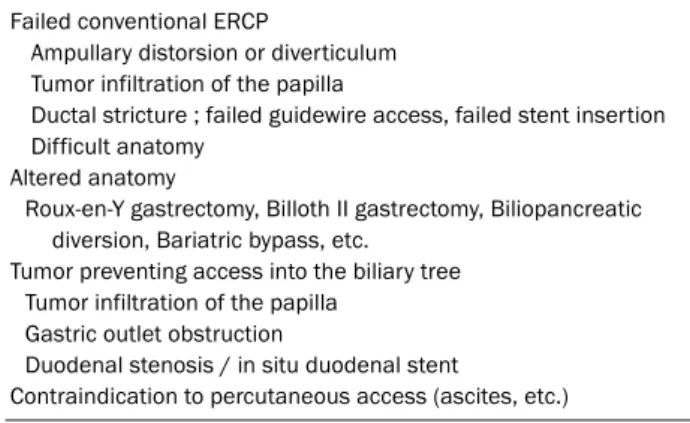
Table 1. Generally Accepted Indications for Endoscopic Ultrasound- guided Biliary Drainage
Failed conventional ERCP
Ampullary distorsion or diverticulum Tumor infiltration of the papilla
Ductal stricture ; failed guidewire access, failed stent insertion Difficult anatomy
Altered anatomy
Roux-en-Y gastrectomy, Billoth II gastrectomy, Biliopancreatic diversion, Bariatric bypass, etc.
Tumor preventing access into the biliary tree Tumor infiltration of the papilla
Gastric outlet obstruction
Duodenal stenosis / in situ duodenal stent
Contraindication to percutaneous access (ascites, etc.) ERCP, endoscopic retrograde cholangiopancreatography.
Table 2. Techniques Approaches to Endoscopic Ultrasound-guided Biliary Drainage
Access
- Intrahepatic : transesophageal, transgastric or transjejunal(in altered anatomy)
- Extrahepatic: gastric antral or duodenal Drainage
- Biliary stent placement Transhepatic
- Transluminal: hepaticogastrostomy (EUS-HGS) - Transpapillary: antegrade procedure (EUS-AG) Transduodenal
- Transluminal: choledochoduodenostomy (EUS-CDS) Biliary access for ERCP completion
- Rendezvous procedure (EUS-RV)
Adapted from Gastrointest Interv 2016;5:203-211.15
EUS-HGS, endoscopic ultrasound-guided hepaticogastrostomy; EUS-AG, endoscopic ultrasound-guided antegrade procedure; EUS-CDS, endo- scopic ultrasound-guided choledocoduodenostomy; ERCP, endoscopic retrograde cholangiopancreatography; EUS-RV, endoscopic ultrasound- guided rendezvous procedure.
빠른 발전을 보이고 있다. EUS를 통해 위나 십이지장에서 담 관과 췌관으로 직접적인 접근이 가능해지면서 ERCP가 불가 능하거나 ERCP를 실패한 경우, 이를 대체하거나 후속 ERCP 를 용이하게 할 수 있는 대안으로 초음파내시경 유도 담도배 액술(EUS-guided biliary drainage, EUS-BD)이 시도되고 있 으며, 이에 대한 연구 또한 활발히 진행되고 있다.
본 론
1996년 Wiersema 등7이 ERCP를 실패한 환자를 대상으로 EUS를 이용한 담도조영술을 처음 보고하였다. 이후 Giovannini 등8이 절제 불가능한 췌장암 환자에 대해 EUS를 이용한 담도 배액술을 처음 소개하였으며, 2004년 Mallery 등9이 EUS 유 도 rendezvous 법(EUS-guided rendezvous procedure, EUS-RV)을 최초 보고하였다. 이후 많은 연구자에 의해 EUS-BD의 다양한 기술적인 접근 및 결과에 대한 보고가 이 루어졌다. EUS-BD의 일반적인 적응증으로는 크게 1) ERCP 가 실패한 경우, 2) 해부학적 변이가 있는 경우, 3) 종양으로 인해 담도계로의 접근이 어려운 경우, 4) 복수와 같은 경피적 시술의 금기사항이 있는 경우 등이 있다(Table 1).10
초음파내시경 유도 담도배액술(EUS-BD) 전 준비
우선 시술을 하기에 앞서 시행하고자 하는 시술이 환자에 게 정말 필요한 것인지, 다른 효과적인 대안은 없는 지에 대한 충분한 사전 검토가 필요하다. 환자의 병력 및 상태뿐만 아니 라 computed tomography나 magnetic resonance imag- ing 검사 등을 통해 담도 폐쇄의 정확한 위치와 췌담도 구조 를 파악하고, 주위 장기와 췌담도의 자세한 해부학적 평가가 선행되어야 한다. 시술 과정에서는 어떤 접근이 가장 이상적 인지 계획해야 한다. 또한 EUS-BD에는 다양한 종류의 보조 기구들이 사용되지만 시술 방법이 표준화되어 있지 않고 확실
한 전용 내시경 기구가 없으므로 성공적인 시술을 위해서는 이러한 보조 기구들의 특징과 한계에 대해 충분히 숙지하여야 한다. 시술은 EUS와 ERCP에 경험이 많은 시술자가 시행하 는 것이 바람직하며, 시술이 실패하거나 합병증이 발생할 경 우에 대비하여 추가적인 대처가 가능한 3차 의료기관에서 시 술하는 것이 바람직하다.
초음파내시경 유도 담도배액술(EUS-BD)의 방법
EUS-BD는 접근 위치에 따라 간내 담도 접근법(intrahepaic bile duct approach)과 간외 담도 접근법(extrahepatic bile duct approach)으로 분류할 수 있다. 또한 담도 배액 방법에 따라 EUS 유도 rendezvous 법(EUS-RV), EUS 유도 경벽 배 액술(EUS-guided transluminal procedure, EUS-TL), EUS 유도 제방향 배액술(EUS-guided antegrade procedure, EUS-AG)로 분류할 수 있으나, 이후 EUS 보조하에 ERCP를 진행하는 rendezvous 법의 경우 EUS가 접근 통로(access)를 제공하는 제한적인 역할로 인해 EUS-BD와는 별도로 분류되 었다(Table 2).11-15 EUS-TL은 다시 위와 좌측 간내 담도를 연 결하는 간위연결술(EUS-hepaticogastrostomy, EUS-HGS) 과 십이지장 구부와 간외 담도를 연결하는 총담관십이지장연 결술(EUS-choledochoduodenostomy, EUS-CDS)로 분류할 수 있다.11-15
1. EUS 유도 rendezvous 법(EUS-RV)
EUS-RV는 내시경적으로 유두부로 접근 가능한 경우 시행 할 수 있으며 보통 일차적으로 ERCP를 실패한 후 시행하게 된다. EUS-RV는 간내 담도 접근법과 간외 담도 접근법으로
Table 3. Comparison of Approach Routes during Endoscopic Ultrasound-guided Rendezvous Technique
Scope position IHBD EHBD
Straight Push (long) Pull (short)
Schema
Puncture site Stomach D1 D2
Scope stability Stable Stable Unstable
Needle maneuverability Easy Difficult Normal
Diameter of bile duct Small Large Large
Needle direction Ampulla Hepatic hilar Ampulla
Distance to papilla Long Short Very short
Adapted from the article of Takuji Iwashita, M.D., Ph.D, Iwashta et al.16 (Clin J Gastroenterol 2014;7:94-102) IHBD, intra hepatic bile duct; EHBD, extra hepatic bile duct; D1, duodenal bulbs; D2, 2nd portion of the duodenum.
A B C
D E F
Fig. 1. Endoscopic ultrasound-guided rendezvous procedure. (A) Cholangiogram revealing a dilated bile duct after common bile duct puncture from the duodenal bulb. (B) A guidewire passed anterograde through the needle across the obstruction and into the duodenum. (C) Withdrawal and remove the endoscopic ultrasound. (D) Stent catheter placed into the bile duct using the duodenoscope. (E, F) Advancement of the stent delivery system across the obstruction and deployment of a metal stent across the obstruction.
분류할 수 있으며 간외 담도 접근법은 내시경 위치에 따라 당김법과 밀기법의 형태로 구분할 수 있다(Table 3).16 시술은 병변 내로 최초 진입을 위한 천자를 시행하게 되며, 성공적인 시술을 위해 이상적인 위치에 내시경을 유지시키는 것이 중요 하다. 우선 EUS를 식도나 위 또는 십이지장에 위치시킨 후 담도를 확인하고 19 또는 22 gauge 천자용 바늘로 천자한
후, 담즙이 흡인되는 것을 확인한 뒤 담도를 조영하고, 방사선 투시하에 유도철선(0.018-0.035 inch)을 십이지장 유두 또는 담도-장관 문합부 출구를 통과하여 십이지장 내로 거치시킨 다. EUS-RV에 있어서 가장 큰 어려움 중의 하나는 유도철선 을 조작하는 것이며 이는 유도철선을 천자용 바늘을 시작으로 담도와 협착부위를 지나 유두부 또는 담도-장관 문합부 출구
A B
C D
Fig. 2. Endoscopic ultrasound-guided choledochoduodenostomy. (A) Puncture of the common bile duct under endoscopic ultrasound. (B) Cholangiogram revealing a dilated bile duct after the needle puncture from the duodenal bulb. (C) Deployment of a metal stent via fistula tract between the bile duct and the duodenal bulb. (D) Endoscopic view of the deployed metal stent.
를 통과해 거치해야 하기 때문이다. 성공적으로 유도철선을 십이지장 내로 유치하였다면 천천히 초음파내시경을 회수하 고, 십이지장경(duodenoscope)으로 바꿔 삽입한 후 주유두 로 내려온 유도철선를 따라 삽관한다. 만일 삽관이 어렵다면 올가미(snare)나 겸자(forceps)로 유도철선 선단부를 잡아 내 시경의 겸자공을 통해 외부로 뺀 후 담도 내 경로를 확보하고, 기본적인 ERCP 및 스텐트 삽입술을 시행한다(Fig. 1).
2. EUS 유도 경벽 배액술(EUS-TL)
EUS-BD는 rendezvous 법을 제외하고 기본적으로 같은 절차를 따른다. 시술은 필수적으로 4단계를 거치며 1) 접근 및 천자(access or needle puncture), 2) 유도철선 조작(wire manipulation), 3) 누공 확장(tract dilatation), 4) 배액관 유 치(stent placement)의 단계가 있다.17
1) EUS 유도 총담관십이지장연결술(EUS-CDS)
EUS-CDS는 EUS 선단을 십이지장 구부에서 밀기법의 형 태로 위치시키고 확장된 간외 담도를 확인한 후, 간문부를 향 해 19(또는 22) gauge 바늘로 천자 후 담즙이 흡인되는 것을 확인하고 조영제를 주입하여 간내 및 간외 담도를 관찰한다.
이후 0.025 또는 0.035 inch의 유도철선을 삽입한 후 ERCP cannula, bougie dilator, balloon, needle knife, stent re- triever 등을 이용하여 누공을 확장하고, 확장된 누공을 통해 스텐트를 삽입한다(Fig. 2).
2) EUS 유도 간위연결술(EUS-HGS)
EUS-HGS는 EUS 선단을 위의 상체부 또는 분문부에 위치 시켜 확장된 좌측 간내 담도를 관찰하고 3번(또는 2번) 담도 를 통해 간문부 방향으로 19 또는 22 gauge 바늘로 천자하고 누공을 확장 후 간내 담관의 직경에 따라 플라스틱 또는 금속
A B
C D E
Fig. 3. Endoscopic ultrasound-guided hepaticogastrostomy. (A) Color flow Doppler of the left liver before puncture. (B) Puncture of the left hepatic duct under endoscopic ultrasound. (C) Fluoroscopic view of puncture of the left hepatic duct by ultrasonography with contrast injection. (D) Deployment of a metal stent across the obstruction. (E) Endoscopic view of the deployed metal stent.
형 배액관을 삽입한다. 간위연결술의 경우 배액관의 이탈을 방지하기 위해 배액관의 말단부가 위 내로 3-4 cm 정도 거치 되도록 한다(Fig. 3).17,18
3. EUS 유도 제방향 배액술(EUS-AG)
EUS-RV의 단점은 시술 중간에 초음파내시경을 회수하고 다시 ERCP를 시행하여 배액관을 삽입하여야 한다는 점이다.
이에 반해 EUS-AG는 초음파내시경을 이용하여 전체 시술을 마칠 수 있다. 우선 EUS 선단을 위 상체부 또는 분문부 소만 부에 위치시키고 EUS-AG를 위해 주로 2번(또는 3번) 담도를 통해 천자한 후 유도철선을 십이지장 유두 또는 담도-장관 문 합부 출구를 통과하여 십이지장 내로 거치시킨다. 이후 다양 한 확장용 기구를 사용하거나 needle knife를 통해 누공을 확장하고 배액관의 말단부를 십이지장 또는 소장 내에 유치시 킨다(Fig. 4).
현재까지 이러한 담도 배액 방법을 결정하는 표준화된 지 침은 마련되어 있지 않다. EUS-RV는 유두부 접근이 가능할 경우 우선 시행하며 EUS-AG는 보통 십이지장 침범이 없으면 서 술 후 해부학적 변이가 있는 경우 시도하게 된다. 일반적으 로 경유두 접근법(EUS-RV 또는 EUS-AG)을 우선 시도하게 되며, 만약 유도철선 조작의 실패로 인해 경유두 접근이 제한 된다면 경벽 배액술(EUS-TL)을 시행하게 된다.16-21 EUS-HGS
는 주로 기존 수술에 의한 위장관 변형이 있는 경우나 종양 침범으로 인해 십이지장 구부가 막혀 있는 경우에 시행하며 EUS-CDS는 간내 담도가 늘어나 있지 않으면서 유두부 종양 침윤에 의한 담도 삽관이 안 되는 경우 시행할 수 있다.21,22
초음파내시경 유도 담도배액술(EUS-BD)의 결과
EUS-BD에 대한 소규모 후향적 연구자료들을 분석한 한 연 구에서는 87%의 시술 성공률과 10-20%의 합병증 발생을 보 고하였다.23 최근 Wang 등24은 42개의 연구를 종합하여 1,192명을 대상으로 EUS-BD의 높은 기술적 성공률과 임상적 성공률을 보고하였으며, 각각 94.71% (1129/1192)와 91.66%
(989/1079)의 결과를 보여주었다. 이 중 일차적으로 ERCP를 실패한 후 구제 요법으로 EUS-BD를 시행하였던 33개의 연구 에서는 672명의 환자를 대상으로 92.41% (621/672)의 기술 적 성공률과 92.57% (548/592)의 임상적 성공률을 보여주었 다.24 29개의 연구에서 EUS-TL의 결과를 보고하였고, 95.68%
(409/532)의 기술적 성공률과 90.32% (401/444)의 기능적 성 공률을 보여주었다.24 Dhir 등17은 환자군이 50명 이상인 14개 의 연구를 분석하였으며 90.6%의 시술 성공률을 보여주었고 합병증의 발생률을 18.8%로 보고하였다. 또한 최근의 연구일 수록 합병증 발생률이 낮아짐을 보여주었다.17,25-27 EUS-RV는 우선적으로 고려하는 배액 방법이지만 유두부를 통과하여 유
A B
C D
Fig. 4. Endoscopic ultrasound-guided antegrade procedure. (A) Cholangiogram revealing a dilated intrahepatic bile duct after the needle puncture from the stomach. (B, C) A guidwire placed into the duodenum via the stenosis and dilation. (D) Antegradely inserted metal stent which is placed into the duodenum via papilla.
도철선을 거치해야 하는 조작의 어려움으로 인해 다른 배액법 에 비해 성공률이 낮았으며 이전 연구에서는 약 81%의 성공 률과 10%의 합병증 발생률을 보고하였다.16,28-32 EUS-CDS와 EUS-HGS를 비교한 연구에서는 EUS-CDS와 EUS-HGS의 시술 성공률을 각각 94%와 87%로 보고하였고, 전반적인 초 기 합병증 발생률은 19%와 27%로 보고하였다.16 EUS-BD와 PTBD를 비교한 2개의 무작위 연구에서 모두 비슷한 성공률 을 보였으며, 한 연구에서는 EUS-BD군에서 합병증의 발생이 낮음을 보여주었다(8.8% vs. 31.2%).33,34
초음파내시경 유도 담도배액술(EUS-BD)의 합병증
EUS-BD은 높은 시술 성공률을 보여줌에도 불구하고 여전 히 합병증의 발생 위험이 상대적으로 높다. 발생 가능한 합병 증은 출혈, 복통, 천공, 담관염, 담즙 유출(bile leakage), 담즙 성 복막염, 기복증(peumoperitoneum), 췌장염, 배액관 이탈, 사망 등이 있으며 이전 연구를 종합하면 약 10-30%에서 합병증 이 발생하였다.22 1,192명을 대상으로 한 Wang 등24의 연구에서 는 EUS-BD로 인한 합병증의 발생률을 23.23% (278/1192)로 보
고하였다. 합병증은 출혈(4.03%), 담즙 유출(4.03%), 기복증 (3.02%), 배액관 이탈(2.68%), 담도염(2.43%), 복통(1.51%), 복막염(1.26%) 순의 빈도로 발생하였으며, 한 연구에서는 시 술과 관련하여 6명의 사망을 보고하였다.35
결 론
EUS를 이용한 중재시술은 경험의 축적과 장비의 발달로 점차 그 적응 범위를 넓혀가고 있다. 최근 시행되는 EUS-BD 는 담도 폐쇄의 치료에 있어 ERCP를 통한 배액술이 실패한 경우 구제요법(rescue therapy)으로 시행되고 있으며, 기존 의 PTBD나 수술을 대체할 수 있는 좋은 대안치료가 될 수 있다. 그러나 시술이 다소 복잡하고 표준화된 방법이 정립되 어 있지 않으며 아직 장기적인 추적관찰 자료가 부족하다. 합 병증 또한 상당한 비율로 보고되고 있으며 간혹 치명적인 합 병증도 발생할 수 있다. 따라서 중재적 방사선 시술이나 수술 적 방법을 비교 검토하여 환자에게 가장 안전하고 효과적인 치료법을 선택하는 것이 중요하며, 향후 대규모의 다기관 전향
적 연구 및 장기 추적 관찰 연구를 통해 표준화된 방법의 정립 과 시술의 안전성 및 유효성을 확보하는 것이 중요하겠다.
감사의 글
이번 종설을 준비하는 데 조언과 사진을 제공해주신 울산대 학교 의과대학 내과학교실, 서울아산병원 이상수 교수님과 조 언을 해주신 박도현 교수님께 깊은 감사의 말씀을 드립니다.
REFERENCES
1. Kedia P, Gaidhane M, Kahaleh M. Endoscopic guided biliary drainage: how can we achieve efficient biliary drainage? Clin Endosc 2013;46:543-551.
2. Perez-Miranda M, de la Serna C, Diez-Redondo P, Vila JJ.
Endosonography-guided cholangiopancreatography as a sal- vage drainage procedure for obstructed biliary and pancreatic ducts. World J Gastrointest Endosc 2010;2:212-222.
3. Weilert F, Binmoeller KF. Endoscopic ultrasound-assisted pan- creaticobiliary access. Gastrointest Endosc Clin N Am 2015;25:
805-826.
4. Nennstiel S, Weber A, Frick G, et al. Drainage-related complica- tions in percutaneous transhepatic biliary drainage: an analysis over 10 years. J Clin Gastroenterol 2015;49:764-770.
5. Crosara Teixeira M, Mak MP, Marques DF, et al. Percutaneous transhepatic biliary drainage in patients with advanced solid ma- lignancies: prognostic factors and clinical outcomes. J Gastrointest Cancer 2013;44:398‐403.
6. van Delden OM, Laméris JS. Percutaneous drainage and stent- ing for palliation of malignant bile duct obstruction. Eur Radiol 2008;18:448‐456.
7. Wiersema MJ, Sandusky D, Carr R, Wiersema LM, Erdel WC, Frederick PK. Endosonography-guided cholangiopancreatography.
Gastrointest Endosc 1996;43(2 Pt 1):102-106.
8. Giovannini M, Moutardier V, Pesenti C, Bories E, Lelong B, Delpero JR. Endoscopic ultrasound-guided bilioduodenal anas- tomosis: a new technique for biliary drainage. Endoscopy 2001;
33:898-900.
9. Mallery S, Matlock J, Freeman ML. EUS-guided rendezvous drain- age of obstructed biliary and pancreatic ducts: report of 6 cases.
Gastrointest Endosc 2004;59:100-107.
10. Kahaleh M, Artifon EL, Perez-Miranda M, et al. Endoscopic ultra- sonography guided drainage: summary of consortium meeting, May 21, 2012, San Diego, California. World J Gastroenterol 2015;21:726-741.
11. Itoi T, Yamao K; EUS 2008 Working Group. EUS 2008 working group document: evaluation of EUS-guided choledochoduodenostomy (with video). Gastrointest Endosc 2009;69(2 Suppl):S8-S12.
12. Savides TJ, Varadarajulu S, Palazzo L; EUS 2008 Working Group.
EUS 2008 working group document: evaluation of EUS-guided hepaticogastrostomy. Gastrointest Endosc 2009;69(2 Suppl):S3-S7.
13. Artifon EL, Safatle-Ribeiro AV, Ferreira FC, et al. EUS-guided ante- grade transhepatic placement of a self-expandable metal stent
in hepatico-jejunal anastomosis. JOP 2011;12:610-613.
14. Nguyen-Tang T, Binmoeller KF, Sanchez-Yague A, Shah JN.
Endoscopic ultrasound (EUS)-guided transhepatic anterograde self-expandable metal stent (SEMS) placement across malig- nant biliary obstruction. Endoscopy 2010;42:232-236.
15. Almandi MA, Pausawasdi N, Ratanchuek T, Teoh AY B, Ho KY, Dhir V. Endoscopic ultrasound-guided biliary drainage. Gastrointest Interv 2016;5:203-211.
16. Iwashita T, Doi S, Yasuda I. Endoscopic ultrasound-guided biliary drainage: a review. Clin J Gastroenterol 2014;7:94-102.
17. Dhir V, Isayama H, Itoi T, et al. EUS-guided biliary and pancreatic duct interventions. Dig Endosc 2017 Jan 24. [Epub ahead of print]
18. Khashab MA, Levy MJ, Itoi T, Artifon EL. EUS-guided biliary drainage. Gastrointest Endosc 2015;82:993-1001.
19. Perez-Miranda M, De la Serna Higuera C, Gil-Simon P, Hernandez V, Diez-Redondo P, Fernandez-Salazar L. EUS guided chol- edochoduodenostomy with lumen-apposing metal stent after failed rendezvous in synchronous malignant biliary and gastric outlet obstruction (with video). Gastrointest Endosc 2014;80:
342; discussion 343-344.
20. Khashab MA, Valeshabad AK, Modayil R, et al. EUS-guided biliary drainage by using a standardized approach for malignant biliary obstruction: rendezvous versus direct transluminal techniques (with videos). Gastrointest Endosc 2013;78:734-741.
21. Park DH, Jeong SU, Lee BU, et al. Prospective evaluation of a treatment algorithm with enhanced guidewire manipulation pro- tocol for EUS-guided biliary drainage after failed ERCP (with vid- eo). Gastrointest Endosc 2013;78:91-101.
22. Hara K, Yamao K, Mizuno N, et al. Endoscopic ultrasonography- guided biliary drainage: who, when, which, and how? World J Gastroenterol 2016;22:1297-1303.
23. Khashab MA, Dewitt J. EUS-guided biliary drainage: is it ready for prime time? Yes! Gastrointest Endosc 2013;78:102-105.
24. Wang K, Zhu J, Xing L, Wang Y, Jin Z, Li Z. Assessment of efficacy and safety of EUS-guided biliary drainage: a systematic review.
Gastrointest Endosc 2016;83:1218-1227.
25. Tyberg A, Desai AP, Kumta NA, et al. EUS-guided biliary drainage after failed ERCP: a novel algorithm individualized based on pa- tient anatomy. Gatrointest Endosc 2016;84:941-946.
26. Dhir V, Itoi T, Khashab MA, et al. Multicenter comparative evalua- tion of endoscopic placement of expandable metal stents for ma- lignant distal common bile duct obstruction by ERCP or EUS-guid- ed approach. Gastrointest Endosc 2015;81:913-923.
27. Kunda R, Pérez-Miranda M, Will U, et al. EUS-guided chol- edochoduodenostomy for malignant distal biliary obstruction using a lumen-apposing fully covered metal stent after failed ERCP. Surg Endosc 2016;30:5002-5008.
28. Maranki J, Hernandez AJ, Arslan B, et al. Interventional endo- scopic ultrasound-guided cholangiography: long-term experi- ence of an emerging alternative to percutaneous transhepatic cholangiography. Endoscopy 2009;41:532-538.
29. Kim YS, Gupta K, Mallery S, Li R, Kinney T, Freeman ML.
Endoscopic ultrasound rendezvous for bile duct access using a transduodenal approach: cumulative experience at a single center. A case series. Endoscopy 2010;42:496-502.
30. Shah JN, Marson F, Weilert F, et al. Single-operator, single-session EUS-guided anterograde cholangiopancreatography in failed ERCP or inaccessible papilla. Gastrointest Endosc 2012;75:
56-64.
31. Iwashita T, Lee JG, Shinoura S, et al. Endoscopic ultrasound-guided rendezvous for biliary access after failed cannulation. Endoscopy 2012;44:60-65.
32. Dhir V, Bhandari S, Bapat M, Maydeo A. Comparison of EUS guid- ed rendezvous and precut papillotomy techniques for biliary ac- cess (with videos). Gastrointest Endosc 2012;75:354-359.
33. Lee TH, Choi JH, Park do H, et al. Similar efficacies of endoscopic ultrasound-guided transmural and percutaneous drainage for
malignant distal biliary obstruction. Clin Gastroenterol Hepatol 2016;14:1011-1019.e3.
34. Artifon EL, Aparicio D, Paione JB, et al. Biliary drainage in patients with unresectable, malignant obstruction where ERCP fails: en- doscopic untrasonography-guided choledochoduodenostomy versus percutaneous drainage. J Clin Gastroenterol 2012;46:
768-774.
35. Poincloux L, Rouquette O, Buc E, et al. Endoscopic ultrasound- guided biliary drainage after failed ERCP: cumulative experience of 101 procedures at a single center. Endoscopy 2015;47:794- 801.