FOCUSED REVIEW SERIES:
Past, present and future of gastrointestinal stents Clin Endosc 2016;49:131-138
http://dx.doi.org/10.5946/ce.2016.044
Print ISSN 2234-2400 • On-line ISSN 2234-2443
Past, Present, and Future of Gastrointestinal Stents: New Endoscopic
Ultrasonography-Guided Metal Stents and Future Developments
Hee Seung Lee and Moon Jae ChungDepartment of Internal Medicine, Institute of Gastroenterology, Yonsei University College of Medicine, Seoul, Korea
Innovations in stent technology and technological advances in endoscopic ultrasonography have led to rapid expansion of their use in the field of gastrointestinal diseases. In particular, endoscopic ultrasonography-guided metal stent insertion has been used for the management of pancreatic fluid collection, bile duct drainage, gallbladder decompression, and gastric bypass. Endoscopic ultrasonography-guided drainage of intra-abdominal fluid collections using a plastic or metal stent is well established. Because of the various limitations—such as stent migration, injury and bleeding in the lumen—recently developed, fully covered self-expanding metal stents or lumen-apposing metal stents have been introduced for those fluids management. This article reviews the recent literature on newly developed endoscopic ultrasonography-guided metal stents and the efficacy thereof. Clin Endosc 2016;49:131-138
Key Words: Endosonography; Metal stent
Open Access
INTRODUCTION
The use of metal stents in the gastrointestinal tract is one of the most influential developments in the interventional endoscopy. The first clinical application of a metal stent in a non-vascular organ was on the tracheobronchial tree by Wallace et al.1 Metal stent technology has evolved rapidly over the past decade. Recent technological developments in inter-ventional endoscopic ultrasonography (EUS) and metal stents have facilitated EUS-guided procedures using metal stents in various gastrointestinal conditions.
Using linear-array echoendoscopes, a needle can be ad-vanced into the biliary or pancreatic duct under real-time ultrasonographic guidance without the risk of injury to the intervening vessel. Because the common bile duct and the pancreatic duct are near the transducer of the EUS probe,
both can be visualized without difficulty, and real-time puncture can be done easily, thus providing ductal drainage using endoscopic retrograde cholangiopancreatography (ER-CP)-based techniques.2,3 Over the past decades, the availability of the linear-array echoendoscope has led to the develop-ment of multiple EUS-related endoscopic techniques, such as pancreatic fluid collection (PFC) drainage, pancreaticogas-trostomy, EUS-guided antegrade cholangiography and hepa-ticogastrostomy (HGS), and choledochogastrostomy.4 In the early 2000s, the availability of a large-channel echoendoscope induced the first reported case of EUS-guided transgastric (EUS-HGS) or transduodenal (choledochoduodenostomy, EUS-CDS) biliary drainage using stents by Giovannini et al.5,6 EUS has since evolved continuously from a only diagnostic imaging method to an invasive therapeutic procedure. We will review new EUS-guided metal stents and future develop-ments in gastrointestinal diseases and other fields.
LITERATURE REVIEW METHODOLOGY
On November 15, 2015, a PubMed search using the EUS in combination with words related to metal stents, such as “metal stent” and “therapeutic EUS,” was done. Next, the inclusionReceived: March 3, 2016 Accepted: March 5, 2016 Correspondence: Moon Jae Chung
Division of Gastroenterology, Department of Internal Medicine, Yonsei Universi-ty College of Medicine, 50-1 Yonsei-ro, Seodaemun-gu, Seoul 03722, Korea
Tel: +82-2-2228-1981, Fax: +82-2-393-6884, E-mail: [email protected]
cc This is an Open Access article distributed under the terms of the Creative Commons Attribution Non-Commercial License (http://creativecommons.org/ licenses/by-nc/3.0) which permits unrestricted non-commercial use, distribution, and reproduction in any medium, provided the original work is properly cited.
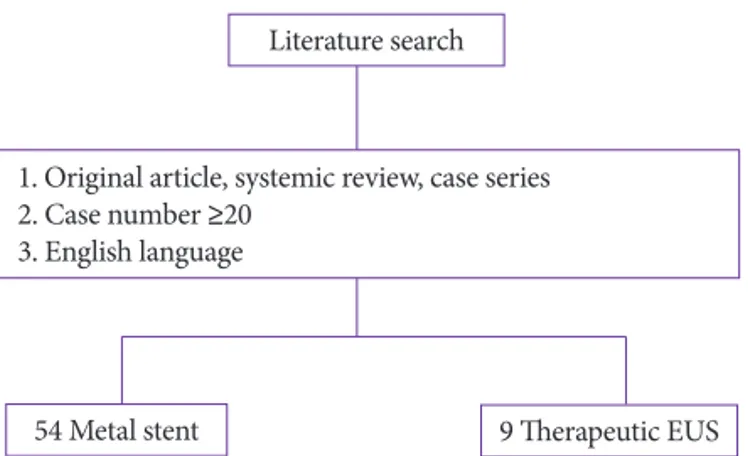
criteria were (1) original research articles (randomized con-trolled trials, prospective and retrospective studies), systemic reviews, and case series; (2) case number ≥20; and (3) En-glish-language publications (Fig. 1).
The publications and their references were reviewed man-ually. References to proper studies and review articles were searched manually to survey additional related studies. The bibliographies of the review articles were scrutinized to iden-tify other references that might have been missed in the initial search. Finally, the two authors of this article jointly reviewed each published paper, and all relevant information was ex-tracted.
PANCREATIC FLUID COLLECTION
PFCs include peri-PFCs, pancreatic pseudocysts, acute ne-crotic collections, and walled-off pancreatic necrosis.7,8 PFCs generally require drainage when they cause symptoms such as pain, gastric outlet obstruction (GOO), and/or bile duct obstruction, or if they are infected.9 The management of PFCs has traditionally been surgical, but surgery may be associated with relatively higher rates of complications and mortality.10 With more recent technological advances and experience, en-doscopic drainage is widely accepted and has replaced surgery as the first-line therapy for PFC drainage, as it is less invasive and has a shorter recovery time, lower cost, and lower compli-cation rates.11
EUS guided-endoscopic transmural drainage of PFCs with plastic or metal stents has become a widely accepted treat-ment modality. Previous studies have reported success rates comparable to those of percutaneous and surgical interven-tions, with significant advantages in terms of invasiveness,
morbidity, length of hospital stay, and cost.12 The procedure
is preferably performed under EUS guidance, which allows access to non-bulging lesions, avoidance of major vessels, and
selection of the number and type of stent to be placed. The clinical success rate of EUS-guided PFCs drainage is directly related to the necrotic content of PFCs, which increases the risk of occlusion of the stents typically used for this type of drainage procedure. Historically, double-pigtail plastic stents have been the mainstay of therapy. However, because of their small caliber, plastic stents can become occluded with the development of secondary infection; thus, increasing the need for re-intervention and surgical treatment when endoscopic re-treatment fails. Moreover, if a necrosectomy is required following drainage by plastic stents, the endoscopist must first remove the stents and then drive the scope through the drain-age tract to extract the necrotic material, which often requires multiple tract dilations and scope insertions into the PFC and increases the risk of procedural morbidity.
To overcome these limitations, fully covered self-expanding metal stents (FCSEMSs) and specially designed lumen-ap-posing metal stents (LAMSs) in which both ends are flared have been proposed, with the rationale of providing a large opening that would theoretically allow for longer patency, reduce rates of stent occlusion and decrease the probability of secondary infections, and provide a lasting approach route to perform multiple tissue sections via necrosectomy.7,9,13,14 In a retrospective cohort study including 230 patients with PFCs, especially pancreatic pseudocysts, EUS-guided drainage using FCSEMSs improved clinical outcomes (complete resolution of pseudocyst, 98% vs. 89%, p=0.01) and reduced adverse event rates (16% vs. 31%, p=0.006) compared with plastic stents.15 A prospective comparative study demonstrated that EUS-guided PFC drainage using a FCSEMS was comparable to that using plastic stents regarding technical feasibility, efficacy, and safe-ty. The median procedure time using a FCSEMS was signifi-cantly shorter than that using plastic stents (15.0 minutes vs. 29.5 minutes, p<0.01).16
Recently, endoscopists have seen the development and commercial release of specially designed, fully covered, transluminal self-expanding metal stents (SEMSs), such as the
AXIOS (Boston Scientific, Natick, MA, USA) and Nagi
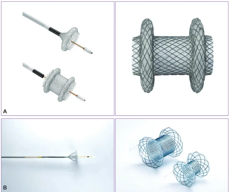
(Tae-woong Medical, Goyang, Korea) stents, which are designed with wide flanges on both ends to prevent migration (Fig. 2). These devices have a wide diameter (10 to 16 mm) and short length, and to some extent, they can actively hold a PFC in the lumen. Endoscopic necrosectomy can be performed directly through these devices owing to their wider diameter. Large cohort studies have demonstrated high technical success rates (91% to 98%) and clinical success rates, defined as resolution of clinical symptoms with a decrease in PFC size to ≤2 cm on imaging (81% to 100%).7,17,18
Few data are available regarding the proper period for
54 Metal stent 9 Therapeutic EUS
1. Original article, systemic review, case series 2. Case number ≥20
3. English language
Fig. 1. Flow diagram of the literature review. EUS, endoscopic ultrasonogra-phy.
Lee HS et al. Endoscopic Ultrasonography-Guided New Metal Stents
stent removal. One prospective, single-center study showed the safety and efficacy of short-duration (3 weeks) FCSEMS placement for symptomatic pseudocysts, along with selective pancreatic ductal stenting in patients with persistent ductal leak. During a median follow-up of 306 days, two recurrences
(4.7%) were detected in 42 patients.19 This study
demonstrat-ed that short-term placement of FCSEMSs with pancreatic ductal stenting for the treatment of pseudocysts is safe and effective.
FCSEMSs have been used with the hope that a large lu-minal diameter would facilitate more effective and lasting drainage. However, unfortunately, SEMSs may migrate and have been reported to cause mucosal injury and bleeding from erosion of the opposite wall of the cavity once the PFC is resolved. A recent study showed a 26% incidence of late
ad-verse events after FCSEMS placement for PFCs.20 A systematic
review including 17 studies and 881 patients who underwent endoscopic treatment of PFCs using plastic versus biliary FCSEMSs did not support routine deployment of metal stents
for drainage of PFCs, reporting similar pooled success rates for metal stents (81.9%) and plastic stents (80.7%) and higher pooled complication rates (such as bleeding, secondary infec-tion and stent migrainfec-tion) with metal stents (23.3%) compared with plastic stents (16.1%).21
Several novel LAMSs specially designed for transmural drainage allow single-step deployment and effective drain-age of cystic fluid with minimal stent migration (Table 1).7,13,14,17,18,22-24 The AXIOS and Nagi stents have high reported treatment success rates of 93% to 100%, but these results are limited to small studies of 10 to 33 patients. Further random-ized trials are needed to justify the use of metal stents for PFC drainage.
Despite advances in metal stent development and the the-oretical advantages of metal stents over plastic stents, there is no universal agreement as to which type of stent should be used for drainage of PFCs. Based on the current literature, one stent type cannot be clearly recommended over other types for EUS-guided drainage of PFCs. Endoscopists should
Fig. 2. Lumen-apposing metal stent. (A) AXIOS stent (Boston Scientific), (B) Nagi stent (Taewoong Medical Co. Ltd.). Adapted from Boston Scientific and Taewoong Medical Co. Ltd.
A
choose a stent type they are comfortable using and one that they feel is most appropriate for each individual case. The AXIOS and Nagi stents may be preferable when necrosecto-my is required, because the endoscopist can drive the scope directly through the stent and into the PFC for removal of solid necrotic material.
PANCREATIC DUCT DRAINAGE
The development of interventional EUS has provided an alternative means of relieving pancreatic duct obstruction that is not possible by ERCP through transpapillary access to the pancreatic duct, especially in pancreaticojejunal stenosis or pancreaticogastric anastomosis after Whipple resection, which can result in recurrent acute pancreatitis, main pancreatic duct (MPD) stenosis due to chronic pancreatitis, or post-pan-creatic damage after failure of ERCP.10 EUS-guided transmural
stenting using plastic stents was performed in cases in which advancing a guidewire across the anastomosis site was impos-sible, when the major papilla was under complete pancreatic obstruction, or when the MPD had a tortuous configuration.25
However, rates of complications, including bleeding, fever, perforation, and hematoma, are typically higher for transmu-ral stenting than for simple rendezvous placement.26 FCSEMSs
have not yet been used for EUS pancreatic duct drainage with transmural stenting because of concerns regarding stent mi-gration and a cross-stream blockage of the MPD that covers the membrane of the FCSEMS.25 Recently, Oh et al.27 showed
that EUS pancreatic duct drainage using a FCSEMS may be technically feasible and relatively safe without adverse events related to FCSEMSs, including stent migration, clogging, and stent-induced ductal stricture. In the future, FCSEMSs should be further modified with an anti-migration structure and appropriate diameter to decrease the risk of branch duct blockage, and a well-designed randomized controlled trial is
also needed.
BILE DUCT DRAINAGE
In cases in which papillotomy and bile duct cannulation are not successful, either because of an altered anatomy by previ-ous gastrointestinal surgery or because of a tight tumor steno-sis, surgery and/or percutaneous transhepatic biliary drainage (PTBD), which have higher morbidity and mortality rates than those of ERCP, have been suggested as alternative meth-ods.28,29 EUS-guided biliary drainage (EUS-BD) is considered
an effective salvage procedure for failed ERCP in patients with unresectable malignant biliary obstruction. In patients with malignant distal common bile duct obstruction requiring SEMS placement, the short-term composite success defined as the ability to complete the intended therapeutic procedure in a single session and resulting in a greater than 50% decrease in bilirubin over 2 weeks using EUS-BD was reported to be comparable to that of ERCP in a retrospective analysis.30
In the early 2000s, a large-channel echoendoscope made possible the first reported case of EUS-BD including trans-gastric or transduodenal biliary drainage, which had been initially described for palliative management of malignant biliary obstruction.5,6,31 The different access routes (transhepatic
or extrahepatic), directions of stent insertion (anterograde or retrograde), or drainage routes (transluminal or transpapil-lary) have been controversial, with varying success rates and complication rates, for EUS-BD.32 Although no randomized
trial has directly compared the rendezvous and transluminal techniques, both techniques were equally effective and safe according to a small retrospective study, and the authors concluded that the transluminal approach is a reasonable approach secondary to rendezvous EUS-BD, because cumber-some wire manipulations are not needed.33
A report by a single endoscopist that included 101 patients
Study Stent type patientsNo. of PFC type PFC size, mm TSR, % CSR, % ADR, n (%)
Y amamoto et al. (2013)24 Nagi 9 Pseudocyst/WON 146a) 100 77 Late 2 (22)
C handran et al. (2015)20 Nagi 47 Pseudocyst/WON 100b) 98.1 76.6 10 (21)/14 (29)c)
Walter et al. (2015)17 Axios 61 Pseudocyst/WON 90b) 98 93/81d) 5 (9)
Siddiqui et al. (2015)18 Axios 82 Pseudocyst/WON 118a) 97.5 98.7 4 (28.5)/9 (13.2)d)
Shah et al. (2015)7 Axios 33 Pseudocyst/WON 90a) 91 93 5 (15)
Bapaye et al. (2015)14 Nagi 19 Pseudocyst/WON 107a) 100 100 2 (10.5)
Itoi et al. (2012)13 Axios 15 Pseudocyst 98a) 100 100 1 (6.6)
PFC, pancreatic fluid collection; TSR, technical success rate; CSR, clinical success rate; ADR, adverse reaction rate; WON, walled-off necrosis.
Lee HS et al. Endoscopic Ultrasonography-Guided New Metal Stents showed that the risk of procedure-related death decreases
with the learning curve, even if EUS-BD is an efficient tech-nique.34 In connection with this, a newly designed stent intro-ducer having a fine gauge metal tip and a 7 F shaft diameter has been evaluated to enable one-step stent placement without a further fistula dilation step, potentially decreasing the risk of adverse events by immediately sealing the transmural fistula.25
There are two main approach routes in EUS-BD in terms of the organ on which a non-anatomic fistula is connected to the biliary tree: EUS-CDS and EUS-HGS. The EUS-HGS and EUS-CDS techniques exhibited similar efficacy and safety in a single-center randomized trial.35 EUS-CDS has several po-tential advantages compared with endoscopic transpapillary stenting (ETS), including a lower risk of post-procedural pan-creatitis, lower risk of tumor ingrowth and overgrowth, and higher technical success rate.35 A comparative cohort study that evaluated the clinical efficacy and safety of EUS-CDS and ETS as first-line treatments for malignant distal biliary obstruction showed that EUS-CDS was associated with short procedure times and no risk of pancreatitis.35
PTBD is the most commonly used alternative drainage method for intrahepatic ducts but has a relatively high rate of complications and is frequently associated with patient dis-comfort related to the external drainage.36 The advantage of EUS-HGS compared with PTBD is the lower risk of bleeding, through the use of color Doppler, which can prevent punc-turing of intervening vessels.3 However, bile leakage is a po-tential significant adverse event. In a multicenter retrospective analysis, EUS-HGS was related to complications including cholangitis, perforation, bile leakage, and bleeding (p=0.031).32 The left hepatic ducts are easily visualized by EUS; thus, a transgastric approach to the left biliary system was initially introduced, and hepatic hilar obstruction and isolated right intrahepatic duct obstruction are not normal indications for EUS-HGS.37 However, recently, sequential deployment of two stacked stents was used in isolated right intrahepatic duct ob-struction to prevent bile leakage from the fistula and bile duct branch occlusion using FCSEMs. This method is known as the locking-stent method and is performed using an uncovered stent initially deployed from the upper common bile duct to B, and then a bare-ended, fully covered stent is inserted into the first stent from B3 to the stomach.38 For standardization of EUS-HGS in cases of failed ERCP, the development of a standard protocol for endoscopic procedures and a more spe-cific and dedicated device for effective and safe deployment of metal stents without complications is required.39
As FCSEMSs offer prolonged stent patency (compared with plastic stents) and easy stent revision without increasing procedural complications such as bile leakage (compared with plastic stents or SEMSs), EUS-BD using FCSEMSs was
sug-gested as the ideal option for failed ERCP cases.40,41 In addi-tion, EUS-BD using the FCSEMS procedure provides a biliary transmural drainage route away from a duodenal SEMS and thus may be expected to prolong the time to dysfunction of a biliary stent compared with transpapillary drainage in cases with an indwelling duodenal SEMS.42 FCSEMSs appear to be a better option for EUS-BD because with full expansion, it effectively seals the puncture tract; thus, preventing leakage, and the larger diameter provides better long-term patency. Furthermore, in cases of stent occlusion, revision was more manageable compared with plastic stents, as a new stent can be placed into the occluded metal stent.10
GALLBLADDER DRAINAGE
Laparoscopic cholecystectomy is the standard approach used for patients with acute cholecystitis.43 However, some patients are improper for cholecystectomy because of ad-vanced age, underlying comorbidities, or malignancies. In these cases, percutaneous transhepatic gallbladder drainage (PTGBD) is the treatment of choice, and shows clinical suc-cess rates of 56% to 100%.44 Nevertheless, the percutaneous approach has many weak points, including bleeding, pneu-moperitoneum, bile leakage, and catheter dislodgement; additionally, long-term percutaneous drainage leads to dis-comfort in patients. Furthermore, high recurrence rates of cholecystitis of up to 41% have been reported after removal of the drainage catheter.45,46
Endoscopic methods for gallbladder drainage (GBD) in-clude the transpapillary approach or EUS-guided transmural GBD. Jang et al.47 showed that EUS-GBD is comparable with PTGBD in terms of technical feasibility and clinical efficacy for the treatment of acute cholecystitis in high-risk surgical patients. However, to date, there are a few data on the safety and feasibility of EUS-guided transmural GBD, because pre-vious tubular-shaped stents have risks, such as bile leakage or migration. Furthermore, the slow flow of bile and the small caliber of plastic stents can result in early malfunction and clogging. Thus, a modified FCSEMS, with enlarged flares (22 mm external diameter) and stents angulated to 90° to prevent migration after placement, was suggested to create a safe fistula tract connecting the bowel and gallbladder without intraperitoneal bile leakage.37 Recently, specifically designed LAMSs have been developed for transenteric drainage and successfully tested in animal models.39 In a small case series, EUS-guided cholecystoenterostomy using a LAMS was found to facilitate internal GBD in non-surgical candidates who have a percutaneous cholecystostomy catheter.48 However, reports on the use of a LAMS for GBD are limited to small case
se-prospective study, EUS-GBD using a newly developed LAMS device showed promising results (technical success rate, 90%; clinical success rate, 96%; stent- or procedure-related mortali-ty rate, 7%) in high-risk surgical patients with acute cholecys-titis.49
FUTURE DEVELOPMENT FOR
GASTROENTEROSTOMY
Surgical gastroenterostomy has been the standard palliative therapy for malignant GOO due to stomach, duodenum, or pancreatic cancer. Endoscopic placement of an enteric SEMS across the malignant stricture is an alternative treatment op-tion. This procedure leads to recovery of oral intake in ~90% of patients; however, it may be complicated by recurrent ob-struction caused by either stent migration or tumor infiltra-tion.50 An alternative approach is the creation of EUS-guided endoscopic gastroenterostomy using metal stents. Endoscopic gastrojejunostomy (GJS) appears to be ideal for malignant GOO because of the short length of anastomosis in surgical GJS and the less invasive nature of endoscopic metal stent placement. Endoscopic GJS is divided into two approaches, GJS using flexible forward-view endoscopy and that using an echoendoscope.51
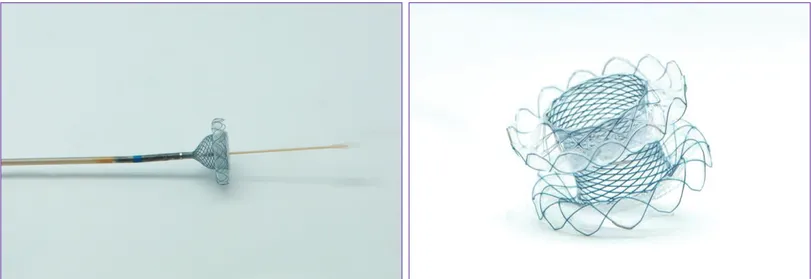
LAMSs such as the Spaxus (Taewoong Medical) or AXIOS stent have been developed, and EUS has been used to guide placement of LAMSs to create gastrogastric fistulas (Fig. 3). Itoi et al.52 showed that creation of an EUS-guided GJS using a novel enteric balloon and metal stent was promising as a minimally invasive treatment in an animal model. This tech-nique involves utilizing a specially created double-balloon enteric tube (Tokyo Medical University type; Create Medic, Yokohama, Japan) to stabilize the small bowel adjacent to the
prospective study was performed on EUS-guided double-bal-loon-occluded GJS bypass using LAMSs in 20 patients in 2015. The technical success rate was 90% (18/20).51,53
We hope that in the near future many patients suffering from malignant GOO can benefit from safe endoscopic gas-troenterostomy using dedicated metal stents, which enable both secure anastomosis and passage of food. In addition, the prevalence of obesity and diabetes has exponentially increased globally, and conventional bariatric surgery is cur-rently regarded as the only treatment that typically results in large, sustained weight losses and diabetes control for patients intractable to medical treatments. However, bariatric surgery also carries perioperative risks. Therefore there is a great need for minimally invasive weight-loss procedures in treatment sequence. In this context, we believe that the endoscopic by-pass procedure using a simple anastomosis method with a LAMS has the potential to be a minimal invasive treatment strategy for metabolic diseases, including obesity and type 2 diabetes.54
CONCLUSIONS
Although no established data support EUS-guided trans-mural drainage of PFC using metal stents over plastic stents, metal stents could be considered at the discretion of the phy-sician, based on operator preference and experience. LAMSs should be considered for walled-off necrosis of the pancreas, because necrosectomy is possible without laborious compli-cated procedures and thus reduces the risk of morbidities. The development of new stents for various procedures has expanded our ability to effectively drain fluids from the pan-creas and gallbladder, approach the bile duct, and perform GJS. Future studies should delineate more clearly which type
Lee HS et al. Endoscopic Ultrasonography-Guided New Metal Stents
of therapeutic procedure using metal stents is best suited to a specific disease entity.
Conflicts of Interest
The authors have no financial conflicts of interest.
REFERENCES
1. Wallace MJ, Charnsangavej C, Ogawa K, et al. Tracheobronchial tree: expandable metallic stents used in experimental and clinical applica-tions. Work in progress. Radiology 1986;158:309-312.
2. Püspök A, Lomoschitz F, Dejaco C, Hejna M, Sautner T, Gangl A. En-doscopic ultrasound guided therapy of benign and malignant biliary obstruction: a case series. Am J Gastroenterol 2005;100:1743-1747. 3. Perez-Miranda M, de la Serna C, Diez-Redondo P, Vila JJ.
Endosonogra-phy-guided cholangiopancreatography as a salvage drainage procedure for obstructed biliary and pancreatic ducts. World J Gastrointest Endosc 2010;2:212-222.
4. Wiersema MJ, Sandusky D, Carr R, Wiersema LM, Erdel WC, Frederick PK. Endosonography-guided cholangiopancreatography. Gastrointest Endosc 1996;43(2 Pt 1):102-106.
5. Giovannini M, Dotti M, Bories E, et al. Hepaticogastrostomy by echo-endoscopy as a palliative treatment in a patient with metastatic biliary obstruction. Endoscopy 2003;35:1076-1078.
6. Giovannini M, Moutardier V, Pesenti C, Bories E, Lelong B, Delpero JR. Endoscopic ultrasound-guided bilioduodenal anastomosis: a new tech-nique for biliary drainage. Endoscopy 2001;33:898-900.
7. Shah RJ, Shah JN, Waxman I, et al. Safety and efficacy of endoscopic ultrasound-guided drainage of pancreatic fluid collections with lu-men-apposing covered self-expanding metal stents. Clin Gastroenterol Hepatol 2015;13:747-752.
8. Smith IB, Gutierrez JP, Ramesh J, Wilcox CM, Mönkemüller KE. Endo-scopic extra-cavitary drainage of pancreatic necrosis with fully covered self-expanding metal stents (fcSEMS) and staged lavage with a high-flow water jet system. Endosc Int Open 2015;3:E154-E160.
9. McVay T, Adler DG. EUS-guided drainage of pancreatic fluid collec-tions: double pigtails, metal biliary, or dedicated transluminal stents? Endosc Ultrasound 2015;4:1-3.
10. Giovannini M, Bories E, Téllez-Ávila FI. Endoscopic ultrasound-guided bilio-pancreatic drainage. Endosc Ultrasound 2012;1:119-129.
11. Varadarajulu S, Bang JY, Sutton BS, Trevino JM, Christein JD, Wilcox CM. Equal efficacy of endoscopic and surgical cystogastrostomy for pancreatic pseudocyst drainage in a randomized trial. Gastroenterology 2013;145:583-590.
12. Seewald S, Ang TL, Teng KC, Soehendra N. EUS-guided drainage of pancreatic pseudocysts, abscesses and infected necrosis. Dig Endosc 2009;21 Suppl 1:S61-S65.
13. Itoi T, Binmoeller KF, Shah J, et al. Clinical evaluation of a novel lu-men-apposing metal stent for endosonography-guided pancreatic pseudocyst and gallbladder drainage (with videos). Gastrointest Endosc 2012;75:870-876.
14. Bapaye A, Itoi T, Kongkam P, Dubale N, Mukai S. New fully covered large-bore wide-flare removable metal stent for drainage of pancreatic fluid collections: results of a multicenter study. Dig Endosc 2015;27:499-504.
15. Sharaiha RZ, DeFilippis EM, Kedia P, et al. Metal versus plastic for pan-creatic pseudocyst drainage: clinical outcomes and success. Gastrointest Endosc 2015;82:822-827.
16. Lee BU, Song TJ, Lee SS, et al. Newly designed, fully covered metal stents for endoscopic ultrasound (EUS)-guided transmural drainage of peripancreatic fluid collections: a prospective randomized study. Endos-copy 2014;46:1078-1084.
17. Walter D, Will U, Sanchez-Yague A, et al. A novel lumen-apposing met-al stent for endoscopic ultrasound-guided drainage of pancreatic fluid collections: a prospective cohort study. Endoscopy 2015;47:63-67. 18. Siddiqui AA, Adler DG, Nieto J, et al. EUS-guided drainage of
peripan-creatic fluid collections and necrosis by using a novel lumen-apposing stent: a large retrospective, multicenter U.S. experience (with videos). Gastrointest Endosc 2015 Oct 26 [Epub]. http://dx.doi.org/10.1016/ j.gie.2015.10.020.
19. Dhir V, Teoh AY, Bapat M, Bhandari S, Joshi N, Maydeo A. EUS-guided pseudocyst drainage: prospective evaluation of early removal of fully covered self-expandable metal stents with pancreatic ductal stenting in selected patients. Gastrointest Endosc 2015;82:650-657.
20. Chandran S, Efthymiou M, Kaffes A, et al. Management of pancreatic collections with a novel endoscopically placed fully covered self-ex-pandable metal stent: a national experience (with videos). Gastrointest Endosc 2015;81:127-135.
21. Bang JY, Hawes R, Bartolucci A, Varadarajulu S. Efficacy of metal and plastic stents for transmural drainage of pancreatic fluid collections: a systematic review. Dig Endosc 2015;27:486-498.
22. Raijman I, Tarnasky PR, Patel S, et al. Endoscopic drainage of pancre-atic fluid collections using a fully covered expandable metal stent with antimigratory fins. Endosc Ultrasound 2015;4:213-218.
23. Song TJ, Lee SS, Park do H, Seo DW, Lee SK, Kim MH. Preliminary re-port on a new hybrid metal stent for EUS-guided biliary drainage (with videos). Gastrointest Endosc 2014;80:707-711.
24. Yamamoto N, Isayama H, Kawakami H, et al. Preliminary report on a new, fully covered, metal stent designed for the treatment of pancreatic fluid collections. Gastrointest Endosc 2013;77:809-814.
25. Park do H, Lee TH, Paik WH, et al. Feasibility and safety of a novel dedicated device for one-step EUS-guided biliary drainage: a random-ized trial. J Gastroenterol Hepatol 2015;30:1461-1466.
26. Khashab MA, Kumbhari V, Grimm IS, et al. EUS-guided gastroenteros-tomy: the first U.S. clinical experience (with video). Gastrointest Endosc 2015;82:932-938.
27. Oh D, Park do H, Cho MK, et al. Feasibility and safety of a fully cov-ered self-expandable metal stent with antimigration properties for EUS-guided pancreatic duct drainage: early and midterm outcomes (with video). Gastrointest Endosc 2016;83:366-373.
28. Puspok A, Lomoschitz F, Dejaco C, Hejna M, Sautner T, Gangl A. En-doscopic ultrasound guided therapy of benign and malignant biliary obstruction: a case series. Am J Gastroenterol 2005;100:1743-1747. 29. Will U, Thieme A, Fueldner F, Gerlach R, Wanzar I, Meyer F.
Treat-ment of biliary obstruction in selected patients by endoscopic ultra-sonography (EUS)-guided transluminal biliary drainage. Endoscopy 2007;39:292-295.
30. Dhir V, Itoi T, Khashab MA, et al. Multicenter comparative evaluation of endoscopic placement of expandable metal stents for malignant dis-tal common bile duct obstruction by ERCP or EUS-guided approach. Gastrointest Endosc 2015;81:913-923.
31. Burmester E, Niehaus J, Leineweber T, Huetteroth T. EUS-cholan-gio-drainage of the bile duct: report of 4 cases. Gastrointest Endosc 2003;57:246-251.
32. Dhir V, Artifon EL, Gupta K, et al. Multicenter study on endoscopic ultrasound-guided expandable biliary metal stent placement: choice of access route, direction of stent insertion, and drainage route. Dig Endosc 2014;26:430-435.
33. Khashab MA, Valeshabad AK, Modayil R, et al. EUS-guided biliary drainage by using a standardized approach for malignant biliary ob-struction: rendezvous versus direct transluminal techniques (with vid-eos). Gastrointest Endosc 2013;78:734-741.
34. Poincloux L, Rouquette O, Buc E, et al. Endoscopic ultrasound-guided biliary drainage after failed ERCP: cumulative experience of 101 proce-dures at a single center. Endoscopy 2015;47:794-801.
35. Kawakubo K, Kawakami H, Kuwatani M, et al. Endoscopic ultra-sound-guided choledochoduodenostomy vs. transpapillary stenting for
endoscopic ultrasonography-guided biliary drainage: results of a pilot study. Endoscopy 2007;39:287-291.
37. Jang JW, Lee SS, Park do H, Seo DW, Lee SK, Kim MH. Feasibility and safety of EUS-guided transgastric/transduodenal gallbladder drainage with single-step placement of a modified covered self-expandable metal stent in patients unsuitable for cholecystectomy. Gastrointest Endosc 2011;74:176-181.
38. Irani S, Baron TH, Grimm IS, Khashab MA. EUS-guided gallbladder drainage with a lumen-apposing metal stent (with video). Gastrointest Endosc 2015;82:1110-1115.
39. Moon JH, Choi HJ, Kim DC, et al. A newly designed fully covered metal stent for lumen apposition in EUS-guided drainage and access: a feasibility study (with videos). Gastrointest Endosc 2014;79:990-995. 40. Park do H, Koo JE, Oh J, et al. EUS-guided biliary drainage with
one-step placement of a fully covered metal stent for malignant bil-iary obstruction: a prospective feasibility study. Am J Gastroenterol 2009;104:2168-2174.
41. Kawakubo K, Isayama H, Kato H, et al. Multicenter retrospective study of endoscopic ultrasound-guided biliary drainage for malignant biliary obstruction in Japan. J Hepatobiliary Pancreat Sci 2014;21:328-334. 42. Hamada T, Isayama H, Nakai Y, et al. Transmural biliary drainage can
be an alternative to transpapillary drainage in patients with an indwell-ing duodenal stent. Dig Dis Sci 2014;59:1931-1938.
43. Kiviluoto T, Sirén J, Luukkonen P, Kivilaakso E. Randomised trial of laparoscopic versus open cholecystectomy for acute and gangrenous cholecystitis. Lancet 1998;351:321-325.
44. Glenn F. Cholecystostomy in the high risk patient with biliary tract dis-ease. Ann Surg 1977;185:185-191.
45. Chang L, Moonka R, Stelzner M. Percutaneous cholecystostomy for acute cholecystitis in veteran patients. Am J Surg 2000;180:198-202.
risk patients. Surg Endosc 2012;26:1343-1351.
47. Jang JW, Lee SS, Song TJ, et al. Endoscopic ultrasound-guided transmu-ral and percutaneous transhepatic gallbladder drainage are comparable for acute cholecystitis. Gastroenterology 2012;142:805-811.
48. Law R, Grimm IS, Stavas JM, Baron TH. Conversion of percutaneous cholecystostomy to internal transmural gallbladder drainage using an endoscopic ultrasound-guided, lumen-apposing metal stent. Clin Gas-troenterol Hepatol 2016;14:476-480.
49. Walter D, Teoh AY, Itoi T, et al. EUS-guided gall bladder drainage with a lumen-apposing metal stent: a prospective long-term evaluation. Gut 2016;65:6-8.
50. Binmoeller KF, Shah JN. Endoscopic ultrasound-guided gastroenteros-tomy using novel tools designed for transluminal therapy: a porcine study. Endoscopy 2012;44:499-503.
51. Itoi T, Ishii K, Tanaka R, Umeda J, Tonozuka R. Current status and perspective of endoscopic ultrasonography-guided gastrojejunostomy: endoscopic ultrasonography-guided double-balloon-occluded gastroje-junostomy (with videos). J Hepatobiliary Pancreat Sci 2015;22:3-11. 52. Itoi T, Itokawa F, Uraoka T, et al. Novel EUS-guided gastrojejunostomy
technique using a new double-balloon enteric tube and lumen-apposing metal stent (with videos). Gastrointest Endosc 2013;78:934-939. 53. Itoi T, Ishii K, Ikeuchi N, et al. Prospective evaluation of endoscopic
ul-trasonography-guided double-balloon-occluded gastrojejunostomy by-pass (EPASS) for malignant gastric outlet obstruction. Gut 2016;65:193-195.
54. Yi SW, Chung MJ, Jo JH, et al. Gastrojejunostomy by pure natural orifice transluminal endoscopic surgery using a newly designed anastomosing metal stent in a porcine model. Surg Endosc 2014;28:1439-1446.