ORIGINAL ARTICLE
J Korean Surg Soc 2011;81:250-256
http://dx.doi.org/10.4174/jkss.2011.81.4.250
JKSS
Journal of the Korean Surgical Society pISSN 2233-7903ㆍeISSN 2093-0488
Received April 1, 2011, Revised June 7, 2011, Accepted June 27, 2011 Correspondence to: Seong Ho Choi
Department of Surgery, Samsung Medical Center, Sungkyunkwan University School of Medicine, 50 Irwon-dong, Gangnam-gu, Seoul 135-710, Korea
Tel: +82-2-3410-3468, Fax: +82-2-3410-1669, E-mail: [email protected]
cc Journal of the Korean Surgical Society is an Open Access Journal. All articles are distributed under the terms of the Creative Commons Attribution Non-Commercial License (http://creativecommons.org/licenses/by-nc/3.0/) which permits unrestricted non-commercial use, distribution, and reproduction in any medium, provided the original work is properly cited.
Role of transduodenal ampullectomy for tumors of the ampulla of Vater
Jieun Kim, Seong Ho Choi, Dong Wook Choi, Jin Seok Heo, Kee-Taek Jang
1Departments of Surgery and 1Pathology, Samsung Medical Center, Sungkyunkwan University School of Medicine, Seoul, Korea
Purpose: Tumors arising from the ampulla of Vater can be benign or malignant. Recently, endoscopic papillectomy has been employed in the management of benign ampulla of Vater tumors; however, surgical resection is the treatment of choice. The aim of this study was to define indications and suggest a role for transduodenal ampullectomy in the management of ampul- la of Vater tumors. Methods: We retrospectively reviewed the medical records of 54 patients treated for ampulla of Vater tu- mors between January 1999 and December 2008. Results: Twenty-two endoscopic papillectomies and 21 transduodenal am- pullectomies were performed. Four patients underwent transduodenal ampullectomy after endoscopic papillectomy due to a recurrent or remnant tumor. Recurrence or a remnant tumor was found in one patient after transduodenal ampullectomy compared to six patients after endoscopic papillectomy. Immediate intraoperative conversion from transduodenal ampullec- tomy to pancreaticoduodenectomy was performed in five patients based on intraoperative frozen biopsy analysis.
Conclusion: Transduodenal ampullectomy should be performed to treat ampulla of Vater tumors that are unsuitable for en- doscopic papillectomy. Transduodenal ampullectomy can serve as an intermediate treatment option between endoscopic papillectomy and pancreaticoduodenectomy in the management of ampulla of Vater tumors.
Key Words: Ampulla of vater, Transduodenal ampullectomy, Ampullary neoplasm, Endoscopic papillectomy
INTRODUCTION
Tumors of the ampulla of Vater have various histo- pathologic characteristics that can be benign or malignant.
It is difficult to achieve an accurate diagnosis and exclude malignancy based on preoperative studies because of the possibility of an invasive carcinoma within an adenoma.
Endoscopic papillectomy has recently been used to man- age periampullary adenomas [1-4], but curative surgical resection remains the treatment of choice [5-7].
Transduodenal ampullectomy was first introduced by Halstead in 1899 [8], and was initially attempted as treat- ment for ampulla of Vater cancer. The use of this proce- dure failed to become widespread because the surgical technique had not been standardized and the recurrence rate was high. Since Whipple introduced pancreatico- duodenectomy in 1935 [9], it has been recommended as the definitive surgery for ampulla of Vater lesions [6,7,10].
The performance of transduodenal ampullectomy has been reported in limited cases and in a small number of
series. No specific consensus has been reached regarding the indications for transduodenal ampullectomy, and there are few studies that address the role of trans- duodenal ampullectomy with respect to its relationship with endoscopic papillectomy [11-14]. The aim of this study was to define indications and suggest a role for transduodenal ampullectomy in the management of am- pulla of Vater tumors based on a review of experience at a single center .
METHODS
The medical records of 54 patients with ampulla of Vater tumors treated at the Samsung Medical Center be- tween January 1999 and December 2008 were retro- spectively reviewed. All patients underwent an esoph- agoduodenoscopic (EGD) exam and biopsy. Computed tomography (CT) or endoscopic ultrasonography (EUS) was performed to assess the extent of the tumor. Patients with a malignancy confirmed by preoperative biopsy were excluded from this study.
Patients were initially treated by endoscopic papillec- tomy, transduodenal ampullectomy, or pancreaticoduo- denectomy, depending on tumor size, the presence of high-grade dysplasia on endoscopic biopsy, and suspi- cions of malignancy on preoperative imaging studies.
Endoscopic papillectomy was performed initially in pa- tients with a benign-looking tumor or a tumor size less than 2 cm. Prior to endoscopic papillectomy, a pancreato- gram and a cholangiogram were obtained by endoscopic retrograde cholangiopancreatography (ERCP) in order to check for ductal involvement. En bloc resection was at- tempted in all patients, and when this failed, piecemeal re- section was performed. After papillectomy, a pancreatic stent and a biliary stent were inserted to prevent post pap- illectomy pancreatitis and stenosis. Chest radiography and laboratory tests were performed for all patients to screen for microperforations, pancreatitis, and bleeding after papillectomy. EGD was performed for stent removal 2 to 5 days after initial endoscopic papillectomy. Follow- up EGD was performed at three to six months intervals for two years, and annual EGD examination was recom-
mended thereafter. If any suspicious mucosal nodularity was found on EGD during follow-up, biopsy was per- formed and histologic results were reviewed. A remnant lesion was defined as a biopsy-confirmed lesion within a suspicious adenomatous lesion at a previous papil- lectomy site on subsequent EGD. Recurrence was defined as a lesion detected six months after treatment with at least one EGD biopsy result showing no residual lesion [15].
When patients were not suitable for endoscopic papil- lectomy (a tumor greater than 2 cm, a tumor with intra- ductal growth, or a lateral spreading tumor) transduo- denal ampullectomy was performed as the initial treat- ment. Transduodenal ampullectomy was attempted as the initial treatment in patients with any suspicious sign of malignancy on preoperative study, such as high-grade dysplasia on preoperative endoscopic biopsy.
Transduodenal ampullectomy was performed as de- scribed in other studies [12,14,16-18]. The operative proce- dure included the Kocher maneuver, longitudinal duode- notomy, full thickness excision of the ampulla, and re-im- plantation of the common bile duct and the pancreatic duct into the duodenal wall. Cholecystectomy was per- formed routinely. Short stents were inserted into the pan- creatic ducts to prevent pancreatitis. Tumors, the bile duct and the pancreatic duct margin were sent to pathology for intraoperative frozen section review. Routine lymph node dissection was not performed.
Ten patients who underwent pancreaticoduodenec- tomy as an initial treatment because of a suspected malig- nancy on preoperative study, but for whom final pathol- ogy revealed a benign tumor, were also included in this study. Patient demographics, symptoms, and preopera- tive endoscopic biopsy and final pathology findings were reviewed. Post-operative outcomes were evaluated with respect to length of hospital stay, and post-operative com- plications and recurrence during follow-up.
RESULTS
A total of 54 patients with an ampulla of Vater tumor were included in this retrospective review; 25 were female and 29 were male. Thirty-seven patients were asympto-
Table 1. Presenting symptoms and signs
Symptoms & signs No. of patients
Screening test 36
Jaundice 8
Abdominal pain 7
Dyspepsia 2
General weakness 1
Total 54
Fig. 1. Diagram of patients flow.
ERCP, endoscopic retrograde cho- langiopancreatography; PPPD, pan- creaticoduodenectomy. a)Remnant tumor, margin (+). b)Remnant tumor.
c)Remnant tumor, Lat. spreading type. d)Recurrence, P-duct stricture.
e)Frozen: 3 adenocarcinoma, 1 car- cinoid very close margin. f)Frozen;
deep mucosal bile duct margin (+).
g)Final pathology: benign.
Table 2.Comparison between results of endoscopic papillectomy vs. transduodenal ampullectomy (TDA)
Endoscopic
papillectomy (n = 22) TDA (n = 21) Hospital stay (day) 5 (3-12) 9 (7-37) Follow up (months) 10 (1-64) 18 (1- 72)
Complications 11 5
Recur/remnant 6 1
Resection margin (+) 3 1
matic and found to have an abnormality on screening tests such as EGD. The median patient age was 58 years (range, 37 to 75 years). Eighteen patients presented with symp- toms including jaundice (8 patients), abdominal pain (7 patients), dyspepsia (2 patients) or general weakness (1 patient) (Table 1). Endoscopic papillectomy was selected as the initial treatment in 22 patients while 32 patients were initially treated with surgery (22 with transduodenal ampullectomy and 10 with pancreaticoduodenectomy) (Fig. 1).
Ten patients had no recurrence after endoscopic papil- lectomy, and nine patients underwent additional endo- scopic papillectomy or ERCP biopsy after endoscopic pap- illectomy for a remnant adenoma or a positive resection margin (Fig. 1). Two patients had remnant adenoma after the second endoscopic papillectomy and subsequently underwent transduodenal ampullectomy (Fig. 1). Imme- diate conversion to pancreaticoduodenectomy was per- formed in one of these two patients because of a positive deep mucosal bile duct margin on intraoperative frozen
section analysis (Fig. 1). One patient was found to have a remnant lateral spreading tumor after endoscopic papil- lectomy. Surgery was recommended, but the patient was unfortunately lost to follow-up (Fig. 1). Two patients even- tually underwent transduodenal ampullectomy for a rem- nant tumor and a pancreatic duct stricture after papil- lectomy (Fig. 1).
Of the 22 patients for whom transduodenal ampullec- tomy was initially attempted, 4 patients were converted to pancreaticoduodenectomy after intraoperative frozen bi- opsy analysis. Of these four patients, three had ad- enocarcinoma and the other had a carcinoid with a very close margin (Fig. 1).
As a result, 22 patients underwent endoscopic papil- lectomy while 21 patients underwent transduodenal ampullectomy. Five patients had immediate intraopera- tive conversion from transduodenal ampullectomy to pancreaticoduodenectomy (Fig. 1). The median hospital stay after endoscopic papillectomy was 5 days (range, 3 to
Table 3. Complications of endoscopic papillectomy and transduodenal ampullectomy (TDA)
Endoscopic
papillectomy (n = 22) TDA (n = 21) Pancreatitis 6 Delayed gastric emptying 1
Bleeding 3 Wound dehiescence 1
Microperforation 1 Passage disturbance1 1 P-duct stricture 1 Pancreatitis
P-duct stenosis 2 Papilla orifice stenosis
Total 11 5
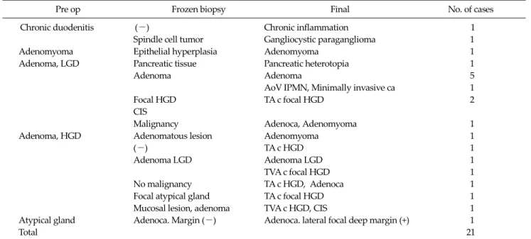
Table 5. Pathology of transduodenal ampullectomy
Pre op Frozen biopsy Final No. of cases
Chronic duodenitis (-) Chronic inflammation 1
Spindle cell tumor Gangliocystic paraganglioma 1
Adenomyoma Epithelial hyperplasia Adenomyoma 1
Adenoma, LGD Pancreatic tissue Pancreatic heterotopia 1
Adenoma Adenoma 5
AoV IPMN, Minimally invasive ca 1
Focal HGD TA c focal HGD 2
CIS
Malignancy Adenoca, Adenomyoma 1
Adenoma, HGD Adenomatous lesion Adenomyoma 1
(-) TA c HGD 1
Adenoma LGD Adenoma LGD 1
TVA c focal HGD 1
No malignancy TA c HGD, Adenoca 1
Focal atypical gland TA c focal HGD 1
Mucosal lesion, adenoma TVA c HGD, CIS 1
Atypical gland Adenoca. Margin (-) Adenoca. lateral focal deep margin (+) 1
Total 21
LGD, low grade dysplasia; HGD, high grade dysplasia; CIS, carcinoma in situ; IPMN, intraductal papillary mucinous neoplasm; TA, tubular adenoma; TVA, tubulovillous adenoma.
Table 4.Pathology of endoscopic papillectomy
Pathology No. of cases
Adenoma, LGD
Tubular 9
Tubulo villous 5
Villous 1
Adenoma, HGD
Tubular 3
Tubulo villous 1
Ampullary adenomyoma 2
Ganglioneuroma 1
Total 22
LGD, low grade dysplasia; HGD, high grade dysplasia.
12 days), which was shorter than the 9 days (range, 7 to 37 days) required after transduodenal ampullectomy (Table 2). The median follow-up period for endoscopic papil- lectomy was 10 months (range, 1 to 64 months), and that of transduodenal ampullectomy was 18 months (range, 1 to 72 months).
Complications
Complications were observed in 11 and 5 patients after endoscopic papillectomy and transduodenal ampullec- tomy, respectively (Table 3); no deaths occurred after ei- ther procedure. The complications that occurred after en- doscopic papillectomy were pancreatitis, bleeding, and
microperforation. Ten patients with complications had a longer hospital stay than average, but all improved with conservative treatment only. One female patient under- went transduodenal ampullectomy eight months after the initial procedure because of a pancreatic duct stricture.
The complications that occurred after transduodenal am- pullectomy were delayed gastric emptying, wound de- hiscence, passage disturbance, p-duct stenosis-induced pancreatitis, and biliary stricture-induced cholangitis.
With the exception of one patient with delayed gastric emptying, the others underwent intervention or surgery
Fig. 2. (A) Microphotograph of specimen of transduodenalampullectomy. Focal minimally invasive carcinoma (<5%) in the background of high grade dysplasia (circle) (H&E, ×10). (B) Magnification view of focal invasive mucinous carcinoma, marked with circle in Fig. 2A (H&E,
×100).
for treatment of their complications.
Pathology
Of the 22 cases that underwent endoscopic papil- lectomy, 15 had a final pathologic diagnosis of low grade adenoma (Table 4). Pathology results after transduodenal ampullectomy ranged from low grade adenoma to ad- enocarcinoma (Table 5). The accuracy of preoperative en- doscopic biopsy versus final pathology was 47.6% (10 of 21 patients), and the accuracy of intraoperative frozen biopsy analysis versus final pathology was 65.2% (15 of 23 pa- tients); frozen section biopsy analysis was not performed in two patients.
Ampulla of Vater cancer was suspected in one patient with liver cirrhosis due to chronic hepatitis B infection.
However, transduodenal ampullectomy was performed instead of pancreaticoduodenectomy because of the risk of postoperative hepatic failure. The ultimate biopsy re- sult in this case was adenocarcinoma with a positive later- al focal deep margin. The patient subsequently underwent adjuvant radiotherapy and there was no evidence of re- currence after 24 months of follow-up.
Five of 22 patients had focal carcinoma or carcinoma in situ according to the final pathology report; all carcinomas were confined to mucosa. One patient with a final patho-
logic diagnosis of adenocarcinoma within adenomyoma with a negative resection margin experienced recurrence at 33 months postoperatively. A metastatic seeding nodule was found, and this patient was treated with concurrent chemo-radiation therapy.
DISCUSSION
Screening esophagogastroduodenoscopy has been per- formed actively over the last decade, and has increased the detection rate of asymptomatic ampulla of Vater tumors [1,2,5,15]. Clinicians are concerned about the proper man- agement of this type of tumor, and radical resection with pancreaticoduodenectomy is widely accepted as the de- finitive treatment; however the perioperative morbidity and mortality associated with this procedure are problem- atic [19,20].
Endoscopic papillectomy is an attractive method for treating ampulla of Vater tumors because it reduces the possibility of laparotomy. However, endoscopic papil- lectomy is inconvenient to patients because frequent en- doscopic examinations are required after the procedure [5,21,22]. Furthermore, specimens obtained after piece- meal resection may have inadequate margins or false neg-
Fig. 3. Flowsheet of recommen- dation for management of AoV tumors. HGD, high grade dysplasia;
LGD, low gradedysplasia; CT, com- puted tomography; EUS, endosco- pic ultrasonogrphy; PD, pancrea- ticoduodenectomy; TDA, transduo- denalampullectomy; EP, endosco- pic papillectomy.
ative results, therefore close monitoring for recurrent or remnant tumors is recommended. In the present study, three positive resection margins and six remnant tumors were found after 22 endoscopic papillectomies, as com- pared with one recurrence and one positive resection mar- gin after 21 transduodenal ampullectomies.
The advantage of transduodenal ampullectomy is that it allows complete circumferential resection of the ampul- la of Vater, which enables precise pathologic examination.
Fig. 2 is a photomicrograph of a specimen obtained after transduodenal ampullectomy with predominantly high- grade dysplasia in the background and a less than 5% focal minimally invasive carcinoma. Had endoscopic papil- lectomy been performed on this patient, the focal carcino- ma would likely have been missed. Accordingly, if it is not possible to obtain a clear resection margin by en bloc re- section in endoscopic papillectomy, the patient should un- dergo transduodenal ampullectomy rather than piece- meal resection. An inadequate resection margin could not only result in tumor recurrence, but may also preclude the chance to diagnosis focal or in situ carcinoma within an adenoma.
The standard procedure for transduodenal ampullec- tomy includes re-implantation of the bile duct and p-duct, and post-operative pancreatitis is therefore rarely encoun- tered [12,14,16,17,23]. Furthermore, intraoperative frozen section examinations provide another opportunity for im- mediate conversion to pancreaticoduodenectomy if in- vasive carcinoma is found. Ten patients underwent pan-
creaticoduodenectomy initially (Fig. 1) due to suspected malignancy on preoperative study, but final pathologic di- agnosis was benign. If transduodenal ampullectomy had been attempted on these patients, pancreaticoduodenec- tomy could have been avoided.
The limitations of this study are 1) Endoscopic papil- lectomy has been actively performed at our institute over recent years; 2) Outcomes may have varied depending on the techniques employed by individual endoscopists; and 3) The mean follow-up duration for patients who under- went endoscopic papillectomy or transduodenal ampul- lectomy was less than 24 months. Further studies with a longer follow-up are required.
In conclusion, transduodenal ampullectomy should be indicated for patients in whom an adequate resection mar- gin was not obtained with endoscopic papillectomy and in patients with high grade dysplasia on endoscopic biopsy.
We recommend that transduodenal ampullectomy should be strongly considered for recurrent or remnant tumors af- ter endoscopic papillectomy in order to obtain an ad- equate resection margin and to provide definite treatment [11-14,24,25]. Because frozen biopsy allows immediate in- traoperative conversion to pancreaticoduodenectomy, transduodenal ampullectomy could act as an intermediate step between endoscopic papillectomy and pancreatico- duodenectomy for the treatment of ampulla of Vater tu- mors (Fig. 3) [26,27]. Use of this technique initially could prevent unnecessary pancreaticoduodenectomy in benign ampulla of Vater tumors [28].
CONFLICTS OF INTEREST
No potential conflict of interest relevant to this article was reported.
ACKNOWLEDGEMENTS
This research was supported by grants from IN-SUNG Foundation for Medical Research in 2009 (CA98101).
REFERENCES
1. Catalano MF, Linder JD, Chak A, Sivak MV Jr, Raijman I, Geenen JE, et al. Endoscopic management of adenoma of the major duodenal papilla. Gastrointest Endosc 2004;59:
225-32.
2. Charton JP, Deinert K, Schumacher B, Neuhaus H. Endo- scopic resection for neoplastic diseases of the papilla of Vater. J Hepatobiliary Pancreat Surg 2004;11:245-51.
3. Cheng CL, Sherman S, Fogel EL, McHenry L, Watkins JL, Fukushima T, et al. Endoscopic snare papillectomy for tu- mors of the duodenal papillae. Gastrointest Endosc 2004;
60:757-64.
4. Yoon SM, Kim MH, Kim MJ, Jang SJ, Lee TY, Kwon S, et al.
Focal early stage cancer in ampullary adenoma: surgery or endoscopic papillectomy? Gastrointest Endosc 2007;66:
701-7.
5. Lee SY, Jang KT, Lee KT, Lee JK, Choi SH, Heo JS, et al. Can endoscopic resection be applied for early stage ampulla of Vater cancer? Gastrointest Endosc 2006;63:783-8.
6. Kobayashi A, Konishi M, Nakagohri T, Takahashi S, Kinoshita T. Therapeutic approach to tumors of the ampul- la of Vater. Am J Surg 2006;192:161-4.
7. Yoon YS, Kim SW, Park SJ, Lee HS, Jang JY, Choi MG, et al.
Clinicopathologic analysis of early ampullary cancers with a focus on the feasibility of ampullectomy. Ann Surg 2005;242:92-100.
8. Halsted WS. Contributions to the surgery of the bile pas- sages, especially of the common bile duct. Boston Med Surg J 1899;141:645-54.
9. Whipple AO, Parsons WB, Mullins CR. Treatment of carci- noma of the ampulla of Vater. Ann Surg 1935;102:763-79.
10. Winter JM, Cameron JL, Olino K, Herman JM, de Jong MC, Hruban RH, et al. Clinicopathologic analysis of ampullary neoplasms in 450 patients: implications for surgical strat- egy and long-term prognosis. J Gastrointest Surg 2010;14:
379-87.
11. Fraguela Mariña JA. Transduodenal ampullectomy in the treatment of villous adenomas and adenocarcinomas of the
Vater's ampulla. Rev Esp Enferm Dig 2004;96:829-34.
12. Dixon E, Vollmer CM Jr, Sahajpal A, Cattral MS, Grant DR, Taylor BR, et al. Transduodenal resection of peri-ampul- lary lesions. World J Surg 2005;29:649-52.
13. Demetriades H, Zacharakis E, Kirou I, Pramateftakis MG, Sapidis N, Kanellos I, et al. Local excision as a treatment for tumors of ampulla of Vater. World J Surg Oncol 2006;4:14.
14. Grobmyer SR, Stasik CN, Draganov P, Hemming AW, Dixon LR, Vogel SB, et al. Contemporary results with am- pullectomy for 29 "benign" neoplasms of the ampulla. J Am Coll Surg 2008;206:466-71.
15. Irani S, Arai A, Ayub K, Biehl T, Brandabur JJ, Dorer R, et al. Papillectomy for ampullary neoplasm: results of a sin- gle referral center over a 10-year period. Gastrointest Endosc 2009;70:923-32.
16. Meneghetti AT, Safadi B, Stewart L, Way LW. Local re- section of ampullary tumors. J Gastrointest Surg 2005;9:
1300-6.
17. Maithel SK, Fong Y. Technical aspects of performing trans- duodenal ampullectomy. J Gastrointest Surg 2008;12:
1582-5.
18. Park JS, Yoon DS, Park YN, Lee WJ, Chi HS, Kim BR.
Transduodenal local resection for low risk group ampulla of Vater cancer patients. J Korean Surg Soc 2004;66:404-8.
19. Böttger TC, Junginger T. Factors influencing morbidity and mortality after pancreaticoduodenectomy: critical analysis of 221 resections. World J Surg 1999;23:164-71.
20. Bakkevold KE, Kambestad B. Morbidity and mortality af- ter radical and palliative pancreatic cancer surgery. Risk factors influencing the short-term results. Ann Surg 1993;217:356-68.
21. Seewald S, Omar S, Soehendra N. Endoscopic resection of tumors of the ampulla of Vater: how far up and how deep down can we go? Gastrointest Endosc 2006;63:789-91.
22. Han J, Kim MH. Endoscopic papillectomy for adenomas of the major duodenal papilla (with video). Gastrointest Endosc 2006;63:292-301.
23. Beger HG, Staib L, Schoenberg MH. Ampullectomy for ad- enoma of the papilla and ampulla of Vater. Langenbecks Arch Surg 1998;383:190-3.
24. Branum GD, Pappas TN, Meyers WC. The management of tumors of the ampulla of Vater by local resection. Ann Surg 1996;224:621-7.
25. Posner S, Colletti L, Knol J, Mulholland M, Eckhauser F.
Safety and long-term efficacy of transduodenal excision for tumors of the ampulla of Vater. Surgery 2000;128:694-701.
26. Clary BM, Tyler DS, Dematos P, Gottfried M, Pappas TN.
Local ampullary resection with careful intraoperative fro- zen section evaluation for presumed benign ampullary neoplasms. Surgery 2000;127:628-33.
27. Kim JW, Hwang YJ, Kim YI, Yun YK. Transduodenal am- pullectomy in ampullary neoplasm. J Korean Surg Soc 2001;60:432-7.
28. Tien YW, Yeh CC, Wang SP, Hu RH, Lee PH. Is blind pan- creaticoduodenectomy justified for patients with ampul- lary neoplasms? J Gastrointest Surg 2009;13:1666-73.