서 론 .
Ⅰ
(endoscope)
. (minimally invasive medicine)
.
, ,
(gastroscope) (colonoscope) (gastrointestinal endoscope)
, .
, ,
.
,
.
.
.
, .
내시경의 종류와 용도에 따른 분류 .
Ⅱ
(endoscope, ) , ‘ (endo-)
(scope) ’ ,
. (rigid
endoscope, ) (flexible endoscope, )
. ,
.
( ) , , ,
.
, .
(rod lens) ,
.
지영준 우지환,
Current and Future Technologies for a Gastrointestinal Endoscopy
Youngjoon Chee, Jihwan Woo
Dept. of Biomedical Engineering, University of Ulsan, Ulsan, Korea (Received September 13, 2010. Accepted September 18, 2010)
This article presents a review of technologies for an endoscope. The classification according to the clinical applications and the imaging modalities are summarized. The major parts are focused on describing the gastrointestinal endoscope’s structures and mechanisms. The details of the image enhanced endoscopic techniques, such as NBI (narrow band imaging), OCT (optical coherence tomography), and EUS (endoscopic ultrasound), are also explained. Finally, the trend of NOTES (natural orifice transluminal endoscopic surgery) which is new fusion technology in the field of endoscopic diagnosis and surgery is introduced.
endoscope, CCD, gastrointestinal endoscope, image enhanced endoscope, electronic endoscope, NOTES, spectral imaging
Corresponding Author : 우지환
울산광역시 남구 대학로102울산대학교 의공학과 Tel : +82-52-259-1308 / Fax : +82-52-259-1306 E-mail : [email protected]
본 연구는 한국연구재단에서 시행하는 일반연구자 지원사업(2009-0064912) 의 지원으로 수행되었음.
(laparoscope, ) ,
(arthroscope, ), (cystoscope, ), (sinuscope, )
. 1.9mm-10mm,
50mm-300 mm . 1
.
, 1990 CCD(charge coupled
device) .
, ( )
, (field of view, angle) ,
( 135 , 2-3 bar, 10-15 ) ,
(autoclavable) .
, , ,
. ,
, , .
.
. ,
.
.
. ,
1980 (image guide using
coherent fiber bundle) .
(light source)
(illumination fiber bundle) [1,2].
(coherent fiber bundle),
. (fiberscope)
. 3-5mm 5
-10
. 1980 CCD
,
CCD( CMOS)
.
,
,
이경(耳鏡, Otocope) 비경(鼻鏡, Sinuscope)
후두경(喉頭鏡, Laryngoscope)
복강경(腹腔鏡, Laparoscope)
방광경(膀胱鏡, Cystoscope)
관절경(關節鏡, Arthroscope)
로드렌즈를 사용한 경성경
소화기 내시경으로서 과거에는Fiber Bundle을 사용하였으나 현대에는 거의 사용하였으나 현대에는 거의 전자식 촬상소자방식으로 바뀌어
전자내시경으로 부르기도 함 대장경(大藏鏡, Colonoscope) 위내시경(衛內視鏡, Gastroscope)
식도와 십이지장을 합하여 :
ESD(esophagus-Stomach-Duodeno) 이라고 부르기도 함 Endoscope
그림1. 인체 사용 부위별 내시경의 명칭과 분류 Fig. 1. Variations and classifications of endoscopes
. , ,
, , ( )
.
, ,
. , 90%
3 .
소화기 내시경의 구조와 이에 필요한 기술 .
Ⅲ
2 [3].
(1) (distal tip) (2) (bending part)
, ,
(3)
(control part), (4)
(connection part) [3].
A. 선단부 (distal tip)
3 (
)
, ( ),
(biopsy) (instru-
ment channel), / [1,3].
9-12mm
. ,
3
. .
9-10mm , ,
, 3-4mm
광섬유다발 물분사튜브
조명용 광섬유 조명용 렌즈 물분자 대물렌즈 물 공기 노즐/
촬상소자 유닛 촬상소자(CCD)
물 공기 튜브/ 기구통로
선단부 플라스틱 캡
그림3.선단부 전면과 내부 구성도
Fig. 3. Front view and cross-section view of the distal tip in a gastrointestinal endoscope
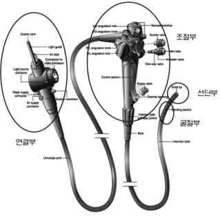
조절부
선단부
굴절부 연결부
그림2. 소화기 내시경의 외관과 부위별 명칭
Fig. 2. Gastrointestinal endoscope and its divisions according to the functions
CCD .
1/6 40 CCD SD(standard
definition) .
,
. (sensitivity)
. CMOS CCD
,
.
, .
3-100mm ,
. (iris) ,
.
(sensitivity), (dynamic
range), , .
굴절부와 방향조절 휠 B.
2 , .
4 (wire)
Up 210°, Down 90°, Left/Right 180°
. (endoscopist)
. , ,
(hand-eye coordination),
.
.
, .
(leakage) .
. ,
. ,
, . ,
, ,
.
,
( )
, .
기타 부위 C.
,
/
. (suction)
.
, .
. 4
.
,
흡인기 제어밸브
물 공기/ 제어밸브
기구 밸브
생검 기구 ( )
통로 노즐
물병연결튜브 광원 연결부 광원장치
램프
공기 펌프
물병 흡인기 연결부
흡인 튜브 공기 통로
물공급채널 물분사채널 물분사 연결
그림4. 내시경 내부의 배관구조
Fig. 4. The internal tubes in a gastrointestinal endoscope
, . CCD
, ,
.
, short arc
Xenon . CCD
, (monochrome CCD)
Red, Green, Blue
Red, Green, Blue
. 5
.
,
.
새로운 영상화 기법 .
Ⅳ
고해상도 내시경
A. (High Definition Endoscope)
100,000-400,000
SD(standard definition) ,
HD(high definition)
100 .
1080i, 720p HDTV
(native resolution)
.
. 16:9
, ,
.
, HD-SDI(high
definition serial digital interface), HDMI(high definition multimedia interface), DVI(digital video interface)
. , HD SD
, HDTV
.
HD HD
[4,7].
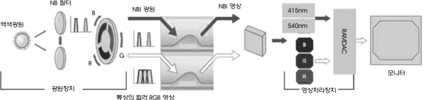
스펙트럼 영상화 B.
(white light) .
(chromoend-
oscopy) .
. NBI (narrow band imaging) ,
CCD
[4,8].
415nm 540nm ,
R/G/B
(pseudo-color) .
,
. NBI
CCD .
NBI .
,
. 6
.
, RGB
, CCD 3
(sequential method). NBI 415nm 540nm
pseudo-color
. Pentax Fujinon
. CCD ,
모니터
소화기 내시경
광원장치와 영상처리장치
기록저장장치
프린터
내시경 카트
그림5.소화기 내시경 시스템
Fig. 5. Endoscope system including light source and visualization equipment
RCB
[5,6]. NBI (spectral imaging)
Pentax i-Scan Fujinon FICE .
, ,
, .
.
, .
. (auto
fluorescence imaging, AFI)
.
multimodal endoscope , .
C. 초음파 내시경 (endoscopic ultrasound) 과 OCT (optical coherent tomography)
, (tomography)
.
.
(instrument channel)
(single element ultrasound transducer) (radial image)
[13]. (endoscopic ultrasound, EUS) 7
.
. (optical coherent tomography, OCT) OCT
, .
필터 NB
광원장치 백색광원
통상의 컬러RGB영상
영상 광원 NBI
NBI
영상처리장치 415nm
540nm RAMDAC
모니터 B
G R
그림6.스펙트럼 영상화를 위한 광원장치와 촬상소자 작동방식
Fig. 6. Light source and CCD operation mechanism for spectral imaging and normal color image
그림7. 내시경용 초음파 트랜스듀서와 획득 영상의 예 Fig. 7. An endoscopic ultrasound transducer and its image
공초점 현미 내시경
D. - (confocal microendoscope)
. (biopsy)
.
.
.
(confocal microscope) (dichromatic mirror)
.
( )
. PENTAX 0-250 m μ
, 500 m × 500 m μ μ .
[12]. 8
.
캡슐형 내시경 E.
10mm
. ,
LED CMOS ,
. ,
.
. ,
.
. ,
,
.
기계 장치를 이용한 새로운 내시경 기술 .
Ⅴ
, ,
, .
.
, .
. (cold light source)
7㎛
250㎛
500×500㎛
그림8.공초점 현미 내시경 좌측 상단은 점막에 선단부를 대어 조직의 횡단면 영상을 얻는 모식도와 공초점 현미 내시경의 선단부 전면을 묘사하고 있으며- . - , 우측에깊이에따른 횡단면획등영상을표현함 좌측하단은일반적인현미경조직검사영상이고 우측이공초점현미 내시경을통해 획득한영상의예. , - Fig. 8. Schematic of a confocal microscope. By probing the mucosal tissue using a confocal micro-endoscope(upper left), each horizontal plane (upper right) can
be acquired. A typical histological image (lower left) and a horizontal section image (lower right) by the microendoscope are shown.
, LED (light emitting diode) . LED
,
. 9 Invendo ,
LED ,
.
내시경 개발 기술의 방향 .
Ⅵ
,
HD .
,
(nasal endoscope) ,
.
. (biopsy) ,
,
. -
. ,
,
. -
(optical biopsy)
. ,
(multi-modal endoscope) [11].
.
.
. ,
.
(endoscopic surgical instrument) .
, ,
NOTES (natural orifice trans-luminal
endoscopic surgery) ,
[9,10].
(hybrid
notes) .
(Minimally Invasive Surgery) , NOTES
. NOTES ,
.
소화기 내시경의 국산화 .
Ⅶ
,
Electrohydraulic deflection
Working channel
Camera head Handheld
device
Driving unit
Inverted sleeve Endoscope Sheath
그림9.Invendo Medical사의 새로운 삽입 및 굴절 방식과 전동화된 조절장치가 구현된 대장내시경(colonoscope) Fig. 9. Colonoscope from Invendo GmbH, Germany
, .
.
3 , , 3
. 3 .
. , IT
,
.
.
, , ,
, . ,
, NOTES, Optical Biopsy , .
.
, ,
, , .
,
. ,
NOTES
.
참고문헌
[1] P.B. Cotton, and C.B. Williams, Practical Gastrointestinal Endoscopy, 5thed., Massachusetts, USA: Blackwell Publishing Ltd., 2003, pp.6-16.
[2] M.V. Sivak, Gastrointestinal Endoscopy, 2nded., Pennsylvania, USA: Saunders Company, 2000, pp, 16-49.
[3] J. Baillie, MB, and W. Salem, “The endoscope”, Gastrointestinal Endoscopy, vol.65, No.6, pp.886-893, 2007.
[4] A. Larghi, P.G. Lecca and G. Costamagna, “High-resolution narrow band imaging endoscopy”, Gut, vol.57, pp.976-986, 2008.
[5] Hiroyuki Osawa, MD, Mitsuyo Yoshizawa, H. Yamamoto, et al.,
“Optimal band imaging system can facilitate detection of changes in depressed-type early gastric cancer”, Gastrointestinal Endoscopy, vol.67, pp.226-234, 2008.
[6] S. Takeuchi, K. Abe, and D. Ayame, “Endoscopic Apparatus”, US2006/0197830, USPTO
[7] ASGE Technology Committee, “High-resolution and high- magnification endoscopes”, Gastrointestinal Endoscopy, vol.69, No. 3, pp.399-407, 2009.
[8] K. Kuznetsov, R. Lambert, and J.F. Rey, ”Narrow-Band Imaging:
Potential and Limitations”, Endoscopy, vol.38, pp.76-81, 2006.
[9] M. Kevin, W. Reavis, and S. Melvin, “Advanced endoscopic technologies”, Surgical Endoscopy, vol.22, pp.1533-1546, 2008.
[10] W.M. White, G.P. Haber, M.J. Doerr, et al., “Natural orifice translumenal endoscopic surgery” Urol. Clin. North Am., vol.36, No.2, pp. 147-55, 2009.
[11] H. Tajiri, “Future perspectives of gastrointestinal endoscopy and joint academic-industrial research following technological innovation in medical and biological engineering”, Digestive Endoscopy, vol.17, pp.S97-S104, 2005.
[12] O. Watanabe, T. Ando, O. Maeda, et al., “Confocal endomicroscopy in patients with ulcerative colitis”, J. Gastroenterol. Hepatol.
vol.23, p.S286-90.2008.
[13] ASGE Technology Committee, “Endoscopic ultrasound probes”, Gastrointestinal Endoscopy, vol.63, No. 6, pp.751-754, 2006.