ORIGINAL ARTICLE
내시경 초음파 세침흡인 결과가 진단불가 혹은 음성인 췌장 고형종괴의 악성 예측인자
정혜원1, 박찬선1, 김기배1, 한정호1,2, 윤순만1,2, 채희복1,2, 윤세진1,2, 박선미1,2
충북대학교병원 내과1, 충북대학교 의과대학 내과학교실2
Predictors of Malignancies in Patients with Inconclusive or Negative Results of Endoscopic Ultrasound-guided Fine-needle Aspiration for Solid Pancreatic Masses
Hyewon Jeong1, Chan Sun Park1, Ki Bae Kim1, Joung-Ho Han1,2, Soon Man Yoon1,2, Hee Bok Chae1,2, Sei Jin Youn1,2 and Seon Mee Park1,2
Department of Internal Medicine, Chungbuk National University Hospital1, Department of Internal Medicine, Chungbuk National University College of Medicine2, Cheongju, Korea
Background/Aims: This study analyzed the diagnostic accuracy of endoscopic ultrasound-guided fine needle aspiration (EUS-FNA) for pancreatic solid masses in patients with or without chronic pancreatitis as well as the clinical parameters relevant to a malignancy when EUS-FNA was negative or inconclusive.
Methods: A total of 97 patients, who underwent EUS-FNA for solid pancreatic masses over 2 years at a single institution, were evaluated. All patients underwent EUS-FNA for 3-5 passes with 22 or 25 G needles without an on-site cytopathologist. The final diag- nosis was obtained by surgery or compatible clinical outcomes for a more than 12 month follow-up. The diagnostic yields in the pa- tients with or without chronic pancreatitis were compared and the histories and laboratory data relevant to pancreatic ductal ad- enocarcinoma (PDAC) or pseudo-tumor were analyzed.
Results: The final diagnoses were adenocarcinoma in 88 patients (90.7%) and inflammatory pseudo-tumor in 9 (9.3%). The results of EUS-FNA were adenocarcinoma (74), suspicious (7), atypical (5), negative (10), and inadequate specimen (1). The diagnostic accu- racies were 76.9% and 91.6% in patients with or without chronic pancreatitis, respectively. Among the 23 cases with non-diagnostic results of EUS-FNA, PDAC was finally diagnosed in 5 out of 7 suspicious, 3 out of 5 atypical, and 5 out of 10 negative cytology cases.
The clinical parameters related to a pseudo-tumor were a history of alcohol consumption and pancreatitis, and normal alkaline phos- phatase levels.
Conclusions: The diagnostic accuracy of pancreatic masses in the background of chronic pancreatitis was low. When EUS-FNA pro- duced inconclusive results, the histories of alcohol consumption, pancreatitis, and serum levels of alkaline phosphatase are useful for making a final diagnosis. (Korean J Gastroenterol 2018;71:153-161)
Key Words: Pancreatic cancer; Chronic pancreatitis; Endosonography; Fine needle aspiration
Received August 1, 2017. Revised December 10, 2017. Accepted December 22, 2017.
CC This is an open access article distributed under the terms of the Creative Commons Attribution Non-Commercial License (http://creativecommons.org/licenses/
by-nc/4.0) which permits unrestricted non-commercial use, distribution, and reproduction in any medium, provided the original work is properly cited.
Copyright © 2018. Korean Society of Gastroenterology.
교신저자: 박선미, 28644, 청주시 서원구 충대로 1, 충북대학교 의과대학 내과학교실
Correspondence to: Seon Mee Park, Department of Internal Medicine, Chungbuk National University College of Medicine, 1 Chungdae-ro, Seowon-gu, Cheongju 28644, Korea. Tel: +82-43-269-6019, Fax: +82-43-273-3252, E-mail: [email protected]
Financial support: None. Conflict of interest: None.
INTRODUCTION
Pancreatic ductal adenocarcinoma (PDAC) is charac-
terized by hypovascular lesions and extensive interstitial fibrosis. These features are helpful for a presumptive diag- nosis by abdominal imaging; however, a pathology diagnosis
is necessary to determine the therapeutic plans. Endoscopic ultrasound (EUS) is useful for specific image features and a definitive diagnosis can be made with EUS-guided fine-nee- dle aspiration (EUS-FNA).1-3 On the other hand, a diagnosis of PDAC in the background of chronic pancreatitis is still chal- lenging because extensive fibrosis leads to inadequate sam- pling and incorrect targeting.1,2 The diagnostic accuracy of EUS-FNA for PDAC was 85-90%1,3 in patients with a normal background, which decreased to 54-74% in those with chron- ic pancreatitis.4,5 Because the diagnostic power of EUS-FNA for pancreatic solid masses was specific, the presence of ma- lignant cells can be diagnosed easily as PDAC. On the other hand, it was difficult to determine whether follow-up or surgi- cal resection were required when the results of EUS-FNA were negative or inconclusive, such as atypical or suspicious.1,3
Various methods, such as lengthening of the passages of FNA, performing a core biopsy instead of cytology, pathology studies by an on-site pathologists, or repeated FNA on different days are recommended to improve the diagnostic accuracy.1,6-8 In addition, the patient’s characteristics are also helpful for clinical decision making, when the cytology data are negative or inconclusive. The weight loss, elevated serum levels of cancer antigen (CA) 19-9, smoking history, no history of pan- creatitis or alcohol intake, and evidence of a bile duct ob- struction were relevant to a pancreatic malignancy. On the other hand, the proportions of inconclusive results, final di- agnosis of these patients, and relevant clinical parameters have been inconsistent in many studies.5,9-13
This study examined the diagnostic yields of EUS-FNA for pancreatic solid masses in patients with or without chronic pancreatitis. In addition, the clinical parameters relevant to a final diagnosis were determined when EUS-FNA was non-diagnostic.
SUBJECTS AND METHODS
1. Patients
Ninety-nine patients were admitted to Chungbuk National University Hospital from May 2013 to May 2015 for solid masses in the pancreas. The patients’ medical records were reviewed retrospectively. The demographics, including age, sex, presenting symptoms such as abdominal pain, jaundice, weight loss, and fever, and habits regarding alcohol intake and smoking, were recorded. Alcohol consumption or smok-
ing habit were positive for patients with current chronic heavy drinkers (average total alcohol intake of ≥60 g/day) or regular current smokers (≥1 pack/day), respectively. Liver function tests, serum glucose, and serum CA 19-9 at admission were collected. All patients underwent an abdominal compu- terized tomography scan before EUS-FNA. Chronic pan- creatitis was diagnosed based on the following criteria, which is applicable to the Korean population: the presence of at least one finding among irregular main duct dilatation and atrophy, the presence of pancreatic duct stones, protein plugs, or pancreatic parenchymal calcification by an abdomi- nal computerized tomography scan.14 A final diagnosis was made by the surgical pathology or by compatible clinical find- ings during a follow-up of more than 12 months or until death.
Stable imaging, benign pathology, or survival for 12 months after EUS-FNA was considered benign. The diagnosis of a ma- lignancy was based on a positive pathology, local invasion, and metastasis on imaging, or death within 12 months from cancer-associated causes.
2. EUS-FNA
Two experienced endoscopists performed this procedure without an on-site cytopathologist. EUS was conducted using linear EUS (GF-UCT240-AL5; Olympus, Tokyo, Japan). FNA was performed for 3-5 passes with 22 or 25 G needles (Echotip;
Wilson-Cook, Winston Salem, NC, USA) for lesions in the body/tail or uncinate/head of the pancreas, respectively.15 The fanning technique and suction method were applied. The stomach and duodenum were sites of needle passage for le- sions in the body/tail and uncinate/head of the pancreas, respectively. The stylet was introduced to the needle, and the extruded material was placed onto glass slides for a primary inspection. The aspirated material was smeared carefully on- to glass slides and fixed in a 96% alcohol solution for a cytol- ogy examination. The smear on slides were performed on- site. Diff-Quik histological staining (Baxter Healthcare, Deerfield, NY, USA), Papanicolaou staining, and cell blocks with hematoxylin and eosin stains were prepared in the cytol- ogy laboratory.
3. Cytology interpretation
One experienced pathologist interpreted all the Papanicolaou staining and cell blocks. The cytological findings were catego- rized as ‘inadequate’, ‘negative’, ‘atypical’, ‘suspicious’, and
Fig. 1. Representative images of the pancreatic cytology (papanicolaou staining, magnification ×400). (A) Positive for a malignancy. (B) Suspicious for a malignancy. (C) Atypical cells. (D) Negative for a malignancy.
‘malignant’.16 The ‘suspicious’ category is defined as atyp- ical cells severe enough to suspect a PDAC, but the quantity of cells is insufficient to prove positive for a malignancy. The
‘atypical’ category is that the level of atypical cells is more than that of benign or reactive, but insufficient for a diagnosis of the ‘suspicious’ category (Fig. 1). The atypical or suspi- cious cytology results were interpreted as negative or neo- plasms, respectively.
4. Clinical parameters for predictors of malignancy Patients with negative or inconclusive results in EUS-FNA for PDAC, were selected. The clinical parameters relevant to PDAC or pseudo-tumors were analyzed. Patients with a PDAC or pancreatic pseudo-tumor were compared using the demo- graphic data, history of alcohol or smoking, presenting symp- toms, and laboratory data (Fig. 2).
5. Statistical analysis
The diagnostic yields of EUS-FNA for pancreatic masses, including sensitivity, specificity, and overall accuracy, were assessed. The data were analyzed with SPSS software ver- sion 18.0 (SPSS Inc., Chicago, IL, USA). The continuous varia- bles are presented as the mean±standard deviation and the two groups were compared using a Mann-Whitney U test. The categorical variables were compared using a chi square test or Fisher’s exact test. p-values less than 0.05 were consid- ered significant.
RESULTS
1. Patient characteristics of pancreatic solid mass Among the ninety-nine patients who underwent EUS-FNA for pancreatic solid masses, two patients, one with neuro- endocrine tumor and the other with a metastasis, were excluded. The final diagnoses were done by surgery in 7
A B
C D
Fig. 2. Flow chart of the diagnostic process. Of the 99 patients who underwent EUS-FNA, each patient with a neuroendocrine tumor and meta- stasis on the pancreas was excluded. Twenty-three patients with negative or non-diagnostic EUS-FNA were included in the analysis of the clinical parameters relevant to pancreatic cancers or pseudo-tumors. EUS-FNA, endoscopic ultrasonography guided fine needle aspiration;
PDAC, percentages of pancreatic ductal adenocarcinoma.
Table 1. Characteristics of Patients with Pancreatic Masses
Parameters Total (n=97) Chronic pancreatitis
p-value
No (n=84) Yes (n=13)
Mean age±SD 67.6±10.6 69.1±9.7 57.9±11.6 0.005
Male:Female 66:31 55:29 11:3 0.214
Symptoms 0.797
Abdominal pain 73 (75.3) 62 (73.8) 11 (84.6)
Weight loss 9 (9.3) 8 (9.5) 1 (7.7)
Jaundice 6 (6.2) 5 (6.0) 1 (7.7)
Fever 6 (6.2) 6 (7.1) 0 (0.0)
Uncontrolled hyperglycemia 3 (3.1) 3 (3.6) 0 (0.0)
Alcohol drinking 15 (15.5) 8 (9.5) 7 (53.8) 0.001
Smoking 18 (18.5) 15 (17.9) 3 (23.1) 0.703
Laboratory data
Glucose ≥126 mg/dL 57 (58.8) 50 (59.5) 7 (53.8) 0.767
CA 19-9 ≥37 UL 69 (71.1) 61 (77.2) 8 (61.5) 0.299
ALT (IU/L) ≥1.5×UNL 24 (24.7) 20 (23.8) 4 (30.8) 0.730
ALP (IU/L) ≥220 U/L 39 (42.9) 35 (44.3) 4 (33.3) 0.545
Total bilirubin (mg/dL) ≥2 12 (12.4) 9 (10.7) 3 (23.1) 0.201
Mass locations 0.002
Pancreatic head 46 (47.4) 34 (40.5) 12 (92.3)
Pancreatic body 23 (23.7) 23 (27.4) 0 (0.0)
Pancreatic tail 28 (28.9) 27 (32.1) 1 (7.7)
Mass size (cm) 3.4±1.3 3.5±1.3 2.7±0.7 0.049
Diameter of CBD ≥7 mm 18 (32.7) 18 (40.0) 0 (0.0) 0.021
Diameter of PD ≥3 mm 43 (51.2) 35 (48.0) 8 (72.7) 0.264
Final diagnosis <0.0001
PDAC 88 (90.7) 82 (97.6) 6 (46.2)
Pseudo-tumor 9 (9.3) 2 (2.4) 7 (53.8)
Values are presented as n (%) unless otherwise indicated.
SD, standard deviation; CA, cancer antigen; ALT, alanine aminotransferase; UNL, upper normal limit; ALP, alkaline phosphatase; CBD, common bile duct; PD, pancreatic duct; PDAC, pancreatic ductal adenocarcinoma.
Fig. 3. Representative images of a pseudo-tumor (A-C) and pancreatic adenocarcinoma (D-F) in the setting of chronic pancreatitis. (A, D) Abdominal computed tomography scan. (B, E) Positron emission tomography-computed tomography scan. (C, F) Endoscopic ultrasound guid- ed fine needle aspiration.
Table 2. Results of EUS-FNA and Final Diagnosis in 97 Patients with Pancreatic Solid Masses
EUS-FNA PDAC (n=88) Pseudo-tumor (n=9) Total (n=97)
Malignancy 74 (84.1) 0 (0.0) 74 (76.3)
Suspicious 5 (5.7) 2 (22.2) 7 (7.2)
Atypical 3 (3.4) 2 (22.2) 5 (5.2)
Negative 5 (5.7) 5 (55.6) 10 (10.3)
Inadequate 1 (1.1) 0 (0.0) 1 (1.0)
Values are presented as n (%).
EUS-FNA, endoscopic ultrasonography guided fine needle aspiration; PDAC, pancreatic ductal adenocarcinoma.
(7.2%) patients and a clinical follow-up in 90 (92.8%) patients. Ninety-seven patients (male [M]:female [F], 66:31;
mean age, 67.6 years) including 13 patients with chronic pancreatitis (13.4%) (M:F, 11:2; mean age, 57 years), and 84 patients without chronic pancreatitis (86.6%) (M:F, 55:29;
mean age, 69 years) were enrolled (Table 1). The presenting symptoms upon admission were abdominal pain (75.3%), weight loss (9.3%), jaundice (6.2%), fever (6.2%), and uncon- trolled hyperglycemia (glycosylated hemoglobin ≥7%, 6.2%).
Patients with chronic pancreatitis had a history of alcohol consumption, were younger, and had a smaller mass located in the pancreas head with a normal common bile duct diame- ter compared to those without pancreatitis. The final diag- nosis was PDAC in 88 (90.7%) patients and inflammatory pseudo-tumor in 9 (9.3%) patients (Fig. 3). PDAC was diag-
nosed in 6 (46.2%) and 82 (97.6%) patients with or without pancreatitis, respectively.
2. Diagnostic accuracy and factors related with diag- nostic yields
EUS-FNA revealed a malignancy in 74 patients, suspicions of a malignancy in 7 patients, atypical in 5 patients, and neg- ative in 10 patients. One sample was inadequate for inter- pretation because of the poor cellularity (Table 2). No compli- cations were encountered during the procedures. The sensi- tivity, specificity, and diagnostic accuracy in the presence or absence of pancreatitis were 83.3%, 71.4%, and 76.9%; and 91.4%, 100%, and 91.6%, respectively (Table 3). The group contained 32 patients with non-diagnostic cytology, 7 pa- tients with ‘suspicious’ findings, 5 patients with ‘atypical’
A B C
D E F
Table 3. Diagnostic Accuracy of EUS-FNA with or without Chronic Pancreatitis
Results Chronic pancreatitis
Total (n=97)
No (n=84) Yes (n=13)
True positive 74 5 79
True negative 2 5 7
False positive 0 2 2
False negative 7 1 8
Indeterminate 1 0 1
Sensitivity 91.4 (83.2-95.8)a 83.3 (43.7-97.0)a 90.8 (82.9-95.3)a
Specificity 100.0 (34.2-100.0)a 71.4 (35.9-91.8)a 77.8 (45.3-93.7)a
Accuracy (%) 91.6 76.9 89.6
aPercent (95% confidence interval).
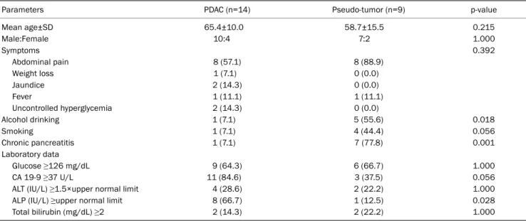
Table 4. Predictors of Malignancy in Patients with Inconclusive or Negative EUS-FNA for Solid Pancreatic Masses
Parameters PDAC (n=14) Pseudo-tumor (n=9) p-value
Mean age±SD 65.4±10.0 58.7±15.5 0.215
Male:Female 10:4 7:2 1.000
Symptoms 0.392
Abdominal pain 8 (57.1) 8 (88.9)
Weight loss 1 (7.1) 0 (0.0)
Jaundice 2 (14.3) 0 (0.0)
Fever 1 (11.1) 1 (11.1)
Uncontrolled hyperglycemia 2 (14.3) 0 (0.0)
Alcohol drinking 1 (7.1) 5 (55.6) 0.018
Smoking 1 (7.1) 4 (44.4) 0.056
Chronic pancreatitis 1 (7.1) 7 (77.8) 0.001
Laboratory data
Glucose ≥126 mg/dL 9 (64.3) 6 (66.7) 1.000
CA 19-9 ≥37 U/L 11 (84.6) 3 (37.5) 0.056
ALT (IU/L) ≥1.5×upper normal limit 4 (28.6) 2 (22.2) 1.000
ALP (IU/L) ≥upper normal limit 8 (66.7) 1 (12.5) 0.028
Total bilirubin (mg/dL) ≥2 2 (14.3) 2 (22.2) 1.000
Values are presented as n (%) unless otherwise indicated.
EUS-FNA, endoscopic ultrasound-guided fine-needle aspiration; SD, standard deviation; PDAC, pancreatic ductal adenocarcinoma; CA, cancer antigen; ALT, alanine aminotransferase; ALP, alkaline phosphatase.
findings, and 10 patients with ‘negative’ findings. PDAC was finally diagnosed in 5 out of 7 ‘suspicious’ cases, 3 out of 5
‘atypical’ cases, and 5 out of 10 ‘negative’ cases.
3. Clinical parameters related with malignancy
PDAC revealed less alcohol consumption (7.1% versus 55.6%, p=0.018), less chronic pancreatitis (7.1% versus 77.8%, p=0.001), and frequent elevated alkaline phospha- tase levels (66.7% versus 12.5%, p=0.028) compared to pseudo-tumors. Elevated CA 19-9 levels and a non-smoking history were related to PDAC; however, the findings were not statistically significant. There were no differences in age, sex, presenting symptoms, and serum bilirubin levels (Table 4).
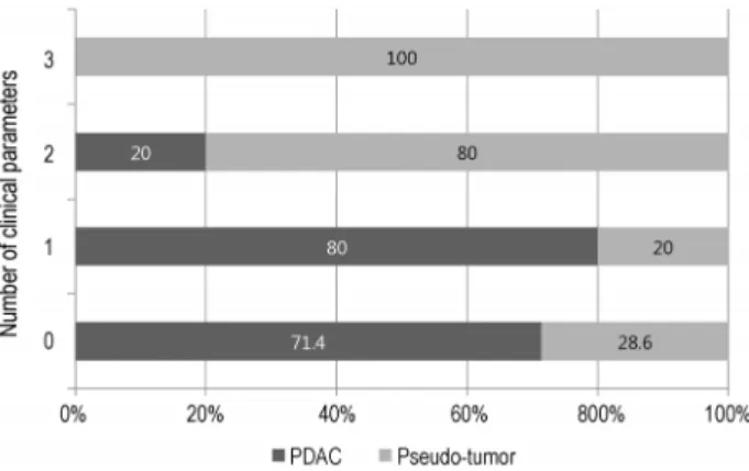
The percentages of PDAC were 71.4%, 80%, 20%, and 0% ac-
cording to the number of clinical parameters of 0, 1, 2, and 3, respectively (Fig. 4). The percentage of PDAC in patients with low risk (≥2) and high risk (<2) malignancies were 16.7%
and 76.5%, respectively.
DISCUSSION
This study showed that the diagnostic accuracy of EUS-FNA for pancreatic solid masses in the presence of coexisting pancreatitis was lower than those without pancreatitis.
‘Suspicious’ or ‘atypical’ categories by EUS-FNA were more frequent in patients with chronic pancreatitis than in those without it. When the EUS-FNA results were negative or incon- clusive, the histories of alcohol consumption, pancreatitis,
Fig. 4. Percentages of PDAC or pseudo-tumors according to the number of clinical parameters relevant to pseudo-tumors (alcohol drinking, history of pancreatitis, normal alkaline phosphatase lev- el). The percentages of PDAC were 71.4%, 80%, 20%, and 0% ac- cording to the number of clinical parameters of 0, 1, 2, and 3, respectively. PDAC, pancreatic ductal adenocarcinoma.
and serum levels of alkaline phosphatase were helpful for di- agnosing pseudo-tumors or PDAC.
EUS-FNA of the pancreatic lesions has been reported to be a reliable and minimally invasive method for evaluating solid masses.3,17 The diagnostic yields of EUS-FNA were improved in recent years in prospective multicenter studies17 com- pared to remote studies.18 EUS-FNA for pancreatic masses in the setting of a normal pancreas showed high sensitivity (85-90%) and high specificity (94.6-96.7%), positive like- lihood ratio of 15.2 (95% confidence interval [CI], 8.5-27.3), and negative likelihood ratio of 0.17 (95% CI, 0.13-0.21).1,3
On the other hand, the sensitivity of EUS-FNA was only 54-74% when sampling solid pancreatic masses in the set- ting of chronic pancreatitis,1,4,5 which was lower than in those without it.2,3 Chronic pancreatitis makes it difficult to target and interpret the cytology.1,2 A conglomeration of pancreatitis- induced lobulations may mimic a pancreatic mass and the presence of acoustic shadowing from a calcified stone may un- dermine the visibility of a neoplasm. Furthermore, the coex- istence of collateral vasculature in patients with severe chronic pancreatitis makes the process of FNA more challenging. In addition, the cytology features that may mimic a malignancy in chronic pancreatitis are occasional atypical cells that include enlarged, single cells with large nuclei, de- generative vacuoles, and occasional mitosis.8 This study showed that the diagnostic accuracy of EUS-FNA of solid le- sions in a normal pancreas was excellent, reaching 91.6%.
Nevertheless, the accuracy was 76.9% in patients with chron-
ic pancreatitis. The sensitivity and specificity of EUS-FNA in diagnosing solid pancreatic masses underlying chronic pan- creatitis were 83.3% and 71.4%, respectively.
The results of EUS-FNA were interpreted as the categories of ‘non-diagnostic’, ‘negative’, ‘atypical’, ‘suspicious’, and
‘positive/malignant’ by the Papanicolaou Society of Cytopathology guidelines.16 ‘Suspicious’ was interpreted as a malignancy and ‘atypical’ as pseudo-tumor.16 If ‘atypical’
and ‘suspicious’ cytology results were included to determine true neoplasms, the sensitivity increases to 91% (95% CI, 90-92); however, the specificity is decreased to 94% (95% CI, 93-96).13
The indeterminate categories of ‘atypical’ and ‘suspicious’
continue to be the most difficult for clinicians to interpret and manage. The proportions of ‘suspicious’ or ‘atypical’ cases were different in various studies. Previous studies reported that the frequency of the ‘atypical’ cases was 1-14%,19,20 and that of ‘suspicious’ cases was 3-5%.13,19 These wide varia- tions between studies were attributed to on-site cytopatholo- gists, reference standards, study type, and mass size and site.20 This study showed similar frequencies of ‘suspicious’
(7.2%) and ‘atypical’ (5.2%) categories compared to previous studies. The frequency of the indeterminate category has been reported to be 20-35% among chronic pancreatitis patients.13,21 This study showed that the proportions of in- determinate categories are higher in patients with chronic pancreatitis than in those without it (30.8% versus 9.5%, p=0.053).
The percentage of pancreatic cancer in patients with suspi- cious or atypical categories showed wide variations. Previous studies reported that 25-100% of the atypical category and 73-96% of the suspicious category had been finally diag- nosed with pancreatic carcinoma.13,20,21 This data was sim- ilar to previous data; 5 out of 7 (71.4%) suspicious and 3 out of 5 (60%) atypical cytology were PDAC.
In addition to EUS-FNA, the clinical characteristics may have a complimentary role to accurately diagnose pancreatic solid lesions. Many studies reported that the clinical parame- ters are useful for determining a malignancy when EUS-FNA showed inconclusive results. Old age, female gender, alcohol abstinence, jaundice, abdominal pain, weight loss, CA 19-9 level, hyperbilirubinemia, presence of a mass, dilation of the pancreatic duct, obstruction of the bile duct, no smoking his- tory, diabetes, and no history of pancreatitis, have been re-
ported as factors relevant to a malignancy. On the other hand, these factors were different in other studies. Weight loss has been reported to be a strong indicator of a malignancy in pa- tients with a mass in the suspicious category.13 In addition, age <50 years, male gender, African race, and the absence of jaundice were significantly associated with chronic pan- creatitis not malignancy.5 Another study of the malignancy risk stratification of pancreatic masses in the setting of chronic pancreatitis reported that female gender, mass loca- tion at the pancreatic body, more than one mass number, hy- perbilirubinemia (>7 µmol/L), and CA 19-9 (>37 U/mL) were associated with a malignancy.12 Increased CA 19-9 was high- ly specific (97%) for a malignancy in older jaundiced patients, or when the preoperative level was greater than 150 U/dL.9,10 Elevated alkaline phosphatase and CA 19-9 were related to a malignancy.5,9-12 These results reflect a higher prevalence of chronic pancreatitis in younger male patients.19 Male gender and alcohol consumption were related to chronic pancreatitis.5 This study showed that a history of alcohol consumption and pancreatitis, and normal levels of alkaline phosphatase were related to pseudo-tumors. There were no differences in tu- mor locations, gender, clinical symptoms, or bilirubin levels between pancreatic cancer and pseudo-tumor. In addition, smoking history was slightly related to pseudo-tumors, not PDAC. Because most patients with chronic pancreatitis have drinking and smoking habits, smoking was more related to a pseudo-tumor. When the parameters were applied to ‘negative’
or ‘inconclusive’ categories, most patients with more than 2 pa- rameters were diagnosed with pseudo-tumors.
This study had some limitations. Although there was a rela- tively long follow-up period, only a small percentage of cases (7.2%) were diagnosed by surgery, and most cases were diag- nosed by a clinical follow-up. In addition, the number of pa- tients with accompanying chronic pancreatitis was in- sufficient to compare the diagnostic yields to those without it. On the other hand, the percentage of patients presenting with pancreatic solid lesions in the background of pan- creatitis was 23.5-25%,5,9 which was similar to this study.
In conclusion, cases categorized as ‘negative’ or ‘indeter- minate’ on EUS-FNA, which showed clinical features relevant to a malignancy, have a high possibility of PDAC. Under these conditions, repeated FNA or surgical resection may be more helpful instead of a close follow-up. Therefore, the clinical pa- rameters play a role in the complimentary data for clinical de-
cision making.
REFERENCES
1. Farrell JJ. Diagnosing pancreatic malignancy in the setting of chronic pancreatitis: is there room for improvement? Gastrointest Endosc 2005;62:737-741.
2. Krishna NB, Mehra M, Reddy AV, Agarwal B. EUS/EUS-FNA for sus- pected pancreatic cancer: influence of chronic pancreatitis and clinical presentation with or without obstructive jaundice on per- formance characteristics. Gastrointest Endosc 2009;70:70-79.
3. Puli SR, Bechtold ML, Buxbaum JL, Eloubeidi MA. How good is en- doscopic ultrasound-guided fine-needle aspiration in diagnos- ing the correct etiology for a solid pancreatic mass?: a meta-anal- ysis and systematic review. Pancreas 2013;42:20-26.
4. Fritscher-Ravens A, Brand L, Knöfel WT, et al. Comparison of en- doscopic ultrasound-guided fine needle aspiration for focal pan- creatic lesions in patients with normal parenchyma and chronic pancreatitis. Am J Gastroenterol 2002;97:2768-2775.
5. Varadarajulu S, Tamhane A, Eloubeidi MA. Yield of EUS-guided FNA of pancreatic masses in the presence or the absence of chronic pancreatitis. Gastrointest Endosc 2005;62:728-736.
6. Eloubeidi MA, Varadarajulu S, Desai S, et al. A prospective evalu- ation of an algorithm incorporating routine preoperative endo- scopic ultrasound-guided fine needle aspiration in suspected pancreatic cancer. J Gastrointest Surg 2007;11:813-819.
7. Sun B, Yang X, Ping B, He Y, Zhang Z. Impact of inconclusive endo- scopic ultrasound-guided fine-needle aspiration results in the management and outcome of patients with solid pancreatic masses. Dig Endosc 2015;27:130-136.
8. Bang JY, Varadarajulu S. Neoplasia in chronic pancreatitis: how to maximize the yield of endoscopic ultrasound-guided fine nee- dle aspiration. Clin Endosc 2014;47:420-424.
9. Bloomston M, Bekaii-Saab TS, Kosuri K, et al. Preoperative car- bohydrate antigen 19-9 is most predictive of malignancy in older jaundiced patients undergoing pancreatic resection. Pancreas 2006;33:246-249.
10. Tessler DA, Catanzaro A, Velanovich V, Havstad S, Goel S.
Predictors of cancer in patients with suspected pancreatic malig- nancy without a tissue diagnosis. Am J Surg 2006;191:191-197.
11. Lee H, Lee JK, Kang SS, et al. Is there any clinical or radiologic feature as a preoperative marker for differentiating mass-form- ing pancreatitis from early-stage pancreatic adenocarcinoma?
Hepatogastroenterology 2007;54:2134-2140.
12. Cai QC, Chen Y, Xiao Y, et al. A prediction rule for estimating pan- creatic cancer risk in chronic pancreatitis patients with focal pan- creatic mass lesions with prior negative EUS-FNA cytology. Scand J Gastroenterol 2011;46:464-470.
13. Alston EA, Bae S, Eltoum IA. Suspicious cytologic diagnostic cat- egory in endoscopic ultrasound-guided FNA of the pancreas: fol- low-up and outcomes. Cancer Cytopathol 2016;124:53-57.
14. Lee HS, Kim TN, Lee DH, et al. Korean guideline for chronic pancreatitis. Korean J Pancreas Biliary Tract 2008;13:38-48.
15. Siddiqui UD, Rossi F, Rosenthal LS, Padda MS, Murali-Dharan V,
Aslanian HR. EUS-guided FNA of solid pancreatic masses: a pro- spective, randomized trial comparing 22-gauge and 25-gauge needles. Gastrointest Endosc 2009;70:1093-1097.
16. Pitman MB, Centeno BA, Ali SZ, et al. Standardized terminology and nomenclature for pancreatobiliary cytology: the Papanicolaou Society of Cytopathology guidelines. Diagn Cytopathol 2014;42:
338-350.
17. Hewitt MJ, McPhail MJ, Possamai L, Dhar A, Vlavianos P, Monahan KJ. EUS-guided FNA for diagnosis of solid pancreatic neoplasms:
a meta-analysis. Gastrointest Endosc 2012;75:319-331.
18. Ardengh JC, Lopes CV, Campos AD, Pereira de Lima LF, Venco F, Módena JL. Endoscopic ultrasound and fine needle aspiration in chronic pancreatitis: differential diagnosis between pseudotu- moral masses and pancreatic cancer. JOP 2007;8:413-421.
19. Savides TJ, Donohue M, Hunt G, et al. EUS-guided FNA diagnostic yield of malignancy in solid pancreatic masses: a benchmark for quality performance measurement. Gastrointest Endosc 2007;
66:277-282.
20. Abdelgawwad MS, Alston E, Eltoum IA. The frequency and cancer risk associated with the atypical cytologic diagnostic category in endoscopic ultrasound-guided fine-needle aspiration speci- mens of solid pancreatic lesions: a meta-analysis and argument for a Bethesda system for reporting cytopathology of the pancreas. Cancer Cytopathol 2013;121:620-628.
21. Layfield LJ, Schmidt RL, Hirschowitz SL, Olson MT, Ali SZ, Dodd LL. Significance of the diagnostic categories “atypical” and
“suspicious for malignancy” in the cytologic diagnosis of solid pancreatic masses. Diagn Cytopathol 2014;42:292-296.