J. Exp. Biomed. Sci. 2012, 18(2): 131~138 pISSN : 1738-3226
Factors to Predict Successful Harvest during Autologous Peripheral Hematopoietic Stem Cell Collection
Mun-Ja Kim1, Soo-Hee Jin2, Duk-Hee Lee2, Dae-Weon Park1, Sung-Ae Koh3, Kyung-Hee Lee3, Myung-Soo Hyun3 and Min-Kyoung Kim3,†
1Hematopoietic Stem Cell Bank, Yeungnam University Medical Center, Daegu 705-717, Korea
2Department of Preventive Medicine School of Medicine Kyungpook National University, Daegu 700-842, Korea
3Department of Internal Medicine, Yeungnam University College of Medicine, Daegu 705-717, Korea
Autologous peripheral blood stem cell transplantation (PBSCT) has been used as a major treatment strategy for hematological malignancies. The number of CD34 positive cells in the harvested product is a very important factor for achieving successful transplantation. We studied the factors that can predict the number of CD34 positive cells in the harvested product of acute myelocytic leukemia (AML), multiple myeloma (MM) and Non-Hodgkin's lymphoma (NHL) patients after mobilizing them with chemotherapy plus G-CSF. A total of 73 patients (AML 19 patients, MM 28 patients, NHL 26 patients) with hematological malignancies had been mobilized with chemotherapy and granulocyte colony-stimulating growth factor from April, 2000 to February, 2012. Group's characteristics, checkup opinion of pre-peripheral blood on the day of harvest & outcome of PBSC were analyzed and evaluated using SPSS statistics program after grouping patients as below; group 1: CD34 cell counts < 2 × 10⁶/kg (n=16); group 2: 2 × 10⁶/kg ≤ CD34 cell counts < 6 × 10⁶/kg (n=32); group 3: CD34 cell counts ≥ 6 × 10⁶/kg (n=25). We analyzed the clinical characteristics, the peripheral blood (PB) parameters and the number of CD34 positve cells in the PB and their correlation with the yield of CD34 positve cells collected from the mobilized patients. The total number of leukapheresis sessions was 263 (mean: 3.55 session per patient), and the mean number of harvested CD34 positive cells per patient was 7.37 × 10⁶/kg.
The number of CD34 positive cells in product was significantly correlated with the number of platelet and CD34 positive cells in peripheral blood (P<0.05). The number of PB CD34 positive cells was the best significant factor for the quantity of harvested CD34 positive cells on the linear regression analysis (P<0.05). Many factors could influence the mobilization of peripheral blood stem cells. Platelet count and PB CD34 positive cells count were the two variables which remained to be significant in multivariate analysis. Therefore, the number of platelet and CD34 positive cells in peripheral blood on the day of harvest can be used as an accurate predictor for successful peripheral blood stem cell collection.
Key Words: Autologous peripheral blood stem cell transplantation, CD34 positive cell, Platelet
서 론
말초혈액 조혈모세포 이식술은 혈액종양 및 고형암의 치료에서 고용량의 항암요법 후 조혈기능의 빠른 회복을
위한 조혈모세포의 공급원으로 다양하게 이용되고 있다.
1979년 이전까지는 고용량 항암화학요법에 따른 골수기 능 억제를 회복시키기 위한 수단으로 주로 골수이식을 사용하였다 (Goldman, 1979). 1980년대 이후 백혈구 분반 술 (leukapheresis)로 말초혈액의 조혈모세포를 채집할 수 있게 되면서 이 분야에서의 발전은 치료와 관련된 사망 률과 이식에 대한 다른 부작용을 현저하게 감소시켰다 (Russell et al., 1993; Schwartzberg et al., 1993).
조혈모세포 이식 (hematopoietic stem cell transplantation, HSCT)은 혈연이나 비혈연 (동종이식) 공여자의 세포 혹 은 환자 본인으로부터 미리 채집된 세포 (자가이식)로 시
*Received: 17 May, 2012 / Revised: 18 June, 2012 Accepted: 25 June, 2012
†Corresponding author: Min-Kyoung Kim. Department of Internal Medicine, Yeungnam University College of Medicine, Daegu 705-717, Korea.
Tel: +82-53-620-4683, Fax: +82-53-654-8386 e-mail: [email protected]
○CThe Korean Society for Biomedical Laboratory Sciences. All rights reserved.
Original Article
행한다. 어떤 방법을 선택할 것인지는 환자의 나이, 대 상 질환, 공여자의 존재 여부 등에 따라 달라진다. 동 종이식은 주조직적합항원 (major histocompatibility antigen, MHA)의 일치가 필요하며 가장 적합한 공여자는 HLA (human leukocyte antigen) 일치 형제이다. 그러나 형제간 에 HLA가 일치할 확률은 30% 내외이기 때문에 비혈연 이식을 위해 골수은행, 제대혈은행이 활성화 되고 있다 (Hwang, 2007). 동종이식은 고용량사이클로포스파미드 (cyclophosphamide)와 전신방사선조사 (total body irradiation, TBI) 등으로 일주일 정도의 전처치를 하고, 이식 당일 공여자로부터 채취한 조혈모세포를 환자의 중심정맥을 통하여 주입한다. 자가이식은 고용량화학요법과 방사선 치료로 종양세포를 제거한 후 세포성장인자 (granulocyte colony-stimulating factor, G-CSF)를 사용하여 골수의 조혈 모세포를 말초혈로 가동화시킨다. 이 시기에 세포분리 반출기를 이용하여 조혈모세포를 채집하여 냉동한 후 이 식하는 방법이다. 동종이식과 과정이 비슷하나 면역억제 가 낮고 회복이 빠르며 이식편대숙주반응 (graft-versus- host disease, GVHD)이 없는 장점이 있다. 자가이식은 동 종이식과 달리 이식관련 사망률은 낮으나 재발이 흔하므 로 이식관련 사망률이 높을 환자, 공여자가 없어 동종이 식을 할 수 없는 환자에게 시행하며 림프종, 고형암 등에 서도 광범위하게 시도되고 있다 (Dicke et al., 2010).
골수이식은 골수 (장골능)에서 직접 흡인하여 조혈모세 포를 채취해야 함으로 공여자에게 마취가 필요하다. 마 취 등 여러 가지 이유에서 최근에는 주로 말초혈로부터 조혈모세포를 채취한다. 말초혈액의 조혈모세포는 골수 모세포보다 이식 후 중성구와 혈소판의 회복이 빠르다 (Bensinger et al., 2001; Couban et al., 2002). 조혈모세포 이 식에서 생착에 필요한 최소한의 조혈모세포수는 CD34 양성 세포수를 기준으로 환자 몸무게당 1.0~2.0 × 10
6이 며 채집된 CD34 양성세포가 5.0~6.0 × 10
6이상일 경우 조혈모세포 이식에 이상적이라고 알려져 있다 (Anguita- Compagnon et al., 2010).
자가 말초혈액 조혈모세포 이식술이 효과적으로 시행 되기 위해서는 가동화요법을 시행하며 말초혈액의 조혈 모세포 채집을 보다 효과적으로 수행하는 것이 중요하다 (Dicke et al., 2010). 성공적인 이식을 위한 조혈모세포의 채집양은 채집 당일 말초혈액의 CD34 양성 세포수가 의 미 있는 예측인자로 알려져 있다. 말초혈액의 백혈구나 단핵구수도 연관이 있다는 보고가 있기는 하지만 그 외 에는 어떤 다른 인자가 연관이 있는지 명확하게 밝혀져
있지 않다 (Lee et al., 2003; Suh et al., 2004; Padmanabhan et al., 2009).
따라서 본 연구의 목적은 성공적인 자가 말초혈액 조 혈모세포 이식을 위하여 조혈모세포 채집술을 시행한 악 성 혈액질환 환자를 대상으로 후향적 조사를 통해 말초 혈액의 CD34 양성 세포수 외에도 환자의 임상적인 소견 이나 채집 당일 말초혈액 검사 소견 등이 조혈모세포 채 집물의 CD34 양성 세포수와 어떤 연관이 있는지 알아보 고 성공적인 조혈모세포 채집을 예측할 수 있는 인자를 알아보고자 하였다.
재료 및 방법
연구대상
2000년 4월부터 2012년 2월까지 자가 말초혈액 조혈 모세포 이식을 위해 조혈모세포를 채집한 환자 73명을 대상으로 263회의 채집술을 시행하였다. 말초혈액 조혈모 세포 산출량은 채집된 CD34 양성 세포수가 총 2 × 10
6/kg 미만인 집단 (n=16)을 Group 1, 2 × 10
6/kg 이상에서 6 × 10
6/kg 미만인 집단 (n=32)을 Group 2, 6 × 10
6/kg 이 상인 집단 (n=25)을 Group 3으로 나누어 조사를 실시하 였다. 본 연구는 Y 대학교병원 임상연구윤리위원회의 승 인을 받은 후 진행되었다 (YUH-12-0373-O39).
방법
가동화요법.
조혈모세포를 채집하기 전 종양세포를 최 대한 제거하기 위해 급성 백혈병 (acute leukemia)의 경우 는 공고요법으로 치료를 하였고, 악성림프종은 화학요 법 DHAP (dexamethasone, cisplatin and cytarabine), ESHAP (etopiside, methylprednisolone, cytarabine and cisplatin) 등의 항암화학요법을 주로 시행하였다. 다발성골수종은 고용 량 싸이톡산 (high dose cytoxan, 3 g/m
2D1)을 사용하여 골 수억제가 회복되는 시점에서 G-CSF (뉴트로진, 중외제약, 한국 ) (10 μg/kg/day)를 투여하여 조혈모세포를 가동화시 켰다 . 가동화요법은 말초혈액의 호중구수가 500/μL 이하 로 감소할 때 투여를 시작하여 백혈구 분반술이 끝날 때 까지 사용하였다.
말초혈액 내 혈구수 측정.
세포성장 촉진인자를 투여하기
시작한 날로부터 채집술 전 말초혈액 총 백혈구수와 단
핵구 (mononuclear cells, MNC) 등의 측정은 말초혈액 3
mL를 채취하여 항응고제 (ethylenediaminetetraacetic acid,
EDTA)로 항응고 처리를 한 후 자동혈구분석기를 이용하 여 산출하였다. 채집된 말초혈액 조혈모세포의 총 유핵 세포수 (total nucleated cell, TNC)와 단핵구 등도 자동혈구 분석기를 이용하여 분석하였다.
말초혈액 CD34 양성 세포 채집술.
말초혈액 조혈모세포 채집술은 말초혈액 백혈구수가 3,000/μL 이상으로 상승 하는 첫째 날부터 총혈액양의 3배 이상의 고용량 백혈구 분반술로서, Fenwal CS-3000 plus (Baxter Healthcare corp., Deerfield, IL, USA)를 이용하여 매일 8~15 L의 혈액을 평 균 4시간에 걸쳐 단핵구층을 분리하였다. 채혈선과 복귀 선은 환자의 경우 대부분 쇄골하정맥으로 이중도관 카테 터를 사용하였다. 분리를 위해서 Granulo chamber를 장착 하고 , 채집을 위해서 A-35 chamber (채집량 200 mL) 또 는 small volume collection chamber (SVCC, 50 mL)를 장착 하였다 . 채집술은 일반적으로 채집하고자 하는 조혈모세 포 양을 얻을 때까지 시작 후 연속적으로 시행하였다.
CD34 양성 세포수 측정.
채집 전 말초혈액 CD34 양성 세 포와 채집된 CD34 양성 세포수의 분석은 튜브 (TruCount tube) 2개를 준비한 다음 말초혈액 100 μl씩을 각각 첨가 하였다. 첫 번째 튜브에는 CD45-fluorescein isothiocyanate (FITC) 10 μl, Mouse IgG1-phycoerythrin (PE) 10 μl를 첨가하 였다 . 두 번째 튜브에는 CD45-FITC 10 μl, CD34-PE 20 μl 를 첨가하였다. 그리고 TruCount tube의 뚜껑을 덮고 실 온 암실에서 15분간 반응시켰다. 각 튜브에 7-AAD 40 μl, BD Pharm Lysing buffer (1X) 2 ml를 첨가하여 실온에서 15분간 두어 적혈구를 용혈을 시킨 후 1시간 이내에 분 석하였다. 분석 시에는 살아있는 세포만 구획을 취하여 CD34 양성 세포수를 측정하였다. 유세포분석 장비는 FACS Calibur (BD Biosciences, San Jose, USA)를 사용하였 고 CellQuest Pro (BD Biosciences) 소프트웨어를 이용하여 분석하였다 .
자료 분석
자료 분석은 SPSS 통계 프로그램 (version 19.0)을 사 용하였다 . 각 그룹간의 특성은 chi-square test, ANOVA를 이용하여 분석하였다. 자료의 표시방법은 % 또는 평균 값 ± 표준편차로 표시하였으며, 유의수준은 0.05 미만으 로 하였다. 채집 완료된 CD34 양성 세포수와 관련이 있 는 요인에 대해서는 선형회귀분석으로 통계학적 의미를 평가하였으며 CD34 양성 세포수와 혈소판수의 기준치를
예측하기 위해서 receiver operating characteristic (ROC)를 이용하였다.
결 과
대상 환자의 일반적 및 임상관련 특성
2000년 4월부터 2012년 2월까지 73명의 환자 (남자: 45, 여자: 28)의 말초혈액 조혈모세포 채집을 하였다. 대상 환 자의 연령은 17세부터 64세까지의 분포를 보였다. 진단 명은 급성골수성백혈병 (acute myelocytic leukemia, AML) 이 26.0% (19명), 악성림프종 (non-hodgkin's lymphoma, NHL)이 35.6% (26명), 다발성골수종 (multiple myeloma, MM)이 38.4% (28명)이었다. 말초혈액 조혈모세포 채집술 을 시행한 환자는 모두 화학치료 후 가동화를 위한 화학 요법을 받았고 가동화 이전 항암화학요법의 평균횟수는 3.9회였다 (Table 1).
조혈모세포의 가동화 (Mobilization)
가동화를 위한 항암화학요법으로 과립구 집락촉진인 자 (G-CSF)를 사용하였으며, 투여경로는 정맥주사 하였 고, 투여일은 1일에서 43일로 평균 7.2일이었다. 투여방 법은 10 μl/kg를 하루 1회 주사하였다.
Table 1. Patient characteristics (n=73)
Age (year) 47 (17~64)
Sex
Male 45 (61.6%)
Female 28 (38.4%)
BSA (m2) 1.67 (1.36~2.2)
G-CSF duration (day)* 7.2 (1~43)
Interval to APBSCT (month)* 4.8 (1~11) Chemotherapies before mobilization
(mean) 3.9 (1~7)
Number of apheresis 3.47 ± 1.68 (1~10)
Diagnosis
AML 19 (26.0%)
NHL 26 (35.6%)
MM 28 (38.4%)
Data were expressed as mean ± standard deviation (SD) or number of case.
Abbreviations: BSA, body surface area; G-CSF, granulocyte colony stimulating factor; AML, acute myelogenous leukemia; NHL, non-Hodgkin's lymphoma; MM, multiple myeloma.
대상 환자의 특성에 따른 말초혈액 조혈모세포 산출량
집단별 일반적 및 임상적 특성.
말초혈액 조혈모세포 산출 량 즉, 채집된 CD34+ 세포수가 2 × 10
6/kg 미만인 집단 (Group 1)과 2 × 10
6/kg 이상에서 6 × 10
6/kg 미만인 집단 (Group 2), 6 × 10
6/kg 이상인 집단 (Group 3)을 비교해 본 결과 나이, 성별에 따른 유의한 차이는 없었다. 진단명에 따른 말초혈액 조혈모세포 산출량은 채집된 CD34 양성 세포수가 2 × 10
6/kg 미만인 집단 (Group 1)에서는 AML 이, 2 × 10
6/kg 이상에서 6 × 10
6/kg 미만인 집단 (Group 2)에서는 NHL, 6 × 10
6/kg 이상인 집단 (Group 3)에서는 MM이 가장 많아 진단명에 따라 말초혈액 조혈모세포 산출량에 유의한 차이가 있었다 (P=0.001, Table 2).
말초혈액 조혈모세포 채집량의 비교.
채집술은 총 263회가 시행되었으며 조혈모세포 채집술로 채집 완료된 CD34 양성 세포수는 평균 2.09 × 10
6/kg (range 0.03~38.40 × 10
6/kg)이었다. 채집회수가 증가함에 따라 CD34 양성 세 포수의 단위 채집량은 감소하였고, 채집 완료된 단핵구수 는 채집회수와 차이가 없었다 (Fig. 1).
말초혈액과 채집물의 관련성 비교.
대상 환자의 말초혈액 소견을 대상으로 채집된 CD34 양성 세포수와의 상관관 계를 분석하였다. 통계학적 의미 (P<0.05)가 있는 상관 계수를 나타내는 요소는 채집 시 말초혈액의 혈수판수와 채집 전 말초혈액의 CD34 양성 세포수가 채집된 CD34 양성 세포수와 유의한 차이가 있었다. 이들을 대상으로
Table 2. The comparison of PBSCs yield among the groups according to general and clinical characteristicsGroup 1
(N=15) Group 2
(N=33) Group 3
(N=25) Characteristics
N (%) or mean ± SD N (%) or mean ± SD N (%) or mean ± SD
P-value
PBSC
TNC (× 108/kg) 1.58 ± 0.85 1.68 ± 0.92 2.53 ± 1.35 0.010
MNC (× 108/kg) 0.74 ± 0.44 0.75 ± 0.37 0.82 ± 0.40 0.775
CD34+ cell (× 106/kg) 0.25 ± 0.23 0.94 ± 0.72 9.47 ± 10.20 0.001 Sex*
Male 8 (17.8) 25 (55.6) 12 (26.7) 0.075
Female 7 (25.0) 8 (28.6) 13 (46.4) 0.075
Age (yrs) 48.33 ± 14.04 45.15 ± 14.47 49.60 ± 12.16 0.450
Diagnosis*
AML 5 (30) 12 (60) 2 (10) 0.001
NHL 5 (20) 17 (64) 4 (16) 0.001
MM 5 (18) 4 (14.3) 19 (67.9) 0.001
BSA 1.64 ± 0.15 1.75 ± 0.16 1.57 ± 0.13 0.001
Prior Radiotherapy*
Yes 5 (35.7) 6 (42.9) 3 (21.4) 0.248
No 10 (18.6) 27 (44.1) 22 (37.3) 0.248
Peripheral blood
WBC (× 103/μL) 5.71 ± 2.88 6.95 ± 5.36 6.01 ± 3.37 0.579
Lym+Mono (%) 20.96 ± 11.04 31.72 ± 21.95 26.57 ± 14.98 0.157
HB (g/dL) 9.39 ± 1.08 9.57 ± 1.32 9.87 ± 1.07 0.426
PLT (× 103/μL) 80.00 ± 55.75 116.79 ± 101.13 129.44 ± 80.81 0.217 CD34+ cell (/μL) 3.99 ± 3.39 10.77 ± 7.53 86.07 ± 99.15 0.001 Data were expressed as mean ± standard deviation (SD) or number of case.
P<0.05 Statistical significance test done by ANOVA, Chi-square test*.
Abbreviation: Group 1, collected product CD34+ cells < 2 × 10⁶/kg; Group 2, 2 × 10⁶/kg ≤ < 6 × 10⁶/kg; Group 3, collected product CD34+ cells ≥ 6 × 10⁶/kg; PBSC, peripheral blood stell cell; AML, acute myelocytic leukemia; NHL, Non-Hodgkin's lymphoma; MM, multiple myeloma; WBC, whole blood cell count; MNC, mononuclear cells; TNC, total nucleated cells.
다시 선형회귀분석을 시행하였다. 채집된 CD34 양성 세 포수에 통계적으로 유의한 영향을 주는 요소로는 채집 시 말초혈액의 혈소판수, CD34 양성 세포수가 유의하였 으며 그 중 CD34 양성 세포수가 가장 통계학적으로 유 의하였다 (Table 3).
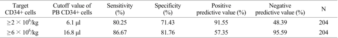
말초혈액 조혈모세포 산출량에 따른 CD34 양성 세포수의 기준치.
성분 채집술 시행 전 시행한 말초혈액의 CD34 양성 세포수가 채집물의 CD34 양성 세포 2.0 × 10
6/kg, 6.0 × 10
6/kg을 얻기 위해 도달해야 하는 기준치를 분 석하였다 . ROC 곡선을 이용하여 분석한 결과 CD34 양 성 세포 2.0 × 10
6/kg 이상 얻기 위한 기준치는 6.1/μL 이며, 6.0 × 10
6/kg 이상 얻기 위해서는 16.8/μL이었으며 각각의 경우 양성예측도는 91.55%, 57.35%, 음성예측도
는 48.39%, 95.59%, 민감도는 80.25%, 86.67%, 특이도는 71.43%, 81.76%였다 (Table 4, Fig. 2).
말초혈액 조혈모세포 산출량에 따른 혈소판 수의 기준치.
성분 채집술 시행 전 말초혈액 내의 혈소판수로 채집물 의 CD34 양성 세포수 6.0 × 10
6/kg을 채집하기 위해 도달 해야 하는 기준치를 알아보기 위해 ROC 곡선을 이용하
Table 3. The efficacy of CD34+ cells harvestLinear regression for CD34+ cells harvest (R2=0.812) Estimated
coefficient (β) Standard
error P-value
PB PLT 0.002 0.001 0.020
PB CD34+ cell 0.096 0.007 0.001 P<0.05 Statistical significance test done by Linear regression.
Abbreviation: PB, peripheral blood. platelet count.
Area under ROC curve=0.657, P=0.001 sensitivity 62.30%; specificity 60.71%
Fig. 2. ROC analysis of pre-mobilization PB CD34+ cells as a predictor of minimum (6 × 10⁶/kg) CD34+ cells count in apheresis product. The best PB CD34+ cells cut-off was 16.80/μL. Positive predictive value 57.35%, negative predictive value 95.59%. ROC analysis of pre-mobilization platelet count as a predictor of mini- mum (6 × 10⁶/kg) CD34+ cells count in apheresis product. The best platelet count cut-off was 82,500/μL. Positive predictive value 33.04%, negative predictive value 83.80%.
Fig. 1. CD34+ cells and MNCs in apheresis products, (A) CD34+ cells, (B) mononuclear cells (MNC).
A
B
PB CD34+ cells.
Area under ROC curve=0.906, P=0.001 sensitivity 86.67%; specificity 81.76%
였다 . 분석한 결과, 6 × 10
6/kg 이상의 CD34 양성 세포 채 집을 예측하기 위한 최소한의 말초혈액 내 혈소판수는 82,500/μL이었다. 이때 영역은 0.657, 민감도는 62.3%, 특 이도는 60.71%였다 (Table 5, Fig. 2).
고 찰
악성 혈액질환에서 자가 말초혈액 조혈모세포 이식은 공고요법의 일환으로 혹은 재발한 경우 치료성적을 향상 시키기 위한 목적으로 동종 조혈모세포 이식과 함께 보 편적으로 이용되고 있다 (Ozkurt et al., 2010). 10년 전까지 만 해도 조혈모세포 이식의 표준은 골수이식이었다. 그러 나 공여자를 구하는데 어려움이 있고 공여자가 있더라도 전신마취 , 채취부위의 통증문제 등으로 새로운 시술법에 대한 연구가 지속되어 왔다. 실제로 자가 말초혈액 조혈 모세포 이식, 동종 말초혈액 조혈모세포 이식, 비혈연이 식, 특히 제대혈이식 등이 그동안 꾸준하게 증가되고 있 다 (Hwang et al., 2007).
일반적으로 말초혈액 조혈모세포 채집은 항암제 투여 후 골수억제가 회복되는 시점에서 G-CSF로 조혈모세포 를 말초혈액으로 가동화시키고 가동화가 최고에 도달하 였다고 예상되는 시점에서 반복적인 백혈구 성분 채집술 을 시행하여 조혈모세포를 채집한다 (Suh et al., 2004). 가 동화 정도를 예측하는 지표로 말초혈액 내 CD34 양성 세포 수치가 흔히 사용되고 있지만 말초혈액 내 CD34 양성 세포수 검사는 비용이 많이 들어 채집기간 동안 매 일 측정하기가 어려우며 경우에 따라서 결과를 얻는데 시간이 많이 걸리는 단점이 있다 (Brecher et al., 1996).
조혈모세포 이식 과정에서 주입된 조혈모세포가 골수 에서 성공적으로 생착되기 위해서는 CD34 양성 세포수 는 환자 몸무게당 1.0~2.0 × 10
6개를 최소 요구량으로 적 용하며, 5.0~6.0 × 10
6개 이상이면 최상인 것으로 알려져 있다 (Sims et al., 1997). 이미 오래전부터 말초혈액 조혈 모세포 채집에 관련된 연구들에서 채집 결과에 영향을 주는 요인으로 환자의 성별이나 나이, 진단, 항암화학요 법의 종류, 방사선 치료력 유무 등이 의미가 있었다고 보 고된 바가 있다. 하지만 대부분의 연구가 소규모의 이질 적인 환자군을 대상으로 하였기 때문에 각 연구 간에 상 반된 결과를 보이는 인자들도 있다 (Demirer et al., 1996).
Lee 등은 G-CSF 가동화에 따른 백혈구수, 단핵구수 및 CD34 양성 절대세포수가 채집된 CD34 양성 세포수를 예측할 수 있는 강력한 예측인자로 가장 상관관계가 있 다고 보고하였으며 이후 다른 연구들에서도 말초혈액의 CD34 세포수가 채집 결과를 예측하는 효과적인 지표로 알려져 왔다 (Lee et al., 2000). 한편 최근 Ozkurt 등은 말 초혈액의 CD34 양성 세포수 외에 말초혈액 내의 혈소판 수치로 조혈모세포의 가동화를 효과적으로 예측할 수 있 다고 보고한 바가 있는데, 본 연구에서도 이와 유사한 결 과를 얻을 수 있었다 (Ozkurt et al., 2010).
저자들은 나이, 성별, 질환명, 가동화요법, 말초혈액의 백혈구수 , 단핵구 비율 및 CD34 양성 세포수와 백혈구 분반술 시행 후 채집된 총유핵세포수, 단핵구수 및 CD34 양성 세포수 등을 비교 분석한 결과 채집 당일 말초혈액 의 혈소판수와 CD34 양성 세포수가 채집 결과를 예측하 는 데 가장 의미 있는 예측인자임을 알 수 있었다. 조혈 모세포 채집술 시행 당일 혈소판수와 CD34 양성 세포수
Table 4. The recommended cutoff value of PB CD34+ cells count for CD34+ cells of collection productTarget
CD34+ cells Cutoff value of
PB CD34+ cells Sensitivity
(%) Specificity
(%) Positive
predictive value (%) Negative
predictive value (%) N
≥ 2 × 10⁶/kg 6.1 μl 80.25 71.43 91.55 48.39 204
≥ 6 × 10⁶/kg 16.8 μl 86.67 81.76 57.35 95.59 204
Data were expressed as ROC analysis.
Abbreviation: PB, peripheral blood.
Table 5. The recommended cutoff value of PB Platelet count for CD34+ cells of collection product Target
CD34+ cells Cut off value of
PB platelet Sensitivity
(%) Specificity
(%) Positive
predictive value (%) Negative
predictive value (%) N
≥ 6 × 10⁶/kg 82,500 62.30 60.71 33.04 83.80 257
Data were expressed as ROC analysis.
Abbreviation: PB, peripheral blood.
와의 상관 분석에서 혈소판수가 많을수록 채집된 CD34 양성 세포수가 많았으며 ROC curve에서 CD34 양성 세 포 2.0 × 10
6/kg 이상을 얻기 위한 말초혈액 내 CD34 양 성 세포 수치의 기준은 6.1/μL, 6.0 × 10
6/kg 이상 얻기 위한 기준치는 16.8/μL이었으며 6 × 10
6/kg 이상의 CD34 양성 세포 채집을 예측하기 위한 혈소판수의 기준치는 말초혈액의 혈소판수가 82,500 /μL였다 (P=0.000).
혈소판은 평균 수명이 10일 정도로 골수에서 끊임없이 생성되어 골수의 조혈기능을 반영하는 지표가 될 수 있 는데, 자가 조혈모세포 이식 후 혈소판수의 회복 정도, 특히 망상 혈소판 수치가 이식 후 골수의 조혈기능 회복 을 반영한다는 연구들이 이를 뒷받침해 준다 (Romp et al., 1994; Figuerres et al., 2001). 말초혈액 내의 백혈구나 적혈구 수치 보다 혈소판 수치가 골수기능을 더 잘 반영 할 수 있는 이유는 평균 수명이 짧고, 외부의 다른 요인 에 영향을 받을 가능성이 적기 때문으로 사료되며 이에 대해 더 연구가 필요하다.
이상의 결과에서 채집물의 CD34 양성 세포수를 예측 하기 위하여 말초혈액의 혈소판수의 기준치와 CD34 양 성 세포의 기준치를 이용하여 조혈모세포 채집 시작 시 기 및 채집 결과를 예측하는데 이용하면 유용할 것으로 사료된다.
REFERENCES
Anguita-Compagnon AT, Dibarrart MT, Palma J, Paredes L, Mosso C, Montalva R, Salas L, Araos D, Delgado I, Majlis A.
Mobilization and collection of peripheral blood stem cells:
guidelines for blood volume to process based on CD34- positive blood cell count in adults and children. Transplant Proc. 2010. 42: 339-344.
Bensinger WI, Martin PJ, Storer B, Clift R, Forman SJ, Negrin R, Kashyap A, Flowers M, Lilleby K, Chauncey TR, Storb R, Blume K, Heimfeld S, Rowley S, Appelbaum FR. Trans- plantation of bone marrow as compared with peripheral- blood cells from HLA-identical relatives in patients with hematologic cancers. N Engl J Med. 2001. 344: 175-181.
Brecher ME, Sims L, Schmitz L, Shea T, Bentley SA. North American multicenter study on flow cytometric enumeration of CD34+hematopoietic cells. J Hematother. 1996. 5: 227 -236.
Couban S, Simpson DR, Barnett MJ, Bredeson C, Hubesch L, Howson-Jan K, Shore TB, Walker IR, Browett P, Messner
HA, Panzarella T, Jeffrey H. A randomized multicenter comparison of bone marrow and peripheral blood in recipients of matched sibling allogeneic transplants for myeloid malignancies. Blood. 2002. 100: 1525-1531.
Demirer T, Buckner CD, Bensinger WI. Optimization of peripheral blood stem cell mobilization. Stem Cells. 1996. 14: 106-116.
Dicke KA, Hood D, Hanks S. Peripheral blood stem cell collection after mobilization with intensive chemotherapy and growth factors. J Hematother. 1994. 3: 141-144.
Figuerres E, Olszewski M, Kletzel M. A flow cytometric technique using thiazole orange to detect platelet engraftment following pediatric stem-cell transplants. Cytotherapy. 2001. 3: 277-283.
Gianni AM, Siena S, bregni M, Tarella C, Stern AC, Pileri A, Bonadonna G. Granulocyte-macrophage colony-stimulating factor to harvest circulating haemopoietic stem cells for autotransplantation. Lancet. 1989. 2: 580-585.
Goldman JM. Autografting cryopreserved buffy coat cells for chronic granulocytic leukaemia in transformation. Exp Hematol. 1979. 7: 389-397.
Hwang TJ. Hematopoietic stem cell transplantation: overview for general pediatrician. Korean J Pediatr. 2007. 50: 613-621.
Kreiger MS, Schiller G, Berenson JR, Stewart K, Noga SJ, Ballester O, Tarantolo S, Stiff P, Kuhn D, Scherzo E, Sing C, Jacobs C, White JM, DiPersio J. Collection of peripheral Blood progenitor cell (PBPC) based on a rising WBC and platelet count significantly increases the number of CD34+
cells. Bone Marrow Transplant. 1999. 36: 160-167.
Lee JL, Kim SB, Lee GW, Ryu MH, Kim EK, Kim S, Kom WK, Lee JS, Park KU, Suh CW. Clinical usefulness of the hematopoietic progenitor cell counts in predicting the optimal timing of peripheral blood stem cell harvest. J Korean Med Sci. 2003. 18: 27-35.
Lee MA, Lee S, Seong CM, Chung WS. The Absolute Number of CD34+ Cells Predicts Optimal Timing of Progenitor Cell Collection and Posttransplant Hematopoietic Recovery.
Korean J Clin Pathol. 2000. 20: 103-109.
Ozkurt ZN, Yegin ZA, Suyani E, Aki SZ, Acar K, Yagci M, Sucak GT. Factors affecting stem cell mobilization for autologous hematopoietic stem cell transplantation. J Clin Apher. 2010.
25: 280-286.
Padmanabhan A, Reich-Slotky R, Jhang JS, Dael S, Crowder T, Colovai AI. Use of the haematopoietic progenitor cell parameter in optimizing timing of peripheral blood stem cell harvest. Vox Sang. 2009. 97: 153-159.
Romp KG, Peters WP, Hoffman M. Reticulated platelet counts in
patients undergoing autologous bone marrow transplantation:
an aid in assessing marrow recovery. Am J Hematol. 1994.
46: 319-324.
Russell NH, Hunter A, Rogers S, Hanley J, Anderson D.
Peripheral blood stem cells as an alternative to marrow for allogeneic transplantation. Lancet. 1993. 341: 1482.
Schwartzberg L, Birch R, Blanco R, Wittlin F, Muscato J, Tauer K, Hazelton B, Wset W. Rapid and sustained hematopoietic
reconstitution by peripheral blood stem cell infusion alone following high-dose chemotherapy. Bone Marrow Transplant.
1993. 11: 369-374.
Suh C, Kim S, Kim SH, Kim EK, Lee JL, Park KU, Park JS, Lee J, Kim MW, Choi HS, Park CJ, Kim SW. Initiation of peripheral blood progenitor cell harvest based on peripheral blood hematopoietic progenitor cell counts enumerated by the Sysmex SE9000. Transfusion. 2004. 44: 1762-1768.