INTRODUCTION
High-dose chemotherapy with autologous stem cell trans- plantation is increasingly used in a wide range of hematolog- ic and solid malignancies and several trials have suggested that this strategy is effective in patients with aggressive lymphoma (1) and multiple myeloma (2). Peripheral blood progenitor cells (PBPC) have almost completely replaced bone marrow as the source of stem cells because of easier accessibility, faster engraftment, and possibly lower tumor contamination (3-5).
As a result of this change in practice, there are currently an increasing number of patients undergoing PBPC mobiliza- tion therapy and subsequent leukapheresis.
Successful autologous peripheral blood stem cell transplan- tation (PBSCT) depends on the infusion of an adequate num- ber of PBPC to achieve rapid and durable hematologic recov- ery. The infusion of≥2.5×10⁶CD34+cells per kg of body weight (/kg) is generally considered to be sufficient to induce stable hematologic engraftment within a reasonable time. The transplantation of ≥5×10⁶CD34+cells/kg results in a faster hematopoietic recovery, particularly of the megakaryocytic lineage (6).
The timing of PBPC collection is important to maximize the number of progenitors harvested with the fewest apheresis procedures. For operational and economical efficiency, it would clearly be beneficial if sufficient PBPC could be obtained from the fewest collections and the optimal timing of these leuka- phereses could be reliably predicted. A further benefit of a higher CD34+cell yield per single apheresis is reduction in the total volume of the progenitor cell component. Besides the decrease in the contaminating RBCs within the graft (7), a reduction in the total volume of the PBPC component may reduce the amount of dimethyl sulfoxide required for cryo- preservation (8). Both factors diminish the risk of adverse side effects related to the graft transfusion.
The most reliable time for harvesting PBPC is yet to be de- termined. Several different predictors for PBPC yield have been used for the timing of apheresis. Circulating progenitor cells (CFU-GM) cannot be used for this purpose because it takes about 2 weeks to form enumerable colonies and is difficult to standardize. Following a chemotherapy nadir, the periph- eral blood (PB) WBC count has been used to indicate when leukapheresis should be commenced. The collections are usu- ally initiated when the WBC count recovers to greater than
27
Clinical Usefulness of the Hematopoietic Progenitor Cell Counts in Predicting the Optimal Timing of Peripheral Blood Stem Cell Harvest
Although enumeration of CD34+cells in the peripheral blood (PB) on the day of apheresis predicts the quantity of those cells collected, the flow cytometric tech- niques used are complex and expensive, and several hours are required to obtain the result in the clinical practice setting. The Sysmex SE-9000 automated haema- tology analyzer provides an estimate of immature cells, called hematopoietic pro- genitor cells (HPC). The aim of this study was to evaluate the clinical usefulness of HPC in predicting the optimal timing of peripheral blood progenitor cells (PBPC) harvest. Studies were performed on 628 aphereses from 160 patients with hema- tologic or solid malignancies. Spearman’s rank statistics was used to assess cor- relation between HPC, WBC, mononuclear cells (MNC), and CD34+cells. A receiv- er operating characteristic (ROC) curve was drawn for cutoff value of HPC, and predictive values of the chosen cutoff value of HPC for different target CD34+cell collections were calculated. The PB HPC had a stronger correlation ( =0.592, p<
0.001) with collected CD34+cells than did PB WBC and PB MNC. The ROC curve showed that the best cutoff value of HPC was 50×106/L for the target CD34+cells
≥1×10⁶/kg with sensitivity of 75%. Positive and negative predictive values of HPC≥50×106/L for CD34+cells≥1×10⁶/kg were 59.7% and 81.1%, respective- ly. In the clinical practice setting, applying variable cutoff values of HPC would be a useful tool to predict the optimal timing of PBPC collection.
Key Words:Hematopoietic Stem Cells; Leukapheresis; Antigens, CD34; Transplantation, Autologous
Jae-Lyun Lee, Sung-Bae Kim, Gyeong-Won Lee, Min-Hee Ryu, Eun-Kyeong Kim, Shin Kim, Woo-Kun Kim, Jung-Shin Lee, Keon Uk Park*,
Cheolwon Suh
Department of Medicine, University of Ulsan College of Medicine, Asan Medical Center, Seoul;
*Department of Internal Medicine, Dongguk University College of Medicine, Gyungju, Korea
Address for correspondence Cheolwon Suh, M.D.
Department of Medicine, University of Ulsan College of Medicine, Asan Medical Center, 388-1 Poongnap-dong, Songpa-gu, Seoul 138-040, Korea
Tel : +82-2-3010-3209, Fax : +82-2-3010-6961 E-mail : [email protected]
Received : 26 August 2002 Accepted : 19 November 2002
1×10⁹/L (9). However, other studies have suggested that wait- ing until the WBC count is greater than 2-10×109/L may be advantageous (10, 11). Furthermore, there is little corre- lation between the PB WBC count and the number of CD34+ cells in a leukapheresis product, resulting in inefficient col- lections and increasing the overall cost of obtaining PBPC for the transplant (12-15). While enumeration of CD34+cells in the PB before apheresis predicts the quantity of those cells collected (16-18), the flow cytometric techniques used are com- plex and expensive. Also, several hours are usually required to obtain the result, which complicates patient management in the clinical practice setting. A simple, faster, and less expensive alternative method should therefore be pursued, especially in the countries with limited medical budget.
Sysmex has developed an automated hematology analyzer (Sysmex SE9000, TOA Medical Electronica Co. Ltd. Japan) that detects a small population of immature white blood cells, hematopoietic progenitor cells (HPC), with the use of special equipment called the immature myeloid information (IMI) channel (19). We have previously reported on the preliminary data, which included 32 patients, of the correlation between the number of PB HPC and collected CD34+cells (20). In the present study, we evaluated potential usefulness of PB HPC count in the clinical practice through the analysis of the data on 160 consecutive patients. We report the kinetics of PB HPC and CD34+cells in apheresis products, the correlation between the PB HPC and collected CD34+cells, sensitivity, specificity, and predictive values of HPC for variable target of CD34+cells collection, and the clinical usefulness of HPC to judge apheresis commencement.
MATERIALS AND METHODS Patients
Between July 1998 and April 2002, 160 consecutive pa- tients with hematologic or solid malignancies who were eligi- ble for autologous PBSCT underwent PBPC harvests at our institution. Prior to beginning PBPC mobilization and har- vest, all patients had given signed informed consent approved by the institutional review board of Asan Medical Center.
PBPC mobilization and harvest
Forty-one patients with multiple myeloma, 31 patients with lymphoma, 20 patients with breast cancer, and 3 patients with other malignancies were mobilized with cyclophosphamide (Cy) 2-4 g/m2with G-CSF (NeutroginTM, Choongwae Ltd, Seoul, Korea). Seven patients were primed with G-CSF alone.
Others were mobilized with various chemotherapy regimens that were chosen both for their efficacy against the patients’
disease and their ability to induce a WBC rebound follow- ing marrow aplasia. For patients mobilized with Cy or chemo-
therapy, the first dose of G-CSF was given subcutaneously at a dose of 5-10 g/kg/day from the day of nadir of the WBC count after Cy or other chemotherapy had ended and contin- ued until the day before the last leukapheresis. The first PBPC harvest was performed on the day when the WBC count ex- ceeded 10.0×10⁹/L or MNC count exceeded 1.0×10⁹/L for patients mobilized with Cy or chemotherapy. It was under- taken on the 4th to 5th day of G-CSF administration if no chemotherapy was used.
PBPC were collected with a continuous-flow blood cell sep- arator (Fenwal CS3000 plus, Baxter healthcare, Deerfield, IL, U.S.A.). Each apheresis procedure was performed for approx- imately 2 to 4 hr, processing 10-14 L of whole blood volume.
The total MNC count and CD34+count of the leukaphere- sis product were monitored daily following each collection.
Leukapheresis was continued until analysis of the component confirmed the collection of≥5.0×10⁶ CD34+cells/kg.
Measurement of HPC
Enumeration of HPC was performed using the Sysmex SE 9000. HPC were detected in the IMI channel. Detection of HPC was made possible by the combination of a special re- agent system and direct current (DC)/radiofrequency (RF) biosensors. The lysis reagent (Stromatolyser-IM) contains deter- gents that are capable of lysing more mature WBC because of their higher membrane lipid content while HPC remains relatively intact. Because various types of immature WBC react differently to the reagent, they also occupy distinct areas on the bivariate matrix of the IMI scattergram (19). Using the purified CD34+cells, the lower DC and RF signal were previously identified for the HPC detection area. HPC were reported both as an absolute number and a percentage of the WBC in the sample.
Measurement of CD34+cells
The quantities of CD34+cells in the leukapheresis compo- nent were determined by flow cytometry using FACScan (Bec- ton Dickinson, Palo Alto, CA, U.S.A.). After lysis of RBCs in an ammonium chloride lysis solution and washing with phosphate-buffered saline with 0.5% human serum albumin, 100 L of cell suspension was stained with phycoerythrin (PE)- conjugated monoclonal anti-CD34 antibody (HPCA-2, Bec- ton Dickinson) and fluorescein isothiocyanate (FITC)-conju- gated anti-CD14 antibody (Leu-M3, Becton Dickinson) at the concentrations recommended by the manufacturer. After incubation for 25 min at 4℃in the dark, additional cells were stained with Leu-M3 and PE-conjugated IgG1 (Becton Dick- inson) as a negative isotope control. Then the cells were washed twice and resuspended and examined with a FACScan. A total of 1.0×10⁵cells were analyzed by the FACScan research soft- ware (Becton Dickinson).
Measurement of nucleated cells in the PB and apheresis products
WBC count in the sample was determined with Sysmex SE9000. Differential counts were done microscopically on a Wright stained smear. The mononuclear cell count was obtain- ed by multiplying the number of leukocytes with the sum of the percentage of lymphocytes and monocytes from the dif- ferential count.
Statistics
Patients’characteristics and apheresis components are de- scribed using summary statistics as median values and ranges.
Correlations between the logarithm of the number of CD34+ cells and HPC cells, WBC, MNC in the PB and apheresis products were assessed using linear regression and Spearman’s rank correlation coefficient ( ). To assess the feasibility of HPC as a diagnostic test for predicting the ability to achieve target CD34+cells of 1×10⁶/kg and 2×10⁶cells/kg, the receiver operating characteristic (ROC) curve was used. We calculat- ed positive and negative predictive values of HPC, utilizing several different cutoff points for HPC. Statistical analysis was performed with SPSS for Windows V.10.0 (SPSS Inc, Chicago, IL, U.S.A.). Significance levels were set at 0.05.
RESULTS Patient characteristics
Patients’characteristics are given in Table 1. There were 80 women and 80 men. The median age was 45 yr ranging from 15 to 68 yr. The patients with non-Hodgkin’s lymphoma comprised 40 percent of the patients (64/160). Other malig- nancies included multiple myeloma in 51 patients, breast can-
cer in 33, acute myelogenous leukemia in 5, medulloblastoma in 4, ovarian cancer in 2, and acute lymphocytic leukemia in 1 patient. Forty-one (29.3%) of 160 patients had been given 7 or more cycles of chemotherapy, and 104 (68%) patients had a history of prior exposure to alkylating agents, which includ- ed melphalan, BCNU, and cyclophosphamide. Twenty-two (14%) of 160 patients had previously undergone axial skele- tal radiotherapy.
Mobilization and collection of PBPC
A total of 628 leukaphereses were done in 160 patients (Table 2). The median number of CD34+cells and MNC in a leukapheresis product was 0.65×10⁶/kg (range, 0.01-25.62) and 1.40×10⁸/kg (range, 0.14-6.74), respectively. The per- centages of leukapheresis products reaching the target yield
Collected CD34+Cells(×106/kg) Peripheral Blood HPC (×106/L)
Days of Apheresis 4.0
3.5 3.0 2.5 2.0 1.5 1.0 0.5 0.0
350 300 250 200 150 100 50 0
1 2 3 4 5 6 7 8
(n=160) (n=160) (n=130) (n=87) (n=47) (n=25) (n=13) (n=5)
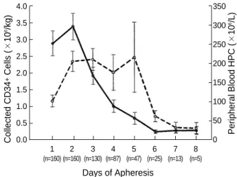
Fig. 1. The kinetics of CD34+cells in an apheresis product and the peripheral blood HPC. Solid line, CD34+cells; dashed line, HPC (mean±SE).
Mobilization therapy
Cyclophosphamide+G-CSF 92
Chemotherapy+G-CSF 61
G-CSF alone 7
Total number of leukapheresis procedures 628
Number of leukapheresis 4 (2-9)*
procedure per person
Number of HPC in PB 46.64×10⁶/L (0-3052.21) Number of WBC in PB 13.10×10⁹/L (0.80-55.50) Number of MNC in PB 1.67×10⁹/L (0.10-10.96) Number of CD34+ 0.65×10⁶/kg (0.01-25.62)
cell in a leukapheresis
Number of MNC in a leukapheresis 1.40×10⁸/kg (0.14-6.74) Number of total CD34+ cell collected 5.43×10⁶/kg (0.03-60.16) Number of total MNC cell collected 6.42×10⁸/kg (0.72-17.73)
*Median (range). G-CSF, granulocyte-colony stimulating factor; HPC, hematopoietic progenitor cell; MNC, mononuclear cell.
Table 2.Mobilization methods, peripheral blood nucleated cell counts, and results of CD34+cell collection
Age (yr), median (range) 45 (15-68)
Male:female 80:80
Disease
Breast cancer 33
NHL 64
MM 51
Others (AML, medulloblastoma, ovary cancer, ALL) 12 Previous axial skeletal RT, No (%) 22 (14%) Cycles of prior chemotherapy, median (range) 5 (2-30) Number of prior chemotherapy regimen
1 83 (53%)
2 56 (35%)
≥3 19 (11%)
Previous exposure to alkylating agents No. (%) 104 (68%) NHL, non-Hodgkin’s lymphoma; MM, multiple myeloma; AML, acute myelogenous leukemia; ALL, acute lymphoblastic leukemia; RT, radio- therapy.
Table 1.Patient characteristics
of CD34+cells≥1×10⁶/kg and 2×10⁶/kg were 42.2% and 26.6%, respectively. The kinetics of HPC in the PB and CD 34+cells in a leukapheresis product over the course of collec- tion for all the patients are depicted in Fig. 1. The kinetics of HPC in PB did not seem to parallel that of CD34+cell in apheresis products. A median of 5.43×10⁶CD34+cells/kg (range, 0.03-60.16) was harvested by a median of 4 (range, 2-9) apheresis procedures. CD34+cells greater than 2.5×10⁶/ kg were harvested from 113 (71%) patients. In 47 patients (29%) who failed to attain 2.5×10⁶CD34+cells/kg with a median of 4 apheresis procedures (range, 2-8), there were no
medical complications that led to discontinuation of apheresis, and harvests were discontinued when they were not likely to yield≥0.2×10⁶/kg CD34+cells per day. Thirty-four (72.3
%) of 47 patients who failed to yield≥2.5×10⁶CD34+cells/
kg underwent≥4 apheresis procedures.
The correlation between cell counts in the PB and the yield of CD34+cells
The median number of PB HPC on the day of leukaphere- sis was 46.6×10⁶/L (range, 0-3052.2, Table 2). The corre-
=0.592, p<0.001 100
10
1
0.1
0.01
0.001
0.1 1 10 100 1,000
Collected CD34+Cells (×106/kg)
A
MNC (×109/L)
=0.316, p<0.001 100
10
1
0.1
0.01
0.1 1 10 100 1,000
Collected CD34+Cells (×106/kg)
C
WBC (×109/L)
=0.168, p<0.001 100
10
1
0.1
0.01
0.1 1 10 100 1,000
Collected CD34+Cells (×106/kg)
B
Collected MNC (×108/kg)
=0.351, p<0.001 100
10
1
0.1
0.01
0.1 1 10 100 1,000
Collected CD34+Cells (×106/kg)
D
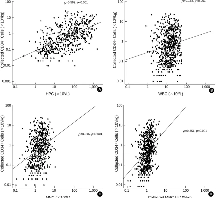
Fig. 2. The relationship between the collected CD34+ cells and peripheral blood nucleated cell counts. Plotted are the yield of CD34+
cells (×106/kg) in an apheresis product (Y-axis) versus: (A) the number of peripheral blood HPC cells (×106/L); (B) the number of peripheral blood WBC counts (×109/L); (C) the number of peripheral blood MNC counts (×109/L); and (D) the number of collected MNC (×108/kg) in an apheresis product (×-axis) in all apheresis procedures.
HPC (×106/L)
sponding median numbers of peripheral blood WBC and MNC on the day of leukapheresis were 13.10×10⁹/L (range, 0.80-55.50) and 1.67×10⁹/L (range, 0.1-10.96), respectively.
The relationship between PB nucleated cell counts on the day of apheresis and the number of CD34+cells in an aphere- sis product is depicted in Fig. 2. The number of HPC in the PB had a stronger correlation ( =0.592, p<0.001) with col- lected CD34+cell counts than did PB WBC ( =0.168, p<
0.001) and PB MNC ( =0.316, p<0.001) counts. There was a weak correlation between collected MNC counts with col- lected CD34+cell counts ( =0.351, p<0.001).
Sensitivity, specificity, and predictive values of PB HPC counts
We calculated the HPC level in PB as a diagnostic tool for predicting a target yield of CD34+cells of at least 1×10⁶/kg and 2×10⁶/kg. To set the cutoff value, we used a ROC curve.
The ROC curve graphically portrays the trade-offs involved between either test’s sensitivity and specificity. We plotted sensitivity as a function of 1-minus-specificity, false positive rate. The ROC curve revealed that the best cutoff point for the most adequate sensitivity and specificity was 50×106/L for target CD34+cells ≥1×10⁶/kg and 55×106/L for tar- get CD34+cells ≥2×10⁶/kg (Fig. 3). The sensitivity and
specificity of that cutoff value are listed in Table 3. The cutoff value of PB HPC and the ROC curves for each target PBPC yield is very close, so the ability of PB HPC to discriminate between PBPC yield ≥1×10⁶CD34+cells/kg and ≥2× 10⁶ CD34+cells/kg is negligible.
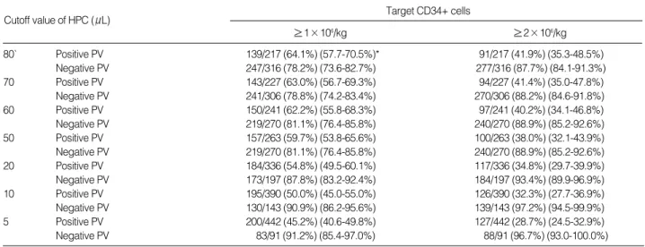
We calculated a positive and a negative predictive value of HPC using several different cutoff values (80, 70, 60, 50, 20, 10, and 5×10⁶/L). These predictive values are shown in Table 4. For patients with HPC counts ≥50×106/L, 59.7% of 263 collections reached the target CD34+cells ≥1×10⁶/kg. The
80` Positive PV 139/217 (64.1%) (57.7-70.5%)* 91/217 (41.9%) (35.3-48.5%)
Negative PV 247/316 (78.2%) (73.6-82.7%) 277/316 (87.7%) (84.1-91.3%)
70 Positive PV 143/227 (63.0%) (56.7-69.3%) 94/227 (41.4%) (35.0-47.8%)
Negative PV 241/306 (78.8%) (74.2-83.4%) 270/306 (88.2%) (84.6-91.8%)
60 Positive PV 150/241 (62.2%) (55.8-68.3%) 97/241 (40.2%) (34.1-46.8%)
Negative PV 219/270 (81.1%) (76.4-85.8%) 240/270 (88.9%) (85.2-92.6%)
50 Positive PV 157/263 (59.7%) (53.8-65.6%) 100/263 (38.0%) (32.1-43.9%)
Negative PV 219/270 (81.1%) (76.4-85.8%) 240/270 (88.9%) (85.2-92.6%)
20 Positive PV 184/336 (54.8%) (49.5-60.1%) 117/336 (34.8%) (29.7-39.9%)
Negative PV 173/197 (87.8%) (83.2-92.4%) 184/197 (93.4%) (89.9-96.9%)
10 Positive PV 195/390 (50.0%) (45.0-55.0%) 126/390 (32.3%) (27.7-36.9%)
Negative PV 130/143 (90.9%) (86.2-95.6%) 139/143 (97.2%) (94.5-99.9%)
5 Positive PV 200/442 (45.2%) (40.6-49.8%) 127/442 (28.7%) (24.5-32.9%)
Negative PV 83/91 (91.2%) (85.4-97.0%) 88/91 (96.7%) (93.0-100.0%)
*95% confidence interval.
Cutoff value of HPC ( L) Target CD34+ cells
≥1×10⁶/kg ≥2×10⁶/kg
Table 4. The positive and negative predictive values (PV) of HPC for different target collection of CD34+cells
≥1×10⁶/kg 50 75.5% (69.9-80.6%)* 68.4% (63.2-73.1%)
≥2×10⁶/kg 55 75.4% (66.9-82.3%) 62.3% (57.8-67.5%)
*95% confidence interval.
Target Cutoff value
Sensitivity Specificity CD34+cells of HPC
Table 3.The recommended cutoff value of HPC for different target collection of CD34+ cells with the best sensitivity and specificity drawn from the ROC curve
1-Specificity
CD34+ cells ≥1×10⁶/kg
CD34+ cells ≥2×10⁶/kg 1.0
0.9 0.8 0.7 0.6 0.5 0.4 0.3 0.2 0.1
0.00.0 0.1 0.2 0.3 0.4 0.5 0.6 0.7 0.8 0.9 1.0
Sensitivity
Fig. 3. ROC curves for the peripheral blood HPC to obtain ≥1×
106CD34+cells/kg (AUC: 0.783, p<0.001) and ≥2×10⁶CD34+ cells/kg (AUC: 0.768, p<0.001). The curves have a similar shape, so the ability to discriminate between PBPC yield ≥1×
106CD34+ cells/kg and ≥2×10⁶CD34+ cells/kg is negligible.
positive predictive values of different cutoff points of PB HPC ranging from 5 to 80×106/L increased from 45.2% to 64.1
%; 16-fold increase in HPC elevated positive predictive value by just 18.9%. For patients with HPC values <50×106/L, 81.1% of 270 patients did not yield target CD34+cell ≥1
×106/kg. When we set 2×10⁶/kg CD34+cells/kg as the target yield, the negative predictive value increased up to 89%, but the positive predictive values decreased below 40%.
Use of PB nucleated cells to decide when to commence apheresis
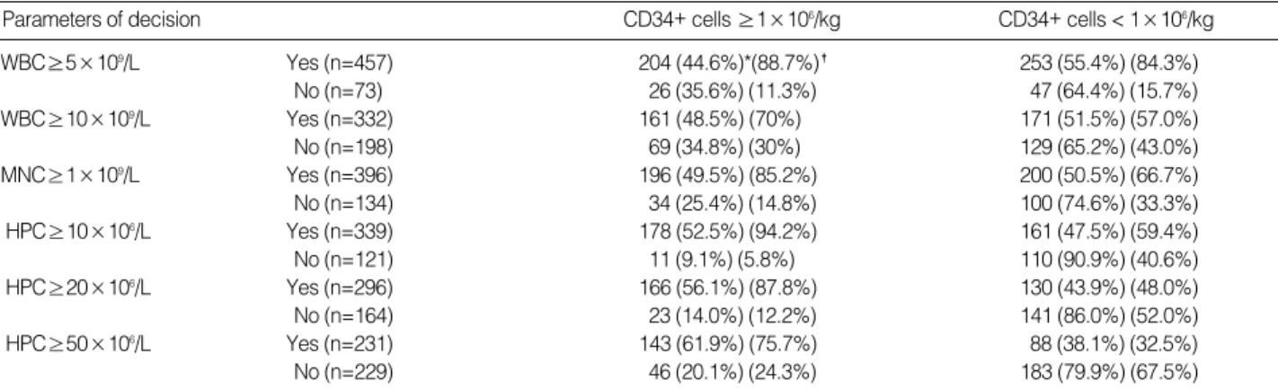
Excluding the patients who were mobilized with G-CSF alone and those whose PB nucleated cell counts were not fully available, 6 options for deciding when to start PBPC collec- tion were considered (Table 5). The first option was to make a decision to start apheresis on the basis of WBC count ≥5
×109/L. Fifty-five percent (253/457) of the patients with WBC count ≥5×10⁹/L had poor CD34+cell yields. Posi- tive predictive value was just about 45%. False negative rate was 35.6%, and only 15.7% of harvests that proved to be inadequate would be avoided. The second option was to start apheresis when WBC counts were above ≥10×109/L. At this threshold, 48.5% of harvests yielded CD34+cells ≥1× 10⁶/kg, and 43% of harvests with poor yield would be avoided. However, 30% of yields greater than 1×10⁶/kg CD34+cells would be missed. The third option was the use of PB MNC counts. When the decision to apherese was taken at the threshold of 1×10⁹/L, 50.5% of the patients who fit- ted the MNC criteria had poor PBPC yields, and 33.3% of harvests that proved to be inadequate would be avoided.
Positive predictive value was 49.5% and false negative rate was 25.4%. The fourth option was to base the decision to commence apheresis on the PB HPC count. At a cutoff value of 10×106/L, positive predictive value was 52.5% with false negative rate of 9.1%, which was the lowest among other options.
At a cutoff value of 20×106/L, 43.9% of the patients who met the criteria had poor PBPC yields, and 52.0% of aphere- ses that proved to be inadequate would be avoided. The last option was to commence apheresis on the day of PB HPC count ≥50×106/L. Although it had the highest positive predictive value, 24.3% of yields greater than 1×10⁶/kg CD34+cells would be missed if aphereses were not performed because of PB HPC level <50×106/L. This option had the lowest false positive rate, and 67.5% of harvests with poor yield would be avoided.
DISCUSSION
The decision when to start leukapheresis is a critical issue for the efficient and cost-effective collection of adequate PBPC for transplantation. Many transplantation centers have used the PB CD34+cell counts to initiate harvesting and predict yields (12, 15, 21, 22). This could be the most precise predic- tor of CD34+cell yields, but the correlation coefficients bet- ween PB CD34+cells and CD34+cells in a leukapheresis product were variable, ranging from 0.57 to 0.95 (12, 23- 25). These relatively inconsistent correlations can be caused by technical difficulties in the harvesting procedure or inac- curacies in the blood CD34+measurements, especially when the PB CD34+percentage is low. In addition, the techniques to enumerate CD34+cells are complex, expensive, require technical skills, and reporting the results may take several hours, often resulting in a 1-day delay before apheresis can be started in the clinical practice setting.
We evaluated the clinical usefulness of the HPC count, enu- merated by Sysmex SE-9000 automated hematology analyz- er, for optimizing stem cell yields. This is rapid and incurs no cost beyond that of a blood count. The detection of HPC is possible through the IMI channel that uses DC and RF to count cells after incubation with a specific lysate designed to lyse red cells and mature white blood cells, leaving only imma-
WBC≥5×10⁹/L Yes (n=457) 204 (44.6%)*(88.7%)� 253 (55.4%) (84.3%)
No (n=73) 26 (35.6%) (11.3%) 47 (64.4%) (15.7%)
WBC≥10×109/L Yes (n=332) 161 (48.5%) (70%) 171 (51.5%) (57.0%)
No (n=198) 69 (34.8%) (30%) 129 (65.2%) (43.0%)
MNC≥1×10⁹/L Yes (n=396) 196 (49.5%) (85.2%) 200 (50.5%) (66.7%)
No (n=134) 34 (25.4%) (14.8%) 100 (74.6%) (33.3%)
HPC≥10×106/L Yes (n=339) 178 (52.5%) (94.2%) 161 (47.5%) (59.4%)
No (n=121) 11 (9.1%) (5.8%) 110 (90.9%) (40.6%)
HPC≥20×106/L Yes (n=296) 166 (56.1%) (87.8%) 130 (43.9%) (48.0%)
No (n=164) 23 (14.0%) (12.2%) 141 (86.0%) (52.0%)
HPC≥50×106/L Yes (n=231) 143 (61.9%) (75.7%) 88 (38.1%) (32.5%)
No (n=229) 46 (20.1%) (24.3%) 183 (79.9%) (67.5%)
*Percent within the row;�Percent within the column.
Parameters of decision CD34+ cells ≥1×10⁶/kg CD34+ cells < 1×10⁶/kg
Table 5. Clinical implications for peripheral blood progenitor cells collection where the decision to commence apheresis is modi- fied by peripheral blood WBC, MNC, or HPC
ture forms to be counted (19, 26, 27). Previous studies report- ed a strong correlation between PB HPC and PB CD34+cell counts (25, 28), and we have reported a strong correlation between HPC enumerated by Sysmex SE-9000 and CD34+ cells in the leukapheresis products from data on 32 patients (20). This strong correlation led us to investigate the clinical usefulness of HPC as a screening test to decide when to com- mence PBPC collection.
In the present study, we confirmed that there are statisti- cally significant but weak correlations between PB WBC or MNC and collected CD34+cell count. The correlation bet- ween PB HPC and CD34+cells in an apheresis product was higher than that between WBC or MNC and CD34+cells.
However, the kinetics of PB HPC and CD34+cells collected did not have a similar configuration. To translate these find- ings into the clinical practice setting, we used a ROC curve to determine the best cutoff value of PB HPC for predicting a target yield of CD34+cells (≥1 or 2×10⁶/kg). The ROC curve revealed that the cutoff point for optimal sensitivity and specificity was 50×106/L for target CD34+cells ≥1×10⁶/kg and 55×106/L for target CD34+cells ≥2×10⁶/kg. The cut- off values for different target values of CD34+cells were very close, just the HPC count difference of 5×10⁶/L. So the abil- ity of PB HPC to discriminate between PBPC yield ≥1× 10⁶CD34+cells/kg and 2×10⁶CD34+cells/kg is negligi- ble. Among those collections with CD34+cell yield ≥1× 10⁶/kg, 75.5% had HPC level ≥50×106/L. In the present study, the positive predictive value of HPC ≥50×106/L was 59.7% and the negative predictive value of that was 81.1%
for target CD34+cell yield ≥1×10⁶/kg. These predictive values rely on the proportion of patients who reach the target PBPC yield: the proportion of harvests reaching the target yield of 1×10⁶/kg was 42.4%. In our series, the majority of patients had been heavily pretreated with alkylating agents, so the proportion of patients who achieved the target CD34+ cells in apheresis was small. In the previous studies, the pos- itive predictive values of different cutoff levels of PB HPC ranging from 5 to 80×106/L changed from 80% to 100%
(25, 29). This is caused by the difference in the study popula- tion. In the study of Yu et al., 16.7% of the patients were healthy stem cell donors, and 80% of the collections reached the target yield of ≥1×10⁶/kg (29). These predictive values of HPC should be applied to a similar population of patients for which those percentages of harvests are expected to reach the target CD34+cells.
We compared the 6 options for deciding when to commence PBPC collection to achieve CD34+cells ≥1×10⁶/kg. The criterion of PB HPC count ≥10×106/L had the lowest false negative rate and that of PB WBC count ≥5×10⁹/L had the highest false positive rates. The PB HPC count ≥50×106/L had better predictive values and the lowest false positive rates compared with those of the PB WBC count ≥5×10⁹/L, ≥ 10×109/L or PB HPC count ≥10×106/L, but the negative predictive value of HPC ≥50×106/L was a little bit inferior
to that of the HPC count ≥10×106/L.
The optimal strategy to prevent unnecessary harvesting while minimizing the risk of missing an adequate harvest would be different according to the patients’premobilization characteristics. For patients who are deemed to have poor PB- PC collection, such as patients who have had prior exposure to alkylating agents and radiation therapy (30, 31), the reduc- tion of the risk of missing an adequate harvest is more impor- tant. So applying a criterion that has the lowest false negative rate, such as HPC count ≥10×106/L, would be appropriate not to miss possible adequate PBPC collection. On the other hand, for patients who seem to be good PBPC yielders, the use of a PB HPC count that had the highest positive predic- tive value and the lowest false positive rate, such as HPC count
≥50×106/L would be better to avoid a harvest with poor PBPC yield.
In our study, we could not check the levels of PB CD34+ cells, which have been reported to be the best predictor of PBPC yields. So, we could not compare the clinical usefulness of PB WBC, MNC, and HPC with that of PB CD34+cell counts to decide when to commence PBPC collection.
In conclusion, the current study suggested that the correla- tion between PB HPC and CD34+cells in an apheresis pro- duct is better than the correlation between PB WBC or MNC and CD34+ cell collection. In the clinical practice setting, incorporating the data on the PB WBC, MNC, and HPC counts and applying variable cutoff values of HPC, depending on patient factors that could affect PBPC mobi- lization, would be a useful and rapid tool to predict the opti- mal timing of PBPC collection.
REFERENCES
1. Shipp MA, Abeloff MD, Antman KH, Carroll G, Hagenbeek A, Loef- fler M, Montserrat E, Radford JA, Salles G, Schmitz N, Symann M, Armitage JO, Philip T, Coiffier B. International Consensus Confer- ence on High-Dose Therapy with Hematopoietic Stem Cell Trans- plantation in Aggressive Non-Hodgkin’s Lymphomas: report of the jury. J Clin Oncol 1999; 17: 423-9.
2. Attal M, Harousseau JL, Stoppa AM, Sotto JJ, Fuzibet JG, Rossi JF, Casassus P, Maisonneuve H, Facon T, Ifrah N, Payen C, Bataille R.
A prospective, randomized trial of autologous bone marrow transplan- tation and chemotherapy in multiple myeloma. N Engl J Med 1996;
335: 91-7.
3. Gratwohl A, Passweg J, Baldomero H, Hermans J. Blood and mar- row transplantation activity in Europe 1997. European Group for Blood and Marrow Transplantation (EBMT). Bone Marrow Trans- plant 1999; 24: 231-45.
4. Chao NJ, Schriber JR, Grimes K, Long GD, Negrin RS, Raimondi CM, Horning SJ, Brown SL, Miller L, Blume KG. Granulocyte colony-stimulating factor “mobilized”peripheral blood progenitor cells accelerate granulocyte and platelet recovery after high-dose chemotherapy. Blood 1993; 81: 2031-5.
5. Sharp JG, Joshi SS, Armitage JO, Bierman P, Coccia PF, Harrington DS, Kessinger A, Crouse DA, Mann SL, Weisenburger DD. Signifi- cance of detection of occult non-Hodgkin’s lymphoma in histologi- cally uninvolved bone marrow by a culture technique. Blood 1992;
79: 1074-80.
6. Weaver CH, Hazelton B, Birch R, Palmer P, Allen C, Schwartzberg L, West W. An analysis of engraftment kinetics as a function of the CD34 content of peripheral blood progenitor cell collections in 692 patients after the administration of myeloablative chemotherapy.
Blood 1995; 86: 3961-9.
7. Alessandrino P, Bernasconi P, Caldera D, Colombo A, Bonfichi M, Malcovati L, Klersy C, Martinelli G, Maiocchi M, Pagnucco G, Varet- toni M, Perotti C, Bernasconi C. Adverse events occurring during bone marrow or peripheral blood progenitor cell infusion: analysis of 126 cases. Bone Marrow Transplant 1999; 23: 533-7.
8. Stroncek DF, Fautsch SK, Lasky LC, Hurd DD, Ramsay NK, McCul- lough J. Adverse reactions in patients transfused with cryopreserved marrow. Transfusion 1991; 31: 521-6.
9. Elias AD, Ayash L, Anderson KC, Hunt M, Wheeler C, Schwartz G, Tepler I, Mazanet R, Lynch C, Pap S. Mobilization of peripheral blood progenitor cells by chemotherapy and granulocyte-macrophage colony-stimulating factor for hematologic support after high-dose intensification for breast cancer. Blood 1992; 79: 3036-44.
10. Ho AD, Gluck S, Germond C, Sinoff C, Dietz G, Maruyama M, Corringham RE. Optimal timing for collections of blood progenitor cells following induction chemotherapy and granulocyte-macrophage colony-stimulating factor for autologous transplantation in advanced breast cancer. Leukemia 1993; 7: 1738-46.
11. Dreger P, Marquardt P, Haferlach T, Jacobs S, Mulverstedt T, Eck- stein V, Suttorp M, Loffler H, Muller-Ruchholtz W, Schmitz N. Effec- tive mobilisation of peripheral blood progenitor cells with‘Dexa- BEAM’and G-CSF: timing of harvesting and composition of the leuka- pheresis product. Br J Cancer 1993; 68: 950-7.
12. Elliott C, Samson DM, Armitage S, Lyttelton MP, McGuigan D, Hargreaves R, Giles C, Abrahamson G, Abboudi Z, Brennan M, Kanfer EJ. When to harvest peripheral-blood stem cells after mobi- lization therapy: prediction of CD34-positive cell yield by preced- ing day CD34-positive concentration in peripheral blood. J Clin Oncol 1996; 14: 970-3.
13. Schots R, Van Riet I, Damiaens S, Flament J, Lacor P, Staelens Y, Steenssens L, van Camp B, De Waele M. The absolute number of circulating CD34+cells predicts the number of hematopoietic stem cells that can be collected by apheresis. Bone Marrow Transplant 1996; 17: 509-15.
14. Jones HM, Jones SA, Watts MJ, Khwaja A, Mills W, Fielding A, Goldstone AH, Linch DC. Development of a simplified single-aphere- sis approach for peripheral-blood progenitor-cell transplantation in previously treated patients with lymphoma. J Clin Oncol 1994; 12:
1693-702.
15. Zimmerman TM, Lee WJ, Bender JG, Mick R, Williams SF. Quan- titative CD34 analysis may be used to guide peripheral blood stem cell harvests. Bone Marrow Transplant 1995; 15: 439-44.
16. Siena S, Bregni M, Brando B, Belli N, Ravagnani F, Gandola L, Stern AC, Lansdorp PM, Bonadonna G, Gianni AM. Flow cytometry for
clinical estimation of circulating hematopoietic progenitors for autol- ogous transplantation in cancer patients. Blood 1991; 77: 400-9.
17. Bender JG, Williams SF, Myers S, Nottleman D, Lee WJ, Unverzagt KL, Walker D, To LB, Van Epps DE. Characterization of chemother- apy mobilized peripheral blood progenitor cells for use in autolo- gous stem cell transplantation. Bone Marrow Transplant 1992; 10:
281-5.
18. Yu J, Leisenring W, Bensinger WI, Holmberg LA, Rowley SD. The predictive value of white blood cell or CD34+cell count in the periph- eral blood for timing apheresis and maximizing yield. Transfusion 1999; 39: 442-50.
19. Takekawa K, Yamane T, Suzuki K, Hino M, Tatsumi N. Identifica- tion of hematopoietic stem cells by the SE-9000 automated hematol- ogy analyzer in peripheral blood stem cell harvest samples. Acta Haematol 1997; 98: 54-5.
20. Park KU, Kim SH, Suh C, Kim S, Lee SJ, Park JS, Cho HJ, Kim KW, Lee K, Kim HJ, Park J, Min YJ, Kim JG, Kim T, Lee JH, Kim SB, Kim SW, Lee KH, Lee JS, Kim WK, Park CJ, Chi HS. Correlation of hematopoietic progenitor cell count determined by the SE-auto- mated hematology analyzer with CD34(+) cell count by flow cytom- etry in leukapheresis products. Am J Hematol 2001; 67: 42-7.
21. Armitage S, Hargreaves R, Samson D, Brennan M, Kanfer E, Navar- rete C. CD34 counts to predict the adequate collection of peripheral blood progenitor cells. Bone Marrow Transplant 1997; 20: 587-91.
22. Schwella N, Siegert W, Beyer J, Rick O, Zingsem J, Eckstein R, Serke S, Huhn D. Autografting with blood progenitor cells: predictive value of preapheresis blood cell counts on progenitor cell harvest and correlation of the reinfused cell dose with hematopoietic reconstitu- tion. Ann Hematol 1995; 71: 227-34.
23. Areman EM, Rohdes PL, Meehan KR, Sacher RA. Cost of autologous peripheral blood stem cell collection can be reduced by PB CD34 screening before apheresis. Blood 1997; 90 (Suppl 2): 322b (abstract 4198).
24. Luider J, Brown C, Selinger S, Quinlan D, Karlsson L, Ruether D, Stewart D, Klassen J, Russell JA. Factors influencing yields of pro- genitor cells for allogeneic transplantation: optimization of G-CSF dose, day of collection, and duration of leukapheresis. J Hematother 1997; 6: 575-80.
25. Pollard Y, Watts MJ, Grant D, Chavda N, Linch DC, Machin SJ. Use of the haemopoietic progenitor cell count of the Sysmex SE-9500 to refine apheresis timing of peripheral blood stem cells. Br J Haema- tol 1999; 106: 538-44.
26. Gowans ID, Hepburn MD, Clark DM, Patterson G, Rawlinson PS, Bowen DT. The role of the Sysmex SE9000 immature myeloid index and Sysmex R2000 reticulocyte parameters in optimizing the timing of peripheral blood stem cell harvesting in patients with lymphoma and myeloma. Clin Lab Haematol 1999; 21: 331-6.
27. Yamane T, Takekawa K, Tatsumi N. Possibility of identification of hematopoietic stem cells using a conventional blood cell counter. Eur J Haematol 1995; 55: 207-8.
28. Peng L, Yang J, Yang H, Peng Z, Xu C, Liu T. Determination of peripheral blood stem cells by the Sysmex SE-9500. Clin Lab Haema- tol 2001; 23: 231-6.
29. Yu J, Leisenring W, Fritschle W, Heimfeld S, Shulman H, Bensinger
WI, Holmberg LA, Rowley SD. Enumeration of HPC in mobilized peripheral blood with the Sysmex SE9500 predicts final CD34+cell yield in the apheresis collection. Bone Marrow Transplant 2000;
25: 1157-64.
30. Dreger P, Kloss M, Petersen B, Haferlach T, Loffler H, Loeffler M, Schmitz N. Autologous progenitor cell transplantation: prior expo- sure to stem cell-toxic drugs determines yield and engraftment of
peripheral blood progenitor cell but not of bone marrow grafts. Blood 1995; 86: 3970-8.
31. Goldschmidt H, Hegenbart U, Wallmeier M, Hohaus S, Haas R. Fac- tors influencing collection of peripheral blood progenitor cells fol- lowing high-dose cyclophosphamide and granulocyte colony-stimu- lating factor in patients with multiple myeloma. Br J Haematol 1997;
98: 736-44.