• 검색 결과가 없습니다.

Right Aortic Arch with a Retroesophageal Left Subclavian Artery and an Anomalous Origin of the Pulmonary Artery from the Aorta

N/A
N/A
Protected

Academic year: 2021

Share "Right Aortic Arch with a Retroesophageal Left Subclavian Artery and an Anomalous Origin of the Pulmonary Artery from the Aorta"

Copied!
3
0
0

로드 중.... (전체 텍스트 보기)

전체 글

(1)

ISSN: 2233-601X (Print) ISSN: 2093-6516 (Online)

− 44 −

Received: February 11, 2016, Revised: March 15, 2016, Accepted: March 23, 2016, Published online: February 5, 2017

Corresponding author: Tae-Gook Jun, Department of Thoracic and Cardiovascular Surgery, Samsung Medical Center, Sungkyunkwan University School of Medicine, 81 Irwon-ro, Gangnam-gu, Seoul 06351, Korea

(Tel) 82-2-3410-1677 (Fax) 82-2-3410-0089 (E-mail) [email protected]

© The Korean Society for Thoracic and Cardiovascular Surgery. 2017. All right reserved.

This is an open access article distributed under the terms of the Creative Commons Attribution Non-Commercial License (http://creativecommons.org/

licenses/by-nc/4.0) which permits unrestricted non-commercial use, distribution, and reproduction in any medium, provided the original work is properly cited.

Right Aortic Arch with a Retroesophageal Left Subclavian Artery and an Anomalous Origin

of the Pulmonary Artery from the Aorta

Chang-Seok Jeon, M.D., Man-shik Shim, M.D., Ji-Hyuk Yang, M.D., Tae-Gook Jun, M.D.

Department of Thoracic and Cardiovascular Surgery, Samsung Medical Center, Sungkyunkwan University School of Medicine

We report the case of a newborn with a rare anatomic variation: a right aortic arch with a retroesophageal left subclavian artery and an anomalous origin of the pulmonary artery from the aorta. This variation was diagnosed using echocardiography and computed tomography, and we treated the condition surgically.

Key words: 1. Congenital heart disease 2. Aortic arch

3. DiGeorge syndrome

Case report

A 6-day-old boy was followed up for a right aortic arch on fetal echocardiography observed at 35 weeks of gestation. His mother underwent a cesarean sec- tion at 36 weeks of gestation due to labor pain and preterm rupture of the membranes. The infant’s birth weight was 2,270 g, and the Apgar scores at 1 and 5 m inutes after birth were 9 and 10, respectively. The infant was referred to Samsung Medical Center due to a right aortic arch with atrial septal defect (ASD) and patent ductus arteriosus (PDA) on postnatal echocardiography. On echocardiography performed at our hospital, severe pulmonary hypertension was ob- served and a double aortic arch, in which the left aortic arch was interrupted, was suspected. The right pulm onary artery was found to em erge from the as- cending aorta. In addition, a large left PDA with a retroesophageal left subclavian artery (LSCA) and right-to-left shunt flow was observed. An intracardiac anomaly (moderate secundum ASD) was also present.

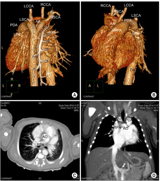
A 3-dimensional computed tomography (CT) heart scan showed an anomalous origin for the right pul- monary artery from the right aorta and retro- esophageal LSCA from the left PDA; the right aortic arch and left PDA formed vascular rings, but tracheal stenosis was not clearly seen (Fig. 1, Fig. 2). The in- fant underwent surgical treatment at 7 days after birth due to aggravated heart failure symptoms. Median sternotomy was performed. The thymus was not visible. Cardiopulmonary bypass (CPB) was initiated with the standard cannulation technique in the as- cending aorta and a bicaval venous cannula, at 34

o

C.

The PDA was ligated and resected at the attachment site of the main pulmonary artery (MPA) and aorta through Kommerell diverticulum resection, followed by LSCA division. The right pulmonary artery, which originated from the posterior side of the ascending aorta, was divided and reimplanted into the MPA, and the ASD was closed with autologous pericardium.

After rewarming, the patient was weaned from CPB.

The CPB time was 144 minutes and the aortic cross-

Korean J Thorac Cardiovasc Surg 2017;50:44-46 □ CASE REPORT □

https://doi.org/10.5090/kjtcs.2017.50.1.44

(2)

A Rare Variant of Right Aortic Arch

− 45 −

Fig. 1. (A–D) Computed tomography angiography showing right AA with retroesophageal LSCA and anom- alous origin of pulmonary artery from aorta. AA, aortic arch; AO, aorta; RPA, right pulmonary artery;

RCCA, right common carotid artery;

LCCA, left common carotid artery;

RSCA, right subclavian artery; MPA, main pulmonary artery; LPA, left pulmonary artery; PDA, patent duc- tus arteriosus; LSCA, left subcla- vian artery; DA, descending aorta;

P, posterior; A, anterior; L, left; H, head; R, right; F, front.

clamping time was 67 minutes. After surgery, nitric oxide ventilation was necessary due to pulmonary hypertension. The patient was extubated on the 11th postoperative day. On postoperative echocardiog- raphy, the right aortic arch flow was good and the right pulmonary artery was found to be normally connected to the MPA. No residual ASD was found.

Chromosomal analysis revealed 22q11 partial dele- tion, also known as DiGeorge syndrome. The infant was discharged on the 28th postoperative day.

Discussion

We diagnosed the neonate with right aortic arch with retroesophageal LSCA and anomalous origin of the pulm onary artery from the aorta using echo-

cardiography and CT, and we treated the condition

surgically. This variant has been reported by Hamzeh

et al. [1], but our case showed some differences in

terms of the presence of an intracardiac anomaly

(ASD) and DiGeorge syndrome. Congenital anomalies

of the aortic arch range from asymptomatic normal

variations in the arch vessel branch pattern to symp-

tomatic vascular rings, stenosis, and arch interrup-

tions. The frequency of aortic arch anomalies ranges

from 0.5% to 3.0%. Right aortic arch occurs in ap-

proximately 0.1% of adults and is strongly associated

with the presence of chromosome 22q11 deletion, al-

so known as DiGeorge syndrome [2,3]. Right aortic

arch with retroesophageal LSCA and left descending

aorta is a rare configuration of the right aortic arch

and may be a variant of the double aortic arch with

(3)

Chang-Seok Jeon, et al

− 46 −

Fig. 2. Diagram. RPA, right pulmonary artery; RCCA, right com- mon carotid artery; LCCA, left common carotid artery; RSCA, Right subclavian artery; MPA, main pulmonary artery; LPA, Left pulmonary artery; PDA, patent ductus arteriosus; LSCA, Left sub- clavian artery.

focal left arch atresia [2]. In this case, there was a left PDA, forming a vascular ring, but the vascular ring was relatively loose due to the anomalous origin of the right pulmonary artery. This rare variant ac- companied by ASD, however, increased left-to-right shunting, resulting in aggravation of pulmonary hy- pertension and heart failure, which required early surgical treatment. Translocation of the retroeso- phageal LSCA to the left carotid artery with Kommerell diverticulum resection, PDA division, right pulmonary artery reimplantation to the MPA, and ASD closure is the preferred surgical approach and can be per- formed in most cases. Occasionally, however, trans- location of the retroesophageal LSCA to the left car- otid artery m ay be a less attractive option because of technical factors related to low-birthweight neonates.

In selected cases of right aortic arch with retroeso- phageal LSCA, especially in neonatal patients, division of the LSCA without reimplantation may be an alter- native [4]. Right aortic arch anomalies are rare, but show diverse variations. Therefore, accurate diagnosis may not be possible by echocardiography alone. A CT scan can support further evaluation and cardiac cath- eterization, and magnetic resonance imaging may al- so be useful [5,6].

Conflict of interest

No potential conflict of interest relevant to this ar- ticle was reported.

References

1. Hamzeh G, Crespo A, Rey E, et al. Right aortic arch with aberrant left subclavian artery and anomalous origin of right pulmonary artery from ascending aorta. World J Pediatr Congenit Heart Surg 2011;2:324-6.

2. Kanne JP, Godwin JD. Right aortic arch and its variants. J Cardiovasc Comput Tomogr 2010;4:293-300.

3. Ryan AK, Goodship JA, Wilson DI, et al. Spectrum of clin- ical features associated with interstitial chromosome 22q11 deletions: a European collaborative study. J Med Genet 1997;34:798-804.

4. Rubay JE, Sluysmans T, Alexandrescu V, et al. Surgical re- pair of coarctation of the aorta in infants under one year of age: long-term results in 146 patients comparing sub- clavian flap angioplasty and modified end-to-end anasto- mosis. J Cardiovasc Surg (Torino) 1992;33:216-22.

5. Ito K, Kogure T, Hayashi S, et al. A case of the incomplete double aortic arch diagnosed in adulthood by MR imaging.

Radiat Med 1995;13:263-7.

6. Schlesinger AE, Krishnamurthy R, Sena LM, et al. Incom-

plete double aortic arch with atresia of the distal left

arch: distinctive imaging appearance. AJR Am J Roentgenol

2005;184:1634-9.

수치

Fig. 1. (A–D) Computed tomography  angiography showing right AA with  retroesophageal LSCA and  anom-alous origin of pulmonary artery  from aorta
Fig. 2. Diagram. RPA, right pulmonary artery; RCCA, right com- com-mon carotid artery; LCCA, left comcom-mon carotid artery; RSCA,  Right subclavian artery; MPA, main pulmonary artery; LPA, Left  pulmonary artery; PDA, patent ductus arteriosus; LSCA, Left

참조

관련 문서

The left subclavian artery, branching from the left brachiocephalic trunk was aberrant between the esophagus and vertebral body but was not severe compressed

Aortiogram image shows the common carotid trunk (long black arrow), left subclavian artery (white arrow) and aberrant right subclavian ar- tery (short black arrow). A

Coronal reformatted image shows early dense enhancement of right common carotid and subclavian arteries and ductus liga- ment (arrowheads) between faintly enhanced left common

Anomalous origin of the left Coronary artery from the pulmonary artery is very rare , and being alleviated surgically , recognition ofthe lesion and associated

BA: brachiocephalic artery, LCCA: left common carotid artery, LSCA: left subclavian artery Table 3.. although conflicting evidence exists in this regard. 15-18) On the other hand,

Postoperative computed tomographic angiography revealed a patent bypass conduit between the left common carotid artery and left subclavian artery.. The patient was discharged

(D) The left pulmonary artery was re-implanted into the main pulmonary artery using an autologous pericardial hood patch. LPA, left pulmonary artery; MPA, main pulmonary artery;

Double aortic arch with atretic left arch distal to the origin of left subclavian artery (LSCA) is a rare type of vascular ring, and it can be easily confused with the more