399
Open Access
Combined Open and Endovascular Repair for Aortic Arch Pathology
Woong Chol Kang, MD1, Eak Kyun Shin, MD1, Tae Hoon Ahn, MD1, Kyung Hoon Lee, MD1, Chan Il Moon, MD1, Seung Hwan Han, MD1, Chul-Hyun Park, MD2, Kook-Yang Park, MD2, Jin Mo Kang, MD3 and Jung Ho Kim, MD4
1Departments of Cardiology, 2Cardiovascular Surgery, 3Vascular Surgery and 4Radiology, Gil Medical Center, Gachon University of Medicine and Science, Incheon, Korea
ABSTRACT
Background and Objectives: We describe our experience with combined open and endovascular repair in patients who have aortic arch pathology. Subjects and Methods: This study is a retrospective analysis of 7 patients who underwent com- bined open and endovascular repair for aortic arch pathology. Medical records and radiographic information were reviewed.
Results: A total of 7 consecutive patients (5 men, 71.4%) underwent thoracic stent graft implantation. The mean age was 59.9±16.7 years. The indication for endovascular repair was aneurysmal degeneration in 5 patients, and rupture or impend- ing rupture in 2 patients. In all 7 cases, supra-aortic transposition of the great vessels was performed successfully. Stent graft im- plantation was achieved in all cases. Surgical exposure of the access vessel was necessary in 2 patients. A total of 9 stent grafts were implanted (3 stent grafts in one patient). The Seal thoracic and the Valiant endovascular stent graft were implanted in 6 patients and 1 patient, respectively. There were no post-procedure deaths or neurologic complications. In 2 patients, bleeding and injury of access vessel were noted after the procedure. Postoperative endoleak was noted in 1 patient. One patient died at 10 months after the procedure due to a newly developed ascending aortic dissection. No patients required secondary inter- vention during the follow-up period. The aortic diameter decreased in 4 patients. In 3 patients, including 1 patient with endole- ak, there was no change in aortic diameter. Conclusion: Our experience suggests that combined open and endovascular re- pair for aortic arch pathology is safe and effective, with few complications. (Korean Circ J 2010;40:399-404)
KEY WORDS: Prostheses and implants; Aortic disease; Aortic aneurysm.
Received: March 17, 2010 Accepted: April 1, 2010
Correspondence: Eak Kyun Shin, MD, Department of Cardiology, Gil Medical Center, Gachon University of Medicine and Science, 1198 Gu- wol-dong, Namdong-gu, Incheon 405-760, Korea
Tel: 82-32-460-3054, Fax: 82-32-469-1906 E-mail: [email protected]
cc This is an Open Access article distributed under the terms of the Cre- ative Commons Attribution Non-Commercial License (http://creativecom- mons.org/licenses/by-nc/3.0) which permits unrestricted non-commer- cial use, distribution, and reproduction in any medium, provided the ori- ginal work is properly cited.
Introduction
Aortic arch aneurysm or dissection repair, requiring car- diopulmonary bypass and hypothermic circulatory arrest, re- mains a surgical challenge with a high rate of mortality (7- 17%) and neurologic complication (4-12%).1-3) Endovascular repair for thoracic and abdominal aortic aneurysm is associ- ated with lower perioperative morbidity and mortality rates than conventional open repair, with similar early and midterm follow-up results.4-8) Recently, combined open and endovas-
cular repair has emerged as an effective adjunct in the treat- ment of various pathologies of the aortic arch, especially in the high-risk patient not suitable for conventional open repair owing to cardiovascular or pulmonary comorbidities.9-13) In the current report, we describe our experience with combined open and endovascular repair in patients who had an aneury- sm or dissection involving the aortic arch.
Subjects and Methods
Patient population
From December 2007 to October 2009, 16 patients were treated at our institution with endovascular repair for thorac- ic aortic disease. This study is a retrospective analysis of the 7 patients who underwent combined open and endovascular repair for aortic disease involving the aortic arch with/without proximal descending aorta. Patient selection for combined repair was based on the length of the proximal landing zone and comorbidities. Patient demographics and clinical risk fac- tors are shown in Table 1. Diagnosis was confirmed by enhanced
computed tomography scan angiography (CTA) and angiog- raphy in all cases. Patients in whom the distal extent of disease was confined to the thoracic aorta were included in this study.
Cases of aortic trauma resulting in pseudoaneurysm or dis- section were also included in the patient cohort. Our indica- tions for endovascular repair were the following: 1) maximum aortic diameter ≥55 mm; 2) rapid aortic enlargement (≥10 mm per year); 3) clinical or radiographic evidence of rupture or impending rupture; 4) intractable chest pain, despite maxi- mal medical therapy.
Medical records and radiographic information were review- ed to determine the operative indications, the repair technique, the peri-procedural complications and the outcomes. Tech- nical success was defined as a successful stent graft deploy- ment without death, conversion to open repair or diagnosis of endoleak before discharge. For each patient, the immedi- ate post-procedural CTA was compared with the most recent CTA. The following parameters were recorded: presence of an endoleak and its nature, and overall maximal aortic diam- eter. Adverse clinical events (mortality, respiratory failure, malperfusion, bleeding, vascular injury, renal failure, stroke and paraplegia) occurring during the peri or post-procedur- al period and during follow-up were recorded.
Preoperative evaluation for proximal landing zone For arterial access, preoperative evaluation was done by CTA scans to exclude major occlusive disease of the aortoili- ac axis. These CT scans were also used as a tool to predict the required length of the intended proximal landing zone. As a prerequisite for successful stent graft placement, a proximal landing zone of at least 1.5 cm along the lesser curvature of the aortic arch was claimed. Furthermore, after supra-aortic trans- position, an additional CTA scan was performed to reconfirm
the effective length of the intended landing zone extension.
Supra-aortic transposition
In all 7 cases, supra-aortic transposition of the great vessels was performed several days prior to endovascular stent grafting.
Left subclavian-to-left carotid artery transposition A standard approach through a skin incision parallel to the left clavicle was chosen. The left subclavian artery (LSCA) was divided at its origin at the level of the aortic arch. The ves- sel was guided dorsal to the left jugular vein, and an end-to- side anastomosis between the LSCA and the left common ca- rotid artery (LCCA) was performed.
Double or triple-vessel transposition
Through an upper hemisternotomy approach, all supra-aor- tic vessels were exposed. Prosthetic graft was used to connect the aorta to the brachiocephalic artery (BA), the LCCA and the LSCA because extensive mobilization of the supra-aortic vessels is not sufficient to accomplish tension free vascular transposition.
Stent graft placement
All procedures were performed in an angiography suite, under general or local anesthesia. The right or left common femoral artery (CFA) was used for access in all the cases to place the endovascular stent grafts. In the majority of patients, stent graft was deployed using a percutaneous approach.
Post-procedure, a suture-mediated closure device (PercloseTM, Abbott Laboratories, Illinois, IL, USA) was used for closure of the access site (Preclose technique).14) Surgical exposure of the access vessel was required in 2 patients who had a severely tor- tuous CFA. Drainage of cerebrospinal fluid was performed in patients who required extensive aortic coverage or had had previous aortic surgery. Stent graft deployment was routinely performed under hypotonic conditions (60 mmHg systolic pressure) by overpacing at 180 beats per minute. Stent grafts from the same manufacturer were used when a patient requir- ed multiple stent grafts. To achieve a satisfactory seal, devices were oversized in diameter by 15-20% in relation to the diam- eter of the proximal landing zone. If an endoleak occurred af- ter stent graft implantation, ballooning was carried out to make the stent graft closely adhere to the blood vessel wall. If the endoleak was caused by stent displacement or angulation, an aortic extending stent graft was implanted.
Definition of endoleak
Type I endoleaks were defined as attachment site leaks, type Ia at the proximal attachment site and type Ib at the dis- tal attachment site. Type II endoleaks were defined as branch leaks without attachment site connection. Type III endoleaks were defined as junctional leaks between stent grafts.
Table 1. Demographics and clinical risk factors of patients (n=7) n (%) Sex
Male 5 (71.4)
Female 2 (28.6)
Age (years) 59.9±16.7
Hypertension 4 (57.1)
Diabetes mellitus 2 (28.6)
Coronary artery disease 2 (28.6)
Chronic obstructive pulmonary disease 0
Chronic renal insufficiency 1 (14.3)
Peripheral vascular disease 0
Prior open aortic surgery 1 (14.3)
Mean time from symptom onset to endovascular
repair (days) 178.4±163.3
Mean time from supra-aortic transposition
to endovascular repair (days) 41.6±38.9
Follow-up period
Patients were followed up according to a follow-up proto- col that required a CTA scan and clinical evaluation. Addi- tional investigations were obtained whenever indicated.
Results
A total of 7 consecutive patients (5 men, 71.4%) were im- planted with a thoracic stent graft for the treatment of aortic disease involving the aortic arch. The mean age at interven- tion was 59.9±16.7 years (range, 28-76) and the mean time be- tween the supra-aortic transposition and endovascular re- pair was 41.6±38.9 days (range, 6-119). The mean time between symptom onset and endovascular repair was 178.4±163.3 days (range, 48-430) (Table 1). Indications for endovascular repair were: aneurysmal degeneration in 5 cases (71.4%), rup- ture or impending rupture in 2 cases (28.6%).
Supra-aortic transposition
In all 7 cases, supra-aortic transposition of the great ves- sels was performed prior to endovascular stent grafting. In 3 patients who had an aneurysm extending from the ascending aorta, Y-shaped bypass surgery with a prosthetic graft to con- nect the aorta to the BA, LCCA and LSCA was performed.
In one patient, Y-shaped bypass surgery with a prosthetic graft connecting the aorta to the BA and LCCA was performed
and followed by end-to-side anastomosis between the LCCA and LSCA (Fig. 1). In another patient who had two localized traumatic aortic dissections of the ascending aorta and the just distal LSCA, graft replacement was performed in the as- cending aorta, followed by graft interposition between the aorta and the LCCA, and end-to-side anatomosis between the LCCA and the LSCA. In the remaining 2 cases, who had relatively limited aneurysms in the aortic arch, transposition of the LSCA onto the LCCA was performed (Table 2).
Procedural details of endovascular repair
Stent graft implantation was achieved in all cases. As de- scribed above, surgical exposure of the access vessel was nec- essary in 2 patients. Two different stent graft systems were used. The Seal thoracic endovascular stent graft (S & G bio- tech, Seoul, Korea) was used in 6 patients and the 3 Valiant en- dovascular stent grafts (Medtronic Inc, Santa Rosa, CA, USA) were implanted in 1 patient who had a long aortic aneurysm (Table 3).
Postoperative results
There were no post-procedure deaths or neurologic com- plications. In one patient who underwent the procedure via per- cutaneous approach, continuous bleeding at the access site was noticed even though a suture-mediated closure device was used. Right CFA injury was noted and surgical repair per-
A
D
B
E
C
F
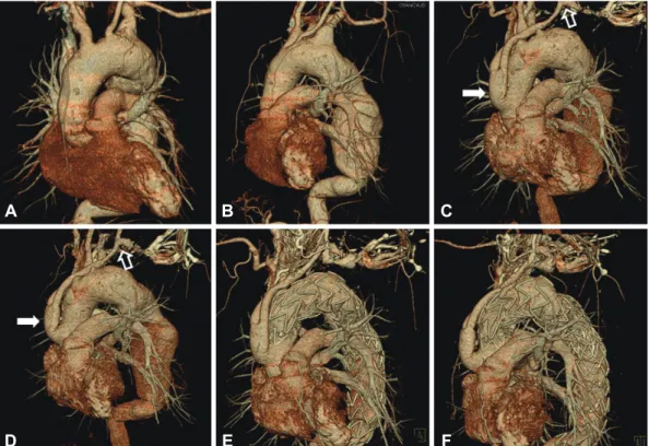
Fig. 1. Reconstructive three-dimensional computed tomography showed extensive aortic aneurysm involving the aortic arch and proximal descending thoracic aorta (A and B). Y-shaped bypass surgery with a prosthetic graft, to connect the aorta to the BA and the LCCA, was per- formed (white arrow) and followed by end-to-side anastomosis of the LCCA and LSCA (open arrow) (C and D). After supra-aortic transposi- tion of the great vessels, 3 Valiant stent grafts were implanted from the ascending aorta to the descending thoracic aorta in a telescopic fash- ion to exclude extensive aortic aneurysm (E and F). BA: brachiocephalic artery, LCCA: left common carotid artery, LSCA: left subclavian artery.
formed. In another patient, a huge hematoma was noted in the groin area after the procedure. CTA showed active bleed- ing from the right deep circumflex iliac artery, which was ma- naged with coil embolization. There were no other postop- erative complications. Postoperative endoleak was noted in 1 patient on the follow up CTA. Therefore, the procedure was considered technically successful in 6 cases (85.7%) (Table 4).
Late survival and secondary intervention
Clinical follow-up was available for all patients and the mean clinical follow-up period was 14.0±4.5 months (range, 4 to 23 months). One patient died at 10 months after the proce- dure due to a newly developed ascending aortic dissection.
The patient had initially undergone stent graft deployment for type B aortic dissection. But type Ia endoleak was noted after the procedure. The follow-up CTA scan showed persis- tent endoleak and slightly progressed aortic dissection. Con- sequently, implantation of an additional stent graft was plan- ned. After the LSCA-to-LCCA bypass surgery, the stent graft was deployed just proximal to the previous stent graft. Post- procedure, the type Ia endoleak disappeared and follow-up CTA showed decreased aortic diameter with complete throm- bosis of the false lumen. At 10 months after the procedure, the patient came to the emergency room with sudden onset chest pain and loss of consciousness. The CTA scan showed newly developed aortic dissection of the ascending aorta and hemo- pericardium and the patient eventually expired. No patients required secondary intervention during the follow-up period.
Endoleak and aortic diameter
Follow up CTA scan showed persistent endoleak without change of aortic diameter in the patient who had showed en- doleak immediately post-procedure. The aortic diameter de- creased in 4 patients. In 3 patients including 1 patient who had endoleak, there was no interval change of aortic diameter.
Discussion
Conventional surgical repair for aortic arch pathology car- ries a high mortality and morbidity, with a particularly signifi- cant incidence of neurologic injury.1-3) Recent advances in st- ent graft technology have enhanced the management of dise- ases of the descending thoracic aorta by avoiding thoracotomy and expanded the group of patients eligible for treatment. Be- cause of the complex anatomy of the aortic arch, however, repair of aortic arch pathology remains a significant endovas- cular challenge as it requires preservation of the supra-aortic great vessels during the procedure. Moreover, insufficient pro- ximal landing zone for stent graft deployment is a major lim- itation of endovascular repair in most cases. Generally, a land- ing zone length of more than 1.5 cm is considered acceptable, Table 2. Category and extent of supra-aortic vessel transposition
N
Success rate of supra-aortic vessel transposition (%) 7 (100)
Type of supra-aortic vessel transposition
Y-shaped bypass surgery with a prosthetic graft to connect the aorta to the BA, the LCCA, the LSCA 3 Y-shaped bypass surgery with a prosthetic graft to connect the aorta to the BA, the LCCA and anastomosis between
the LCCA and the LSCA 1
Graft replacement for ascending aorta followed by graft interposition between aorta and the LCCA, anastomosis between
the LCCA and the LSCA 1
Anastomosis between the LCCA and the LSCA 2
BA: brachiocephalic artery, LCCA: left common carotid artery, LSCA: left subclavian artery Table 3. Endovascular repair (n=7)
Indication for procedure (%)
Increased aneurysm 5 (71.4)
Rupture or impending rupture 2 (28.6)
Approach (%)
Percutaneous 5 (71.4)
Surgical exposure of access vessel 2 (28.6) Type of stent graft (%)
Seal thoracic 6 (85.7)
Valiant 1 (14.3)
Table 4. Results of procedure (n=7) In-hospital outcomes (%)
Primary success 6 (85.7)
Endoleak 1 (14.3)
Bleeding 1 (14.3)
Vascular injury 1 (14.3)
Neurologic deficit 0
Malperfusion 0
Renal failure 0
Paraplegia 0
Respiratory failure 0
Death 0
Follow up outcomes (%)
Reintervention 0
Death 1 (14.3)
although conflicting evidence exists in this regard.15-18) On the other hand, a sufficient proximal landing zone can be achieved by transposition of the supra-aortic vessels. Im- portantly, this procedure, which avoids the need for cardiopul- monary bypass and aortic cross-clamping, may have advan- tages for high-risk patients.19) In addition, second-stage en- dovascular repair may be performed much sooner than a se- cond open procedure for patients requiring a two-stage ap- proach. Bergeron et al.20) reported results of combined open and endovascular repair for 15 aortic arch pathologies. The success rates of transposition of the great vessels and endo- vascular repair were 97% and 92%, respectively. In our study, supra-aortic transposition was successful in all patients and the technical success rate of endovascular repair was 85.7%.
No deaths related to supra-aortic transposition occurred in our study, thereby emphasizing the safety of these procedures.
In 2006, Saleh and Inglese21) successfully treated 15 aortic an- eurysms using combined transposition of the supra-aortic gr- eat vessels and endovascular stent graft deployment. The suc- cess rate of these procedures was 100%. All stent grafts and by- pass vessels were patent without endoleaks, stent displacement, or neurological deficits in the early postoperative period. One patient died 2 months postoperatively because of a pulmonary complication. In our study, except for one case in which endo- leak was noted immediately after the procedure, all stent gr- afts and bypass vessels were patent without stent displacement in the early postoperative period. Each case in which endovas- cular stent grafting was performed after supra-aortic transpo- sition of the great vessels was successful not only in effectively sealing or excluding the aortic arch lesions, but also in preserv- ing the blood supply to the brain. There were no peri- or post- operative neurological complications, indicating that a combi- nation of open supra-aortic transposition of the great vessels fol- lowed by endovascular stent grafting is an effective and safe me- thod for treating aortic arch pathology.
Persistent primary endoleak was noted in one patient. Type I endoleak occurring after stent graft implantation can usu- ally be treated by performing balloon angioplasty of the stent graft to make the stent tightly adhere to the blood vessel wall.22) This patient had diffuse extensive aortic aneurysm from the ascending aorta to the suprarenal abdominal aorta. After supra-aortic transposition (LSCA to LCCA anastomosis), a stent graft (Seal Thoracic 38×125 mm) was deployed. The problem, however, was that the proximal landing zone in- cluded aneurismal change with a diameter of around 36 mm.
The stent graft was therefore not able to adhere to the aortic wall completely by balloon angioplasty. Immediate postop- erative and follow-up CT scans showed persistent endoleak at the proximal site of the stent graft. To achieve sufficient proximal landing zone at the ascending aorta, Y-shaped by- pass surgery with a prosthetic graft to connect the aorta to the supra-aortic vessels might have been helpful in this pa-
tient. Follow-up CT scan revealed no change in aortic diam- eter despite persistent endoleak, so secondary intervention was not indicated.
In one patient who underwent stent graft placement th- rough a percutaneous approach, bleeding was not controlled at the access site after removal of the sheath. Emergent oper- ation revealed the puncture site to be located around the bi- furcation site of the superficial and deep femoral arteries, sh- owing that the suture-mediated closure device had not worked properly. In this technique, care should be taken to puncture the CFA along its anterior aspect at least 1 cm proximal to the origin of the deep femoral artery. This can be confirmed by femoral angiography using an ipsilateral oblique projection.
In summary, the use of combined open and endovascular repair in the treatment of aortic arch pathology appears safe and effective at perioperative, postoperative and early midterm follow-up and offers several advantages over conventional repair, including the potential to offer therapy to patients who are not candidates for open repair and a shorter time between stages for patients requiring two-stage repair.
Conclusions
Our experience suggests that combined open and endovas- cular repair for aortic arch pathology is safe and effective, with few complications. In cases without sufficient landing zone for stent graft at the proximal end, complete or partial supra- aortic transposition of the great vessels can be performed to ensure both cerebral blood supply and sufficient landing zone for the stent graft. Given the small population and rela- tively short follow-up period of our study, a much larger clini- cal study with a longer follow-up period may be warranted to further demonstrate the efficacy of this procedure.
REFERENCES
1) Bachet J, Guilmet D, Goudot B, et al. Antegrade cerebral perfusion with cold blood: a 13-year experience. Ann Thorac Surg 1999;67:1874- 8; discussion 1891-4.
2) Harrington DK, Walker AS, Kaukuntla H, et al. Selective antegrade cerebral perfusion attenuates brain metabolic deficit in aortic arch sur- gery: a prospective randomized trial. Circulation 2004;110(11 Suppl 1):II231-6.
3) Westaby S, Katsumata T, Vaccari G. Arch and descending aortic an- eurysms: influence of perfusion technique on neurological outcome.
Eur J Cardiothorac Surg 1999;15:180-5.
4) Gottardi R, Funovics M, Eggers N, et al. Supra-aortic transposition for combined vascular and endovascular repair of aortic arch pathol- ogy. Ann Thorac Surg 2008;86:1524-9.
5) Joung B, Kang WC, Lee SH, et al. Favorable late outcome of endo- vascular abdominal aortic aneurysm repair. Korean Circ J 2003;33:
797-804.
6) Kim BK, Park S, Ko YG, et al. Immediate and mid-term outcomes of the endovascular stent-graft treatment of abdominal aortic aneurysm.
Korean Circ J 2005;35:583-90.
7) Stone DH, Brewster DC, Kwolek CJ, et al. Stent-graft versus open- surgical repair of the thoracic aorta: mid-term results. J Vasc Surg 2006;
44:1188-97.
8) Bavaria JE, Appoo JJ, Makaroun MS, Verter J, Yu ZF, Mitchell RS.
Endovascular stent grafting versus open surgical repair of descending thoracic aortic aneurysms in low-risk patients: a multicenter compar- ative trial. J Thorac Cardiovasc Surg 2007;133:369-77.
9) Buth J, Penn O, Tielbeek A, Mersman M. Combined approach to st- ent-graft treatment of an aortic arch aneurysm. J Endovasc Surg 1998;
5:329-32.
10) Czerny M, Fleck T, Zimpfer D, et al. Combined repair of an aortic arch aneurysm by sequential transposition of the supra-aortic branches and endovascular stent-graft placement. J Thorac Cardiovasc Surg 2003;
126:916-8.
11) Czerny M, Zimpfer D, Fleck T, et al. Initial results after combined repair of aortic arch aneurysms by sequential transposition of the su- pra-aortic branches and consecutive endovascular stent-graft place- ment. Ann Thorac Surg 2004;78:1256-60.
12) Dambrin C, Marcheix B, Hollington L, Rousseau H. Surgical treat- ment of an aortic arch aneurysm without cardio-pulmonary bypass:
endovascular stent-grafting after extra-anatomic bypass of supra-aor- tic vessels. Eur J Cardiothorac Surg 2005;27:159-61.
13) Gottardi R, Seitelberger R, Zimpfer D, et al. An alternative approach in treating an aortic arch aneurysm with an anatomic variant by supra- aortic reconstruction and stent-graft placement. J Vasc Surg 2005;42:
357-60.
14) Lee WA, Brown MP, Nelson PR, Huber TS, Seeger JM. Midterm out- comes of femoral arteries after percutaneous endovascular aortic re- pair using the Preclose technique. J Vasc Surg 2008;47:919-23.
15) Antona C, Vanelli P, Petulla M, et al. Hybrid technique for total arch repair: aortic neck reshaping for endovascular-graft fixation. Ann Tho- rac Surg 2007;83:1158-61.
16) Czerny M, Cejna M, Hutschala D, et al. Stent-graft placement in ath- erosclerotic descending thoracic aortic aneurysms: midterm results. J Endovasc Ther 2004;11:26-32.
17) Czerny M, Grimm M, Zimpfer D, et al. Results after endovascular stent graft placement in atherosclerotic aneurysms involving the de- scending aorta. Ann Thorac Surg 2007;83:450-5.
18) Wheatley GH, Gurbuz AT, Rodriguez-Lopez JA, et al. Midterm out- come in 158 consecutive Gore TAG thoracic endoprostheses: single center experience. Ann Thorac Surg 2006;81:1570-7.
19) Hughes GC, Nienaber JJ, Bush EL, Daneshmand MA, McCann RL.
Use of custom Dacron branch grafts for “hybrid” aortic debranching during endovascular repair of thoracic and thoracoabdominal aortic aneurysms. J Thorac Cardiovasc Surg 2008;136:21-8.
20) Bergeron P, Mangialardi N, Costa P, et al. Great vessel management for endovascular exclusion of aortic arch aneurysms and dissections.
Eur J Vasc Endovasc Surg 2006;32:38-45.
21) Saleh HM, Inglese L. Combined surgical and endovascular treatment of aortic arch aneurysms. J Vasc Surg 2006;44:460-6.
22) Kato N, Shimono T, Hirano T, et al. Midterm results of stent-graft re- pair of acute and chronic aortic dissection with descending tear: the com- plication-specific approach. J Thorac Cardiovasc Surg 2002;124:
306-12.