좌측 무명동맥 분리(isolation)는 아주 드물며, 주로 우측 대동맥궁과 동반된다(1, 2). 소아에서 주로 보고되고 있지만 (3-6) 선천성 심장병이 없이 성인에서 발견되기도 한다(7). 대 부분이 좌측 무명동맥 분리의 혈관조영술 소견들을 보고 하였 고 다면검출 컴퓨터 단층촬영 (Multi-Detector Computed Tomography, 이하 MDCT) 소견에 관한 보고는 드물다. 저 자들은 76세 남자에서 우연히 발견된 우측 대동맥궁과 좌측 무명동맥 분리의 MDCT 소견을 보고하고자 한다.
증례 보고
76세 남자 환자는 농약중독으로 내원하였으며 특별한 병력 은 없었다. 내원 당시 시행한 흉부 사진에서 우측 대동맥궁을 보였고 양측 폐야에 불규칙하게 증가한 음영을 보여 흡인성 폐 렴으로 진단하였다. 흉부 CT에서 우측 대동맥궁에서 좌측 무 명동맥의 기시부위를 찾을 수 없었고 우측 총경동맥이 상대적 으로 커져 있었다. 그 다음날 CT 혈관조영술을 시행하였는데 CT 기종은 64-MDCT scanner(Lightspeed VCT, GE Healthcare, Milwaukee, WI, U.S.A.)를 이용하여 영상 획 득 조건은 120 kvP, 300 mA, 두께 5 mm, pitch 1.375로 폐 동맥에서 덩어리 추적(bolus tracking)을 실시하여 100 HU 가 되는 시점부터 스캔을 시작하였다. 스캔은 하부경부에서 대 동맥궁까지 시행하였다. 초기영상을 얻은 7초 후, 14초 후에 같은 부위의 영상을 다시 얻었다. 비이온성 조영제(Iomeprol, Iomeron; Ilsung, Bracco s.p.a. Milano, Italy) 130 mL를 4 mL/sec로 주입하였다. 1.25 mm로 재구성하여 기초영상을 얻었으며, 다평면 재구성(multiplanar reconstruction)과 용 적재구성 영상(volume rendering image, VRI)을 얻었다.
초기영상에서 우측 대동맥궁에서 기시하는 우측 총경동맥과
쇄골하동맥의 조영증강을 볼 수 있으나 좌측 경동맥, 쇄골하동 맥, 추골동맥의 희미한 조영증강을 볼 수 있었다(Fig. 1A). 초 기영상으로 얻은 용적재구성 영상에서 우측 두 혈관은 잘 보이 는데 대동맥궁에서 기시하는 좌측 혈관들은 볼 수 없었다(Fig.
1B). 7초 후의 영상에서 좌측 총경동맥과 쇄골하동맥의 강한 조영증강은 볼 수 있었으며(Fig. 2A) 용적재구성 영상에서 좌 측 무명동맥의 일부가 보이면서 근위부가 점차 가늘어지는 모 양을 보였으며 대동맥궁과의 연결은 볼 수 없었다(Fig. 2B).
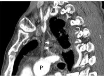
다평면 재구성한 관상면, 시상면 영상에서 좌측 폐동맥과 좌측 무명동맥의 원위부를 연결하는 동맥관 인대(ductus ligament)를 볼 수 있었으며 내부에 혈류는 보이지 않았다 (Fig. 3).
환자의 혈압은 우측 팔에서 120/80 mmHg, 좌측 팔에서 110/80 mmHg였다. 우측 팔에서 측정한 동맥혈의 산소분압 은 106 mmHg, 산소포화도는 97.6%, 좌측 팔의 산소분압은 101.5 mmHg, 산소포화도는 97.7%였다. 양측 팔의 맥압 (pulse pressure)은 거의 같았다.
고 찰
대동맥궁 기형 중에 드문 기형으로 대혈관 분리가 있는데 쇄 골하동맥, 총경동맥, 그리고 무명동맥에 분리가 생긴다. 이러 한 대혈관 분리는 대동맥궁의 발생과정에서 두 군데에서 단절 이 생기면 이들 혈관들이 대동맥궁과 분리되면서 동맥관을 통 해 좌측 폐동맥과 연결된다. 그래서 태생기와 신생아시기에 이 들 혈관이 폐동맥으로부터 혈류공급을 받게 된다(8, 9). 하지 만, 이러한 연결은 동맥관이 퇴화하면서 우측 혈관들을 통해 혈류공급을 받게 된다. 이러한 대혈관의 분리는 쇄골하동맥에 좀 더 많이 생기는데 대동맥궁의 반대에 위치한다. 즉 우측 대 동맥궁에서는 좌측 쇄골하동맥 분리가 동반되고 좌측 대동맥 궁에 우측 쇄골하동맥 분리가 동반된다. 우측 대동맥궁은 약
─ 321 ─ 대한영상의학회지 2009;60:321-323
76세 남자의 우측 대동맥궁와 좌측 무명동맥 분리:
증례 보고
1김 영 통∙조 성 식∙배 원 경
좌측 무명동맥 분리는 아주 드문 기형으로, 대부분이 우측 대동맥궁과 동반된다. 대부분 환자 들이 심장병이 있는 신생아 혹은 소아들이다. 저자들은 선천성 심장병이 없는 76세 남자에서 우 측 대동맥궁과 좌측 무명동맥 분리의 MDCT 소견을 보고하고자 한다.
1순천향대학교 천안병원 영상의학과
이 논문은 2008년 12월 3일 접수하여 2009년 2월 19일에 채택되었음.
0.1%의 빈도를 보이고, 이 중 우측 하행대동맥은 95%를 차지 하며 좌측 하행대동맥은 5%를 차지한다. 우측 하행대동맥을 가진 우측 대동맥궁에서 좌측 쇄골하동맥을 동반하는 경우는 약 1%로 알려져 있다. 반면에 좌측 대동맥궁에 우측 쇄골하동 맥 분리에 관한 보고는 극히 드물다. 역시 우측 대동맥궁과 좌 측 무명동맥 분리가 주로 보고되고 좌측 대동맥궁과 우측 무명 동맥 분리에 관한 보고도 극히 드물다(1, 2).
대혈관 분리는 대부분이 심장 결손을 동반하는데 동맥관 개 존증, 폐동맥 협착, 팔로사징, 심실 중격 결손, 심방 중격 결손 등과 연관이 있을 수 있다. 그리고 디죠지(DiGeorge)증후군 과 연관이 있을 수 있다(9).
쇄골하동맥 분리는 반대편 순환을 통해 혈류공급 받는 쇄골 하동맥도류증후군(subclavian steal syndrome)과 추골기저 동맥부전증(vertebrobasilar insufficiency)을 유발하여 현기 증, 두통, 실신과 같은 증상을 보일 수 있다. 무명동맥 분리의
경우, 종격동과 추골동맥을 통해 분리된 무명동맥으로 혈류공 급이 이루어지는데, 본 증례도 대동맥궁에서 좌측 경동맥과 쇄 골하동맥으로 혈류공급하는 혈관을 볼 수 없었고 우측 총경동 맥이 커져 있으면서, 초기영상에서 좌측 혈관들이 우측 혈관보 다 희미한 조영증강을 보였기 때문에 우측 총경동맥을 통하여 좌측 총경동맥과 쇄골하동맥으로 혈류가 가는 것으로 생각하 였다.
좌측 무명동맥 분리는 동반된 기형이 없다면 증상으로는 진 단이 어렵다. 증상은 좌측 팔과 경동맥 맥박이 약하거나 좌측 팔의 혈압이 낮을 수 있다. 하지만, 본 증례의 환자는 양측 팔 의 산소분압과 산소농도, 그리고 혈압과 맥박의 차이가 없었고 76세의 나이로 농약 중독 때문에 우연히 발견된 증례로 생각 된다. Hara 등(10)이 보고한 우측 개동맥궁과 좌측 쇄골하동 맥 분리를 가진 52세 여자 환자는 본 증례와 같이 심장 잡음도 없고 증상도 거의 없었다.
─ 322 ─
김영통 외: 76세 남자의 우측 대동맥궁와 좌측 무명동맥 분리
A B
Fig. 1. Early phase of CT angiography.
A. Coronal reformatted image shows early dense enhancement of right common carotid and subclavian arteries and ductus liga- ment (arrowheads) between faintly enhanced left common carotid artery (arrow) and left pulmonary artery (P). A; aortic arch.
B. Volume rendering image shows right common carotid and subclavian arteries arising from right aortic arch, but left common carotid and subclavian arteries are still not identified.
A B
Fig. 2. CT scans obtained 7 seconds later.
A. Coronal reformatted image shows contrast filling of left common carotid and subclavian arteries, and ductus ligament (arrow- heads) connecting between these vessels and left pulmonary artery. A; aortic arch
B. Volume rendering image shows common trunk of left common and subclavian arteries without connection with aortic arch.
결론적으로 우측 대동맥궁에서 좌측 무명동맥의 기시부위가 명확하지 않을 때 뚜렷한 증상이 없다 하더라도 무명동맥 분리 의 가능성을 생각하고 MDCT를 이용한 CT 혈관조영술을 시 행하면 진단에 도움을 줄 수 있다.
참 고 문 헌
1. Amplatz K, Moller JH. Radiology of congenital heart disease. St.
Louis: Mosby Year Book, 1993;1016-1017
2. Moes CA, Freedom RM. Rare types of aortic arch anomalies.
Pediatr Cardiol 1993;14:93-101
3. Harrington DP, Brennan T, Varghese JP. Right aortic arch with iso- lation of the left innominate artery. Cardiovasc Intervent Radiol 1981;4:24-26
4. Fong LV, Venables AW. Isolation of the left common carotid or left innominate artery. Br Heart J 1987;57:552-554
5. Martin EC, Mesko ZG, Griepp RB, Haller JO, Gordon DH.
Isolation of the left innominate artery, a right arch, and a left patent ductus arteriosus. AJR Am J Roentgenol 1979;132:833-835 6. Park MK. Right aortic arch with isolation of left innominate artery.
Chest 1979;76:106-108
7. Boren EL Jr, Matchett WJ, Gagne PJ, McFarland DR. Isolation of the left innominate artery in an elderly patient without congenital heart disease. Cardiovasc Intervent Radiol 2000;23:63-65
8. Fu YC, Hwang B, Chang Y, Chi CS. Anomalous origin of one pul- monary artery from the innominate artery: a report of two cases.
Pediatr Cardiol 2001;22:63-65
9. Miyaji K, Hannan RL, Burke RP. Anomalous origin of innominate artery from right pulmonary artery in DiGeorge Syndrome. Ann Thorac Surg 2001;71:2043-2044
10. Hara M, Kitase M, Satake M, Miyagawa H, Ogino H, Itoh M, Ohba S. A case of right-sided aortic arch with isolation of the left subcla- vian artery: CT findings. Radiat Med 2001;19:33-36
─ 323 ─ 대한영상의학회지 2009;60:321-323
J Korean Soc Radiol 2009;60:321-323
Address reprint requests to : Young Tong Kim, M.D., Department of Radiology, Soonchunhyang University, Cheonan Hospital 23-20 Bongmyung-dong, Cheonan 330-721, Korea
Tel. 82-41-570-3515 Fax. 82-41-579-9026
Isolation of the Left Innominate Artery with a Right Aortic Arch in a 76 year-old Man: A Case Report1
Young Tong Kim, M.D., Sung Shick Jou, M.D., Won Kyung Bae, M.D.
1Department of Radiology, Soonchunhyang University Cheonan Hospital
Isolation of the left innominate artery is a rare anomaly and is usually combined with a right side aortic arch.
Most patients are neonates or children with congenital heart disease. We report the MDCT findings of a right aortic arch and isolation of the left innominate artery in a 76-year-old man without congenital heart disease.
Index words :Brachiocephalic trunk
Tomography, X-ray computed Aorta, thoracic
Fig. 3. CT scan obtained 14 seconds later.
Sagittal reformatted image shows ductus ligament (arrow- heads) between distal portion of left innominate artery and left pulmonary artery (P). There is no blood flow from innominate artery into pulmonary artery.