대동맥궁 차단증을 동반한 동맥간의 일차 완전교정
-1예 보고-
One-stage Repair of Truncus Arteriosus with Interrupted Aortic Arch
Truncus arteriosus with interrupted aortic arch is a very rare congenital cardiac anomaly that has an unfavorable natural course. We report a successful one-stage repair of truncus arteriosus with interrupted aortic arch through median sternotomy in a 25-day-old neonate weighing 3.1 kg. We reconstructed the aortic arch with direct side-to-end anastomosis between ascending and descending aortas. The right ventricular outflow reconstruction was performed with untreated autologous pericardial conduit without valve following Lecompte maneuver. The patient has been grown-up in good condition (25 50 percentile of body weight) and shows the right ventricular outflow tract wide 1 year after the operation.
(Korean J Thorac Cardiovasc Surg 2003;36:759-765) Key words
:1. Truncus arteriosus
2. Aortic arch interruption
3. Congenital heart disease, cyanotic
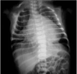
Fig. 1. Preoperative chest roentgenogram.
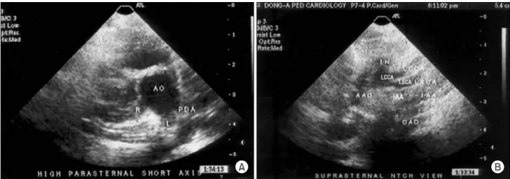
Fig. 2. Preoperative 2D echocardiography. (A) High parasternal short axis view (B) Suprasternal notch view. Ao=Ascending aorta;
R=Right pulmonary artery; L=Left pulmonary artery; PDA=Patent ductus arteriosus; AAo=Ascending aorta; IN=Innominate artery;
LCCA=Left common carotid artery; LSCA=Left subclavian artery; IAA=Interrupted aortic arch; DAO=Descending aorta.
Fig. 3. Schematic drawings of operative procedure (A, B, C, D, E).
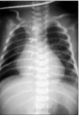
Fig. 4. Postoperative chest roentgenogram at discharge.
Fig. 5. Postoperative 2D echocardiogram at discharge. Sup- rasternal notch view. AAo=Ascending aorta; INN=Innominate artery; LSCA=Left subclavian artery; DAO=Descending aorta.
Fig. 6. Postoperative chest roentgenogram 1 year after ope- ration shows a convexity of the pulmonary artery segment.
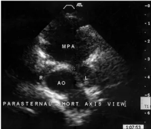
Fig. 7. Postoperative 2D echocardiogram 1 year after opera- tion. Parasternal short axis view. Ao=Ascending aorta; R=Right pulmonary artery; L=Left pulmonary artery; MPA=Main pulmo- nary artery.
=국문 초록=
중심 단어