414 www.kjtcvs.org
KJTCVSThe Korean Journal of Thoracic and Cardiovascular Surgery
CaseReport
Left Hemitruncus Treated Along with Ventricular Septal Defect in a Neonate
Jun Hee Lee, M.D., Hong Ju Shin, M.D., Ph.D., Jae Seung Shin, M.D., Ph.D.
Department of Thoracic and Cardiovascular Surgery, Korea University Ansan Hospital, Korea University College of Medicine, Ansan, Korea
ARTICLE INFO
Received March 30, 2020 Revised June 15, 2020 Accepted June 19, 2020 Corresponding author Hong Ju Shin
Tel 82-31-412-4925 Fax 82-31-412-5060 E-mail [email protected] ORCID
https://orcid.org/0000-0002-0731-3523
Hemitruncus arteriosus is a rare cardiovascular malformation in which one of the pulmo- nary arteries anomalously originates from the aorta. Left hemitruncus arteriosus, defined as the origination of the left pulmonary artery from the aorta, is less common than right hemitruncus arteriosus. In this study, we report the case of a neonate diagnosed with left hemitruncus arteriosus, ventricular septal defect, and atrial septal defect who underwent successful surgical treatment.
Keywords: Truncus arteriosus, Heart septal defects, Ventricular heart septal defects
Copyright©The Korean Society for Thoracic and Cardiovascular Surgery. 2020. All right reserved.
This is an Open Access article distributed under the terms of the Creative Commons Attribution Non-Commercial License (http://creativecommons.org/licenses/
by-nc/4.0) which permits unrestricted non-commercial use, distribution, and reproduction in any medium, provided the original work is properly cited.
Case report
In a female neonate, prenatal ultrasonography showed a parallel course of 2 great arteries and other malformations, including left first toe polydactyly and hemivertebra. The neonate was delivered at 38 weeks and weighed 3.6 kg.
The patient was admitted to the neonatal intensive care unit at birth due to respiratory distress. Chest radiography revealed segmentation anomalies in the vertebral bodies, from T10 to the sacrum. Echocardiography showed atrial septal defect, ventricular septal defect (VSD), and suspect- ed patent ductus arteriosus (PDA) between the right pul- monary artery and the descending aorta. A 3-dimensional
reconstruction computed tomography (CT) scan was per- formed on postnatal day 7 to evaluate the great vessels, and this scan confirmed the anomalous origin of the left pul- monary artery (LPA) from the ascending aorta (Fig. 1A).
The patient underwent surgical repair on day 18 after birth. The operation was carried out through a median sternotomy. After pericardial tenting, careful dissection was performed to encircle the aorta, LPA, and PDA (Fig.
1B). After routine ascending aortic bicaval cannulation im- mediately prior to the initiation of cardiopulmonary by- pass, PDA ligation and LPA division were performed, with the aortic end repaired with 6-0 polypropylene suture and the other end ligated with black silk.
A B C D
Fig. 1. (A) Preoperative computed tomography image and (B) operative findings showing the left pulmonary artery originating from the aorta. Postoperative computed tomography image showing (C) the reimplanted left pulmonary artery and (D) no anastomotic site steno- sis. The white arrow indicates the left pulmonary artery.
https://doi.org/10.5090/kjtcs.20.020 pISSN: 2233-601X eISSN: 2093-6516
Korean J Thorac Cardiovasc Surg. 2020;53(6):414-416
415
Jun Hee Lee, et al. Left Hemitruncus Treated with VSD in a Neonate
www.kjtcvs.org
KJTCVS
After aortic cross-clamping, VSD closure with an autolo- gous pericardial patch and direct atrial septal defect clo- sure were performed via right atriotomy. The divided LPA was then re-implanted into the main pulmonary artery with an autologous pericardial hood patch to avoid anasto- motic stenosis (Fig. 2).
The operation was finished with sternal closure. Howev- er, the chest was opened due to low cardiac output at post- operative 6 hours, and delayed sternal closure was per- formed on postoperative day 3. Follow-up echocardiography 2 weeks after the operation showed a patent LPA without anastomotic stenosis. Follow-up CT scanning also verified the presence of normal vascularization without anastomot- ic stenosis (Fig. 1C, D). The patient recovered smoothly but had to remain in the hospital for 25 additional days after surgery for treatment of other anomalies. At the follow-up visit 10 months later, she was doing well, and her echocar- diogram showed no evidence of stenosis at the LPA re-im- plantation site. However, to carefully evaluate LPA growth, we plan to perform a lung perfusion scan, CT scan, and catheter examination to quantify the aortopulmonary col- lateral vessels in the near future.
The patient’s parents provided written informed consent for the publication of the patient’s clinical details and im- ages.
Discussion
Hemitruncus arteriosus is a rare congenital cardiovascu- lar malformation in which 1 pulmonary artery anomalous- ly originates from the aorta. Left hemitruncus is less com- mon than right hemitruncus [1].
The anomaly was first described by Fraentzel [2] in 1868.
Since then, several case reports and series of hemitruncus have been published, but reports of left hemitruncus are scarce. Early diagnosis is essential for prompt surgical re- pair to prevent death following congestive heart failure and the development of irreversible pulmonary vascular ob- structive disease [3]. Nathan et al. [4] reported that early hemitruncus repair resulted in excellent hemodynamic and anatomic outcomes.
The anatomic correction of hemitruncus by translocat- ing the anomalous pulmonary artery to the pulmonary trunk was first described by Kirkpatrick et al. [5] in 1967.
Since then, direct re-implantation has become the treat- ment of choice. We also attempted direct reimplantation;
however, due to the lack of tissue, we used an autologous pericardial patch hood to prevent anastomotic site stenosis.
With regard to the timing of surgery, we believe that sur- gery during the neonatal period is ideal for favorable re- sults. If possible, we recommend performing the operation within 2 weeks of birth.
After a CT examination on postnatal day 7 (Fig. 1A), we initially thought that re-implantation of the LPA would be simple. However, by more than 2 weeks after birth, de- creased pulmonary vascular resistance together with the increased PDA and VSD shunt had led to enlargement of the left and main pulmonary arteries (Fig. 1B); thus, dis- secting and encircling the aorta, PDA, and LPA before starting cardiopulmonary bypass were difficult. Thus, if this rare disease can be detected early, it is important not to delay surgery.
With regard to postoperative myocardial swelling result- ing in low cardiac output syndrome, staged repair with
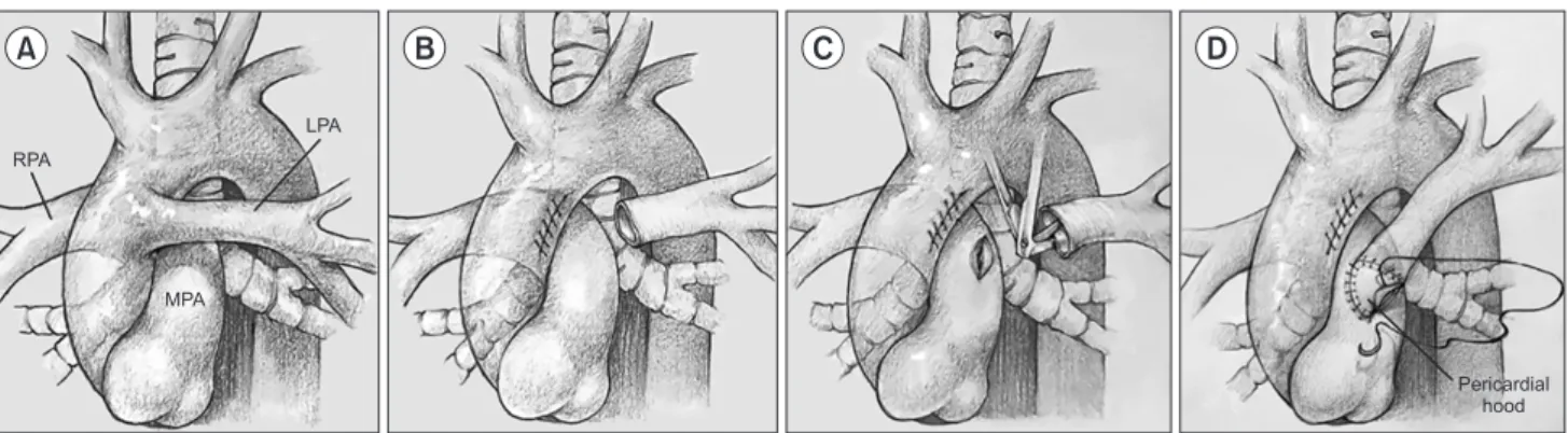
Fig. 2. Reimplantation technique for the left pulmonary artery. (A) Abnormal anatomy of left hemitruncus arteriosus: anomalous origin of the left pulmonary artery from the ascending aorta. (B) The left pulmonary artery was divided, and the aortic wall was repaired with a 6-0 polypropylene running suture. (C) The main pulmonary artery and the posterior wall of the left pulmonary artery were excised.
(D) The left pulmonary artery was re-implanted into the main pulmonary artery using an autologous pericardial hood patch. LPA, left pulmonary artery; MPA, main pulmonary artery; RPA, right pulmonary artery.
A B C D
RPA
LPA
MPA
Pericardial hood
416 www.kjtcvs.org
KJTCVS https://doi.org/10.5090/kjtcs.20.020
LPA reimplantation and external pulmonary artery band- ing is a possible option to avoid needing to open the ster- num in the neonatal period.
Overall, early preoperative detection of this rare congen- ital cardiovascular malformation and postoperative detec- tion of restenosis requiring re-intervention are important.
Three-dimensional reconstruction CT images can be use- ful diagnostic tools [6].
Conflict of interest
No potential conflict of interest relevant to this article was reported.
ORCID
Jun Hee Lee: https://orcid.org/0000-0002-6592-6483 Hong Ju Shin: https://orcid.org/0000-0002-0731-3523 Jae Seung Shin: https://orcid.org/0000-0001-8147-6665
References
1. Kutsche LM, van Mierop LH. Anomalous origin of a pulmonary ar- tery from the ascending aorta: associated anomalies and pathogene- sis. Am J Cardiol 1988;61:850-6.
2. Fraentzel O. Ein fall von abnormer communication der aorta mit der arteria pulmonalis [A case of abnormal communication of the aorta with the pulmonary artery]. Virchows Arch Pathol Anat Physiol Klin Med 1868;43:420-6.
3. Penkoske PA, Castaneda AR, Fyler DC, van Praagh R. Origin of pul- monary artery branch from ascending aorta: primary surgical repair in infancy. J Thorac Cardiovasc Surg 1983;85:537-45.
4. Nathan M, Rimmer D, Piercey G, et al. Early repair of hemitruncus:
excellent early and late outcomes. J Thorac Cardiovasc Surg 2007;
133:1329-35.
5. Kirkpatrick SE, Girod DA, King H. Aortic origin of the right pulmo- nary artery: surgical repair without a graft. Circulation 1967;36:777- 82.
6. Kim TH, Kim YM, Suh CH, et al. Helical CT angiography and three- dimensional reconstruction of total anomalous pulmonary venous connections in neonates and infants. AJR Am J Roentgenol 2000;
175:1381-6.