www.labmedonline.org 215 eISSN 2093-6338
사한다. 평활근항체가 양성인 혈청은 혈관벽, 위의 점막근층(mus- cularis mucosa)과 점막고유층(lamina propria), 선사이수축섬유 (interglandular contractile fibers), 간문맥구역(hepatic portal tract) 과 신장의 기질에 형광 양성을 보인다[4]. 평활근항체의 형광 형은 신장 기질의 형광양상에 따라 혈관벽(vessel wall)에만 형광을 띄 는 평활근항체 V형(SMA-V형), 혈관벽과 사구체(glomerulus)에 형 광을 보이는 평활근항체 G형(SMA-G형) 그리고 세뇨관(tubule)에 도 형광을 보이는 평활근항체 T형(SMA-T형)으로 나누어진다[4].
평활근항체와 연관성을 보이는 임상상은 제1형 자가면역성 간염 이외에도 여러 간질환, 바이러스 감염 그리고 다양한 악성종양 등 이 있다. 악성 흑색종, 신경모세포종, 림프종, 폐암, 간암, 자궁경부 암 등 다수의 악성종양 환자에서 평활근항체가 발견되어 보고된 연구[5, 6]들이 있으나, 갑상선암 환자에서는 평활근항체가 보고된 바가 없다. 저자들은 본원에서 진단된 갑상선 유두암종 환자에서 평활근항체를 확인하여 보고하고, 평활근항체의 형광양상 아형의 질환특이성의 가능성을 제시하는 바이다.
서 론
평활근항체(smooth muscle autoantibodies, SMA)는 제1형 자가 면역성 간염의 혈청학적 진단 표지자로 알려져 있다[1-3]. 다양한 세포 골격 구성요소(cellular cytoskeleton component)가 평활근항 체의 표적항원으로 밝혀져 있고, 평활근항체의 검출은 일반적으 로 쥐의 간, 신장 그리고 위 조직을 이용한 간접 면역형광법으로 검
갑상선 유두암종 환자에서 확인된 평활근항체 V형 1예
A Case of Smooth Muscle Autoantibody V Pattern in a Patient with Papillary Thyroid Carcinoma
한송희·김신규
Song Hee Hahn, M.D., Think-You Kim, M.D.
한양대학교병원 진단검사의학과
Department of Laboratory Medicine, Hanyang University Medical Center, Seoul, Korea
증례보고
Lab Med Online
Vol. 5, No. 4: 215-218, October 2015
http://dx.doi.org/10.3343/lmo.2015.5.4.215 진단면역학
Corresponding author: Think-You Kim
Department of Laboratory Medicine, Hanyang University Medical Center, 222 Wangsimni-ro, Seongdong-gu, Seoul 04763, Korea
Tel: +82-2-2290-8975, Fax: +82-2-2298-1735, Email: [email protected] Received: September 11, 2014
Revision received: November 7, 2014 Accepted: January 31, 2015
This article is available from http://www.labmedonline.org 2015, Laboratory Medicine Online
This is an Open Access article distributed under the terms of the Creative Commons Attribution Non-Commercial License (http://creativecommons.org/licenses/by-nc/3.0/) which permits unrestricted non-commercial use, distribution, and reproduction in any medium, provided the original work is properly cited.
Smooth muscle antibodies (SMAs) are diagnostic markers for the serological diagnosis of type 1 autoimmune hepatitis. SMA that is restricted to staining of the stomach muscle and blood vessel walls was referred to as “SMA-V”. In addition, SMAs are classified into the peritubular (SMA-T) and glomerular (SMA-G) patterns. SMAs are occasionally present in patients with malignancies, but have not yet been reported in thyroid cancer.
We came across the first case of SMA positivity in a patient with papillary thyroid carcinoma (PTC). A 31-yr-old male was admitted to our hospital for evaluation of incidentally detected thyroid cancer. He had been diagnosed with PTC based on pathological results following fine-needle aspira- tion biopsy. The patient underwent total thyroidectomy followed by radio-iodine treatment. The serum levels of AST and ALT were increased before radiotherapy. Tests were conducted for the evaluation of liver disease. SMA was positive at a titer of 1:320, showing positive results for the vessel walls but negative results for the glomerulus and tubules in the kidney (SMA-V pattern). The association of SMA with malignancies and the classifi- cation of SMA immunofluorescent subtypes have been previously reported. However, these studies have not clearly established the ability of SMA subtype to predict a specific disease. Therefore, evaluation of an association of SMA pattern with specific diseases in SMA-positive patients may provide additional and useful information for the rapid diagnosis and accurate treatment of patients with autoimmune diseases or malignancies.
This case report could serve as a great resource for further studies on SMA.
Key Words: Smooth muscle autoantibodies, Thyroid cancer, SMA-V
한송희 외: SMA-V in a Thyroid Cancer Patient
http://dx.doi.org/10.3343/lmo.2015.5.4.215 216 www.labmedonline.org
증 례
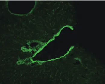
31세 남자가 건강검진 중 시행한 갑상선 초음파 검사의 이상소 견으로 내원하였다. 흡인생검에서 갑상선 유두암종으로 진단되어 갑상선전절제술 후 방사성 요오드 치료를 예정하였다. 내원 초 29/49 IU/L였던 아스파라진산 아미노전이효소(AST)/알라닌 아미 노전이효소(ALT)가 방사성 요오드 치료 전 외래에서 시행한 검사 에서 80/141 IU/L로 증가하여 추가적인 검사를 시행하였다. B형 간염 바이러스 표면항원 및 항체는 각각 음성과 양성이었다. 자가 면역표적(autoimmune target, AIT)검사에서 양성(diffuse granular, 1:320; skeleton pattern, 1:80), 항미토콘드리아항체 음성, 항 liver- kidney microsomal 항체 음성, 평활근항체는 양성(1:320)으로 확 인되었다. 환자의 혈청으로 시행한 평활근항체 검사에서 혈관벽 (vessel wall)과 주변 간문맥구역에 형광반응을 나타내었다(Fig. 1).
또한 위 근육층, 점막고유층, 점막근층 그리고 선사이수축섬유의 형광양상을 확인할 수 있었다(Fig. 2). 한편 신장의 사구체와 세뇨 관을 제외한 혈관에만 형광 양성(Fig. 3)을 보여 평활근항체의 형 광양상의 3가지 아형 중 V형으로 분류할 수 있었다. 약 한 달 후 시 행한 추적검사에서 AST와 ALT는 정상으로 회복되어 평활근항체 에 대한 추가적인 추적검사는 이루어지지 않았다.
고 찰
평활근항체는 제1형 자가면역성 간염의 진단기준에 포함되어 있는 대표적인 장기특이 자가면역항체 중 하나이다[1-3]. 자가면역
성 간염 외에도 바이러스를 비롯한 다양한 원인에 의한 감염 환자 에서 일시적인 검출이나, 간을 침범하지 않는 여러 악성종양 환자 에서의 존재가 보고되었다[7]. 이전의 연구들[5, 6]에서 악성흑색종, 신경세포종, 유방암, 폐암, 림프종 등 다양한 악성종양 환자의 평활 근항체는 이미 보고되었지만 갑상선암 환자에서 평활근항체의 확 인은 보고된 바가 없었다. 평활근항체가 검출되는 모든 환자에서 임상증상이 나타나는 것은 아니고, 무증상의 환자 혹은 정상인에 서도 낮은 역가의 평활근항체가 확인되는 경우가 있지만[4], 환자
Fig. 1. Smooth muscle autoantibodies stain hepatic portal tracts with the walls of vessels in an indirect immunofluorescence assay of the mouse liver substrate (×200, fluorescein isothiocyanate).
Fig. 3. Immunofluorescent pattern of smooth muscle antibodies (SMA) showing positively stained vessel walls and negative glomerulus and tubules, which indicates an SMA-V pattern (×200, fluorescein isothio- cyanate).
Fig. 2. Immunofluorescent pattern of smooth muscle autoantibodies (SMA) on a mouse stomach section. SMA stains muscularis mucosa, lamina propria and interglandular contractile fibers (×200, fluoresce- in isothiocyanate).
한송희 외: SMA-V in a Thyroid Cancer Patient
http://dx.doi.org/10.3343/lmo.2015.5.4.215 www.labmedonline.org 217
는 명확한 AST와 ALT의 상승소견을 동반하였으며 평활근항체의 역가도 1:320으로 의미가 있다고 볼 수 있었다. Whitehouse와 Hol- borow [5]는 악성종양 환자에서 관찰되는 항체의 형광양상이 혈 관벽, 간과 신장 조직에 양성을 보이지만 위의 점막근층 혹은 선사 이수축섬유에는 음성인 것이 일반적인 평활근항체와 다르다고 보 고하였다. 하지만 본 증례의 환자에서는 신장과 간 조직의 혈관과 주변 간문맥구역, 위 근육층, 위 점막고유층, 위 점막근층과 선사 이수축섬유에 형광 양성을 보이는 SMA-V형으로 확인되어 White- house와 Holborow [5]의 연구에서 확인된 악성종양 환자의 항체 와는 또 다른 형광양상을 보이는 것으로 판단되었다. 한편 평활근 항체의 주된 표적항원은 실모양가는근육미세섬유(filamentous actin)로 알려져 있고[8, 9], 그 외에도 중간미세섬유(intermediate filaments)인 vimentin, desmin 등 다양한 세포골격 구성성분이 평 활근항체의 표적항원에 포함된다[2, 4]. 이러한 표적 항원들은 AIT 검사에서 skeleton 형태를 보이는 것으로 알려져 있는데 환자의 AIT 검사에서도 이에 부합하는 결과(Fig. 4)를 확인할 수 있었다.
자가항체의 검출이 새롭게 발병된 또는 아직 진단되지 않은 자 가면역 질환을 의미하기도 하지만 일부에서는 일시적으로 자가면 역 현상이 일어나는 경우도 존재한다. 이전의 많은 연구들[10-12]
에서 악성종양 또는 만성 질환을 갖는 환자를 대상으로 인터페론 또는 인터루킨 등의 치료제를 사용한 후에 일시적으로 다양한 자 가면역 항체가 발생할 수 있는 것으로 보고하였고, 장기 이식 후에 도 자가면역 증상들이 발생할 수 있는 것으로 알려져 있다[13, 14].
본 증례의 환자의 경우 이후에 평활근항체에 대한 추가적인 검사 를 시행하지 않아 그 정확한 의미를 확인할 수 없었다. 이와 같이 치료 후에 관찰된 자가항체가 실제 질환의 존재를 의미하는 것인
지 일시적인 현상으로 나타나는 것인지를 구분하는 것은 환자의 조기 치료와 불필요한 검사나 치료를 줄이는 데 도움이 될 것으로 생각된다.
1976년 Bottazzo 등[4]은 평활근항체의 형광양상에 따라 SMA-V 형과 SMA-G형, SMA-T형으로 그 아형을 구분하였다. SMA-V형이 가장 흔하게 관찰되는 아형이었고 SMA-G형과 SMA-T형은 비슷한 빈도로 확인되었다. SMA-G형과 SMA-T형이 자가면역성 간염의 진 단에 조금 더 높은 특이성을 보이고, SMA-V형은 자가면역성 간염 외에 감염이나 악성종양 환자에서 비교적 자주 관찰되는 것으로 알려져 있다. 하지만 평활근항체 형광양상의 아형이 뚜렷한 질병 특이성을 보이는 것은 아니었다. 평활근항체의 아형과 질환 특이 성에 대한 연구 자체가 많지 않고 특히 국내 환자를 대상으로는 평 활근항체의 형태 아형에 대한 연구가 전혀 없는 상황이다. 평활근 항체의 형광양상에 대하여 이전의 분류 기준을 이용하거나 혹은 새로운 분류 기준을 이용하여 질환과의 연관성을 밝히려는 연구 가 진행된다면, 그 존재가 일시적인 것인지 치료로 인해 새롭게 발 병한 질환을 의미하는 것인지 또는 잠재적인 자가면역 질환을 의 미하는 것인지에 대한 좀 더 정확한 정보를 제공할 수 있을 것으로 생각된다. 평활근항체 검사는 간접 면역형광법으로 시행되는 것으 로 그 역가뿐 아니라 형태에 대한 판독도 동시에 가능하다. 그러므 로 향후 더 많은 국내의 증례를 통해 평활근항체의 형광 형태 혹 은 아형과 질병과의 연관성에 대한 연구가 광범위하게 진행된다면 평활근항체 양성의 의미를 더 정확하게 판단하여 환자의 진단과 치료에 도움을 줄 수 있을 것으로 생각된다.
요 약
평활근항체(smooth muscle autoantibodies, SMA)는 제1형 자가 면역성 간염의 혈청학적 진단 표지자로 알려져 있다. Bottazzo 등 은 위의 근육, 혈관벽에 국한되어 염색되는 SMA를 SMA-V형으로 명명하였고, 그 외에 SMA-T형과 SMA-G형으로 구분하였다. 악성 종양 환자에서 평활근항체의 발견이 종종 보고되었지만, 갑상선암 환자에서 평활근항체는 보고된 경우가 없다. 저자들은 최초로 갑 상선유두암 환자에서의 평활근항체 양성을 확인하였다. 31세 남자 가 우연히 발견된 갑상선암의 검사를 목적으로 입원하였다. 흡인 생검에서 갑상선 유두암으로 진단되어 갑상선전절제술 시행 후 방 사성 요오드 치료를 예정하였다. 방사성 요오드 치료 직전 시행한 검사에서 AST와 ALT가 증가하여 간질환에 대한 검사를 시행하였 다. 평활근항체는 1:320 역가로 양성이었다. 신장의 사구체와 세뇨 관을 제외한 혈관에만 형광 양성을 보여 SMA-V형으로 분류할 수 있었다. 이전의 연구들은 평활근항체와 악성종양 사이의 연관성 과 평활근항체 형광양상의 아형을 보고한 바 있다. 하지만 이 연구 Fig. 4. Skeleton pattern in the autoimmune target (AIT) test using an
indirect immunofluorescense method with IT-1 cells at 1:80 dilution (×
400).
한송희 외: SMA-V in a Thyroid Cancer Patient
http://dx.doi.org/10.3343/lmo.2015.5.4.215 218 www.labmedonline.org
들에서 평활근항체의 아형이 특정 질환을 예측할 수 있는지에 대 한 것은 명확하지 않은 상태이다. 따라서 평활근항체 양성 환자를 대상으로 평활근항체 양상의 질병특이성에 대한 연구는 자가면역 질환이나 악성종양 환자의 신속한 진단과 정확한 치료에 추가적인 정보를 제공할 것이며, 본 증례가 이후의 평활근항체의 연구에 좋 은 자료가 될 수 있을 것이다.
REFERENCES
1. Liberal R, Mieli-Vergani G, Vergani D. Clinical significance of autoan- tibodies in autoimmune hepatitis. J Autoimmun 2013;46:17-24.
2. Liberal R, Grant CR, Longhi MS, Mieli-Vergani G, Vergani D. Diagnos- tic criteria of autoimmune hepatitis. Autoimmun Rev 2014;13:435-40.
3. Vergani D, Alvarez F, Bianchi FB, Cançado EL, Mackay IR, Manns MP, et al. Liver autoimmune serology: a consensus statement from the committee for autoimmune serology of the International Autoimmune Hepatitis Group. J Hepatol 2004;41:677-83.
4. Bottazzo GF, Florin-Christensen A, Fairfax A, Swana G, Doniach D, Groeschel-Stewart U. Classification of smooth muscle autoantibodies detected by immunofluorescence. J Clin Pathol 1976;29:403-10.
5. Whitehouse JM and Holborow EJ. Smooth muscle antibody in malig- nant disease. Br Med J 1971;4:511-3.
6. Tannenberg AE, Muller HK, Cauchi MN, Nairn RC. Incidence of auto- antibodies in cancer patients. Clin Exp Immunol 1973;15:153-6.
7. Toh BH. Smooth muscle autoantibodies and autoantigens. Clin Exp Im- munol 1979;38:621-8.
8. Lidman K, Biberfeld G, Fagraeus A, Norberg R, Torstensson R, Utter G, et al. Anti-actin specificity of human smooth muscle antibodies in chronic active hepatitis. Clin Exp Immunol 1976;24:266-72.
9. Granito A, Muratori L, Muratori P, Pappas G, Guidi M, Cassani F, et al.
Antibodies to filamentous actin (F-actin) in type 1 autoimmune hepa- titis. J Clin Pathol 2006;59:280-4.
10. Ioannou Y and Isenberg DA. Current evidence for the induction of au- toimmune rheumatic manifestations by cytokine therapy. Arthritis Rheum 2000;43:1431-42.
11. Okanoue T, Sakamoto S, Itoh Y, Minami M, Yasui K, Sakamoto M, et al. Side effects of high-dose interferon therapy for chronic hepatitis C.
J Hepatol 1996;25:283-91.
12. Rönnblom LE, Alm GV, Oberg K. Autoimmune phenomena in patients with malignant carcinoid tumors during interferon-alpha treatment.
Acta Oncol 1991;30:537-40.
13. Holbro A, Abinun M, Daikeler T. Management of autoimmune dis- eases after haematopoietic stem cell transplantation. Br J Haematol 2012;157:281-90.
14. Aubert O, Sberro-Soussan R, Scemla A, Casadevall N, Teyssandier I, Martinez F, et al. Autoimmune neutropenia after kidney transplanta- tion: a disregarded entity of posttransplant neutropenia. Transplanta- tion 2014;97:725-9.