83
Received:August 16, 2018, Revised: August 28, 2018, Accepted:September 11, 2018 Corresponding to:Jinseok Kim http://orcid.org/0000-0001-7518-3284
Division of Rheumatology, Department of Internal Medicine, Jeju National University School of Medicine, 15 Aran 13-gil, Jeju 63241, Korea. E-mail:[email protected]
Copyright ⓒ 2019 by The Korean College of Rheumatology. All rights reserved.
This is a Open Access article, which permits unrestricted non-commerical use, distribution, and reproduction in any medium, provided the original work is properly cited.
Clinical Image
pISSN: 2093-940X, eISSN: 2233-4718
Journal of Rheumatic Diseases Vol. 26, No. 1, January, 2019 https://doi.org/10.4078/jrd.2019.26.1.83
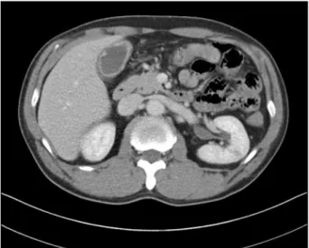
Figure 1. Abdominal and pelvic computed tomography scan showing diffuse wall thickening of the gallbladder with some irregularity.
Eosinophilic Granulomatosis with Polyangiitis Diagnosed by Gallbladder Tissue
WooSeong Jeong1, Changlim Hyun2, Jinsoek Kim1
Departments of 1Internal Medicine and 2Pathology, Jeju National University School of Medicine, Jeju, Korea
In eosinophilic granulomatosis with polyangiitis (EGPA), the incidence of gastrointestinal involvement is reported to range from 17% to 59% [1]. Gallbladder involvement is a rare comorbid condition in EGPA [2]. We present an atypical case of EGPA diagnosed on the basis of histo- logical findings of the gallbladder after cholecystectomy.
The study was approved by the Institutional Review Board of the Jeju National University Hospital (IRB no.
2018-07-009).
A 47-year-old man visited the hospital with progressive weakness and sensory deterioration in both the lower legs for 9 days. He had been diagnosed with asthma 6 months ago and had a history of surgery for sinusitis 5 months ago. Physical examination showed decreased muscle strength and right sided foot drop. On blood test- ing, leukocytosis with a marked increase in eosinophils was observed (white blood cell count 21,300/μL; seg- mented neutrophils 33.7%, lymphocytes 7.5%, mono- cytes 2.1%, eosinophils 56.5%). Further laboratory ex- amination revealed an increase in C-reactive protein to 4.68 mg/dL, erythrocyte sedimentation rate of 45 mm/hr, immunoglobulin E level of 2,500.0 IU/mL). Also, rheu- matoid factor (27 IU/mL) and myeloperoxidase antibody (150.5 IU/mL) were positive.
Muscle weakness progressed gradually, with left sided foot drop developing on the night of admission, followed by right wrist drop, which presented the following day.
On the third day, a nerve conduction study was per- formed, which showed multiple mononeuropathy. A 3 cm long sural nerve was biopsied from the lateral aspect of the left ankle, and high dose corticosteroid treatment (1
mg/kg prednisolone) was initiated immediately.
Abdominal and pelvic computed tomography (APCT) and chest CT were performed to rule out the possibility of peripheral neuropathy associated with malignancy.
Diffuse irregular gallbladder wall thickening was seen on APCT (Figure 1). However, positron emission tomog- raphy-computed tomography revealed no findings suspi- cious for gallbladder cancer. Cholecystectomy was per- formed as recommended by the surgeon, in order to rule out malignancy. Nerve biopsy results showed no in- flammatory cell infiltration or vasculitis. However, eosi- nophilic granulomatosis with polyangiitis was diagnosed from the gallbladder tissue due to presence of chronic ac-
WooSeong Jeong et al.
84 J Rheum Dis Vol. 26, No. 1, January, 2019
Figure 2. Granuloma surrounds the blood vessel (blue ar- rows) and granulomatous inflammation is also noted in and around the blood vessels (H&E, ×40).
Figure 3. The vessel wall (yellow arrows) is destructed by in- flammatory infiltrates in the right upper area of the vessel (vas- culitis with fibrinoid necrosis, red arrow). Many eosinophils which have bright red colored cytoplasm infiltrates into and around the vessel (black arrows) (H&E, ×200).
tive inflammation with granulomatous vasculitis and eo- sinophilic infiltration (Figures 2 and 3). The patient was started on cyclophosphamide and high dose cortico- steroid treatment, after which muscle strength gradually improved.
Histopathologic analysis still remains the gold standard for diagnosis of antineutrophil cytoplasmic anti- body-associated vasculitis. Most cases are diagnosed by performing a biopsy at the symptomatic site, but charac- teristic EGPA findings may be seen in a biopsy performed at the symptom-free site, as observed in this patient.
CONFLICT OF INTEREST
No potential conflict of interest relevant to this article was reported.
REFERENCES
1. Ebert EC. Gastrointestinal manifestations of churg-strauss syndrome. J Gastroint Dig Syst 2011;1:1-3.
2. Ye L, Lu X, Xue J. Eosinophilic granulomatosis with poly- angiitis complicated by cholecystitis: a case report and re- view of the literature. Clin Rheumatol 2016;35:259-63.