INTRODUCTION
Lung cancer is the leading cause of cancer death in most countries. Although much effort has been made over the last few decades to improve the survival of lung cancer patients, overall five-year survival rates remain unsatisfactorily low (14% in the U.S.A.) (1).
Brain metastasis (BM) is a major cause of the low survival rate and poor quality of life of cancer patients. The progno- sis for patients with BM who go untreated is extremely poor (about one month following diagnosis) (2), whereas patients with non-small cell lung cancer (NSCLC) who are treated with radiation therapy survive for about 8 months (3). The incidence of BM in patients with locally advanced NSCLC is 12-28% over the entire course of the disease. In most cases of NSCLC, BM are diagnosed only after the development of symptoms, which is partly responsible for the poor progno- sis of patients with NSCLC. In patients with small cell lung cancer (SCLC), the incidence of detectable BM at the time of initial diagnosis is 10-14%; the cumulative incidence at three years for patients that are in complete remission and that have few signs of disease is 59% (4). For patients with
NSCLC, the incidence of BM at the time of initial diagnosis is about 6% according to a prospective study in which com- puterized axial tomography (CT) was used (5). The prognoses of patients with symptomatic BM are substantially worse than those in which the metastases are asymptomatic (6-8). Clear- ly, improvements in systemic and local therapies can improve the long-term survival of cancer patients, which means that early and accurate diagnosis of BM has become crucial to im- proving the quality of life and poor survival rates of cancer patients.
The use of imaging to detect extrathoracic metastasis, par- ticularly BM, at the time of initial staging in asymptomatic patients is the subject of debate (8-10). Imaging is usually recommended in patients who form part of a population in which the overall incidence of BM is particularly high, such as in cases with adenocarcinomas (6, 11-13). Of the different imaging methods, MRI is more sensitive than CT and is the method of choice with which to screen for intracranial metas- tasis (12, 14-17). However, a major drawback to the routine use of brain MRI is its high cost. Therefore, we carried out the modified standard MRI or limited MRI procedure to detect BM at lower cost and without loss of sensitivity (18). In our
Sun-Young Kim*,‖, Jae-Sung Kim�,‖, Hee-Sun Park*, Moon-June Cho�,‖, Ju-Ock Kim*,‖, Jin-Whan Kim�,‖, Chang-Jun Song�,‖, Seung-Pyung Lim�,‖, Sung-Soo Jung*
Departments of Internal Medicine*, Therapeutic Radiology�, Diagnostic Radiology�, Thoracic and Cardiovascular Surgery�, College of Medicine, Cancer Research Institute‖, Chungnam National University, Daejon, Korea
Address for correspondence Sung Soo Jung, M.D.
Department of Internal Medicine, Division of Pulmonary Medicine, College of Medicine, Chungnam National University Hospital, 640 Daesa-dong, Jung-gu, Daejon 301-721, Korea Tel : +82.42-259-8103, Fax : +82.42-257-5753 E-mail : [email protected]
121
Screening of Brain Metastasis with Limited Magnetic Resonance Imaging (MRI): Clinical Implications of Using Limited Brain MRI During Initial Staging for Non-small Cell Lung Cancer Patients
The purpose of this prospective study was to determine whether using magnetic resonance imaging (MRI) for early screening for brain metastases (BM) can improve quality of life, survival in patients with non-small cell lung cancer (NSCLC). The study group comprised 183 patients newly diagnosed with NSCLC. All patients underwent limited brain MRI and routine workups. The control group comprised 131 patients with NSCLC who underwent limited brain MRI only if they had neuro- logic symptoms. The incidence of BM was 20.8% (38/183) in the study group and 4.6% (6/131) in the control group. The rate of upstaging based on the MRI data was 13.5% (15/111) overall and 15.9% (11/69) in patients that had been consid- ered initially to be resectable surgically. There was no significant difference in sur- vival outcome between the groups. Patients who had BM alone had a greater overall survival time (49 weeks) than those who had multiple systemic metastases (27 weeks; p=0.0307). In conclusions, limited brain MRI appears to be a useful, cost-effective method to screen for BM at the time of initial staging. And it may facilitate timely treatment of patients with NSCLC and improve their survival and quality of life.
Key Words : Carcinoma, Non-Small-Cell Lung; Neoplasm Staging; Neoplasm Metastasis; Magnetic Reso- nance Imaging; Diagnosis; Radiography
Received : 21 May 2004 Accepted : 20 September 2004
pilot study limited MRI showed no difference with conven- tional MRI for detecting BM (sensitivity 97.67%, specifici- ty 100%) (18). The cost of the modified MRI was US$ 180, which is substantially lower than the cost of conventional brain MRI (US$ 480). Based on these results, we conducted the present study to screen for BM at the time of initial stag- ing and validated the clinical significance of the early detec- tion of BM using limited brain MRI. The results have impli- cations for the quality of life and survival of patients with NSCLC.
MATERIALS AND METHODS
Between May, 2001 and April, 2002, 183 patients were newly diagnosed with primary NSCLC at Chungnam Natio- nal University Hospital. All patients underwent the following initial staging procedures: clinical examination; routine blood
tests; chest radiography; chest CT (including liver and adrenal glands); whole-body bone scan; and limited brain MRI. The limited brain MRI was modified from conventional MRI by omitting T2-weighted axial, proton density axial, and con- trast-enhanced T1-weighted images (Table 1) to decrease the cost from US$ 480 to US$ 180 without any decrease in sen- sitivity (18). As a control group, we used 131 patients who were newly diagnosed with NSCLC between May, 2000 and April, 2001. Control patients underwent the initial staging procedures described above, except that a brain MRI was car- ried out only in cases that exhibited neurologic symptoms related to BM (e.g. headaches, seizures, or unexplained weak- ness) at the time of initial diagnosis. The incidence of BM in the initial staging of patients with primary NSCLC and the incidence of metastases to other systemic sites were com- pared. We compared the clinically relevant characteristics of both groups (age, sex, cell type, performance status, clinical stage, and incidence of brain metastasis). Patients were fol- lowed up until the 30th of April, 2003 (i.e., a minimum peri- od of one year) and we analyzed the clinical significance of early detection of BM and the relationships among the vari- ous prognostic factors. The statistical analysis was done using SPSS (version 11.0). Differences between the two groups were evaluated for statistical significance using a chi-squared test.
Survival data were calculated using the Kaplan-Meier method and statistical differences were confirmed by means of the log-rank test.
RESULTS
There were no statistically significant differences between the groups with respect to gender, age, performance status, tumor subtype, or clinical stage (excluding brain metastasis) (Table 2).
WI, weighted image; CE, contrast-enhanced.
Image type Conventional Limited
T1-WI axial O O
T2-WI axial O X
Proton density axial O X
CE T1-WI axial O O
CE T1-WI coronal O O
CE T1-WI sagittal O X
Cost (US$) 480 180
Table 1.Differences between conventional and limited brain magnetic resonance imaging (MRI)
Sq, squamous cell carcinoma; Ad, adenocarcinomas; La, large cell car- cinoma; Un, undifferentiated; NS, not significant.
Study group (n=183)
Control group
(n=131) p value
Sex (male:female) 139:44 105:26 NS
Median age (yr) (range) 67 (40-79) 66 (31-85) NS ECOG performance:
0-2 171 123 NS
>3 12 8 NS
Cell type:
Non-squamous 92 70 NS
Ad/La/Un 78/6/8 53/8/9 NS
Sq 91 61 NS
Stage I 13 9 NS
II 13 14 NS
IIIA 32 31 NS
IIIB 38 36 NS
IV 87 (47.6%) 42 (32.1%) <0.05
Brain 38 (20.8%) 6 (4.6%) <0.001
Bone 39 (21.3%) 12 (9.2%) NS
Lung 35 (19.1%) 22 (16.8%) NS
Liver 6 (3.3%) 7 (5.3%) NS
Other 8 (4.4%) 4 (3.1%) NS
Table 2.Patients’ characteristics
Sq, squamous cell carcinoma; Ad, adenocarcinomas; La, large cell car- cinoma; Un, undifferentiated. *male vs. female: p=0.0124, �squamous vs. nonsquamous: p<0.0001. BM; brain metastasis.
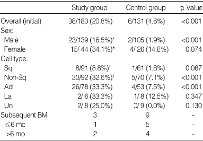
Study group Control group p Value Overall (initial) 38/183 (20.8%) 6/131 (4.6%) <0.001 Sex:
Male 23/139 (16.5%)* 2/105 (1.9%) <0.001 Female 15/ 44 (34.1%)* 4/ 26 (14.8%) 0.074 Cell type:
Sq 8/91 (8.8%)� 1/61 (1.6%) 0.067
Non-Sq 30/92 (32.6%)� 5/70 (7.1%) <0.001
Ad 26/78 (33.3%) 4/53 (7.5%) <0.001
La 2/ 6 (33.3%) 1/ 8 (12.5%) 0.347
Un 2/ 8 (25.0%) 0/ 9 (0.0%) 0.130
Subsequent BM 3 9 -
≤6 mo 1 5 -
>6 mo 2 4 -
Table 3.Incidence of brain metastasis according to the patients’
characteristics
BM was detected in 38 of 183 (20.8%) and 6 of 131 (4.6%) patients in the study and control groups, respectively (p<
0.001). Twenty-three of 139 (16.5%) males and 15 of 44 (34.1%) females had BM. Patients with non-squamous-type cells (32.6%) had a higher frequency of BM than those with squamous-type cells (8.8%). During the follow-up period, subsequent BM developed in 3 of study group, and 9 of con- trol group. In these patients, 1 of study group and 5 of con- trol group had synchronous BM (within 6 months after ini- tial diagnosis) (Table 3).
Eighteen patients (47.4%) had a single focus of metastasis within the brain, 17 patients (44.7%) had multiple intracra- nial foci, and 3 patients (7.9%) had a leptomeningeal pat- tern of metastasis (Table 4). Only 7 of 38 patients (18.4%) in which BM was observed using limited MRI also exhibit- ed clinical symptoms related to BM (Table 4). In 15 of 38 patients (39.5%) in which BM was observed using limited MRI, the brain was the only site of systemic metastasis, and these 15 patients were upstaged to stage IV based solely on the diagnosis of BM. This number included 11 of 68 patients (15.9%) that were considered initially to be resectable sur- gically in the absence of BM. Two stage I, three stage II, six stage IIIA, and four stage IIIB patients were upstaged to stage IV (Table 5).
For the survival analysis, 10 of 183 patients in the study group were excluded due to insufficient follow-up data. The survival analysis was carried out on 173 patients. The overall
median survival time and one-year survival rate of patients with BM were 43 weeks and 39.5% and 31 weeks and 16.7%
for the study and control groups, respectively. The number of patients in the control group was too small to determine whether there were any statistically significant differences between the two groups. The one-year survival rate and medi- an survival time of patients who had BM (46.7% and 49 weeks, respectively) was greater only than patients who had multiple systemic metastases that included the brain (34.8%
and 27 weeks, respectively). Eighteen patients had between one and three metastatic nodules in the brain that could be accessed for stereotactic radiosurgery and 20 patients (includ- ing three with leptomeningeal involvement) had multiple (>3 nodules) foci within the brain. The one-year survival rate and median survival time for the former and latter type of patient was 44.4% and 45 weeks and 35.0% and 24 weeks, respectively. Of the 38 patients who had BM and were strong- ly recommended to receive radiation therapy to the brain with systemic chemotherapy, 14 were treated with whole brain irradiation only and 23 were treated with both radiation and chemotherapy. One patient refused any further treatment.
The one-year survival rate and median survival time of irra- diation-treated patients was 21.4% and 21 weeks, respec- tively, and the one-year survival rate and median survival time of those treated with both radiation and chemotherapy was 52.2% and 49 weeks, respectively. The one-year survival rate and median survival time for symptomatic vs. asymptomatic patients was 42.9% and 45 weeks and 38.7% and 43 weeks, respectively (Table 6).
Study group (n=38)
Control group (n=6) Number of metastatic foci:
1-3 18 (47.4%) 2 (33.3%)
Multiple (>3) 17 (44.7%) 4 (66.6%)
Leptomeningeal 3 (7.9%)
Metastasis site:
Brain only 15 (39.5%) 2 (33.3%)
Brain plus other systemic sites 23 (60.5%) 4 (66.7%) Symptoms related to brain metastasis:
Symptomatic 7 (18.4%) 6 (100%)
Asymptomatic 31 (81.6%) -
Table 4.Characteristics of brain metastasis
Median survival (weeks)
One-year survival Overall:
Study group 43 39.5% (15/38)
Control group 31 (p=0.44) 16.7% (1/6)
(p=0.2804) Systemic metastasis or not:
Brain metastasis only 49 46.7% (7/15)
Systemic metastasis 27 (p=0.0307) 34.8% (8/23) (p=0.4638) Number of metastatic foci in brain:
1-3 45 44.4% (8/18)
Multiple (>3) 24 (p=0.0835) 35.5% (7/20) (p=0.6456) Symptoms related to brain metastasis:
Symptomatic 45 42.9% (3/7)
Asymptomatic 43 (p=0.5660) 38.7% (12/31) (p=0.3689) Treatment modality:*
Radiation only 21 21.4% (3/14)
Radiation plus chemotherapy 49 (p=0.0196) 52.2% (12/23) (p=0.0647)
*No treatment: 1 case.
Table 6.Median survival period and one-year survival rate of patients with brain metastasis detected by limited brain MRI screening
Before the detection of brain metastasis
Stage After the detection of
brain metastasis
I 15 13 (-2)
II 16 13 (-3)
IIIA 38 32 (-6)
IIIB 42 38 (-4)
IV 72 87 (+15)
Overall upstaging rate: 15/111 (13.5%).
Upstaging rate in patients initially considered resectable surgically: 11/69 (15.9%).
Table 5.Impact of limited brain MRI screening on stage deter- mination
The one-year survival rate and median survival time over- all for stage IV patients were 36.1% and 37 weeks, respective- ly. The median survival of 12 patients in whom BM was diag- nosed during the follow-up treatment period was 8 weeks.
DISCUSSION
Even though locoregional control rates of 50% and higher have been achieved after neoadjuvant chemoradiotherapy and definitive resection (multimodal therapy) in patients with locally advanced NSCLC, the brain is the most frequent site of initial treatment failure and is the site of approximately one third of all relapses that occur (11, 13, 19, 20). Therefore, treatment strategies that reduce the risk of BM are needed to optimize the efficacy of multimodal therapy and to improve survival.
Between 25 and 30% of all patients with lung cancer devel- op BM at some stage, and this incidence is likely to increase if survival is prolonged by means of aggressive multi-modal treatment. The increasing frequency of treatment failure, the development of new imaging techniques and treatments, as well as the reports of prolonged survival of patients that have received aggressive treatment, suggest that approaches to treat- ing patients who have BM should be reassessed. Specifically, novel treatment paradigms should be developed to ensure that such patients receive optimal therapeutic intervention. To this end, prophylactic cranial irradiation in NSCLC and SCLC patients has been introduced and has led to several reports of positive outcomes, although a large-scale study is needed to confirm this (11, 13, 21). An alternative means to the same end is the introduction of brain imaging when the initial workup is carried out. This approach would facilitate the early detection of BM and promote the timely application of appro- priate treatments, which would improve the survival rates of cancer patients. However, such early imaging should be considered only if it is practicable and available at a reason- able cost. Considering each of the aforementioned factors, we designed the present study to assess the clinical effectiveness of using limited brain imaging at the time of initial screen- ing of lung cancer patients.
The rate of BM in patients with locally advanced NSCLC has been estimated previously to be between 17 and 32%
(median: 22%) with a median time-to-brain recurrence of 7.5-9.3 months (8, 11, 13). It has been shown previously that most recurrences of metastases within the central nervous sys- tem occur within 2 yr of the initial diagnosis (13). Further- more, BM is a direct cause of death in between one third and one half of affected patients (12).
The incidence of BM in the initial screening of patients with primary lung cancer has been reported to be between 12 and 18% (22, 23). In the present study, the incidence was 18.9%. We suspect that this slightly higher incidence was the result of using the limited brain MRI procedure, which
is more sensitive than brain CT (5, 17). There are several risk factors that are related to BM, including non-squamous his- tological characteristics (particularly adenocarcinomas), bulky lymph nodes in the mediastinum, younger age, stage IIIB, etc. (5, 6, 11-13, 24, 25). Our data supported the same trend and, in addition, suggested that the incidence of such risk factors is greater in females than in males. Specifically, in the present study, the incidence of BM and non-squamous tumors in females was 30.6 and 26.2%, respectively, which was sta- tistically higher than in males (15.5 and 8.8%, respectively).
The incidence of BM in all patients with adenocarcinomas was 33.3%, whereas the incidence in females with adenocar- cinomas was 50%. In clinical stage IV patients, the brain was the second most common site of metastasis (38/87 patients or 43.7%), following bone (39/87, 44.8%), and was a more common site of metastasis than lung, liver, and the adrenal glands.
Most patients with BM were asymptomatic, even those with multiple foci. Symptomatic metastasis was observed in only 7 of 38 patients (18.4%). Although neurological symp- toms have been reported to be associated with a poor prog- nosis (7, 8, 14), the survival rates in the present study were unrelated to the presence of clinical symptoms. We found that patients who had a single focus of BM, and isolated BM, and who were treated with chemotherapy had better survival, as reported by others. The survival rate of patients who had BM was equivalent to those who did not have any clinical symptoms of BM; this was due to early treatment using whole brain radiotherapy, stereotactic radiosurgery, and chemother- apy. Fujita et al. reported that the use of chemotherapy to con- trol not only BM but also extracranial lesions prolonged the survival of patients with BM due to NSCLC (26). In the pre- sent study, the survival rates of patients who had BM and who received both chemotherapy and radiotherapy were similar to those of patients in stage IIIB (data not shown), which would appear to support our hypothesis that early detection of BM and subsequent application of appropriate treatment may improve overall survival.
In the present study, 15 patients (15/111, 13.5%) were upstaged from clinical stage I (n=2), II (n=3), IIIA (n=6), or IIIB (n=4) to stage IV, because of the presence of BM, includ- ing 11 patients (11/68, 15.9%) who had been considered initially to be suitable for surgical resection in the absence of evidence for the presence of BM. All of the aforementioned patients were treated with either radiotherapy alone (n=7) or both radiotherapy and chemotherapy (n=8). The median survival time and one-year survival rate of the former and latter patients was 43 weeks and 28.6% and 72 weeks and 66.7%, respectively. That was greater than the survival rate of all patients with BM (median survival time and one-year survival rate for radiotherapy only and radiotherapy plus che- motherapy for all patients with BM were 21 weeks and 21.4%, and 49 weeks and 52.2%, respectively) (Table 6). During a follow up period of at least one year, 12 patients (3 of study
group and 5 of control group) subsequently developed BM.
All of these patients (except one) were treated with chemo- therapy after the diagnosis of BM. Initially, these patients responded to chemotherapy such that four exhibited partial responses, five exhibited a stable disease state, and three exhib- ited progression of the disease. The median time to relapse in treated patients (n=11) was 38 weeks, which is similar to the time reported by Ceresoli et al. (9 months) (13). These 11 patients were treated with radiation only without any further chemotherapy; their median survival time after the diagno- sis of BM was 8 weeks, which was shorter than that of patients in whom metastasis was detected during the initial screen- ing and who were treated with radiation therapy alone (21 weeks). Their overall median survival time and one-year sur- vival rate (after the diagnosis of lung cancer) was 38 weeks and 27.3%, respectively, worse than the study group that had BM at the time of the initial screening (43 weeks and 39.5%) and worse than patients who received whole-brain radiation and chemotherapy (49 weeks and 52.2%) following the initial screening. Nevertheless, these outcomes were slight- ly better than those of the control patients, who were diag- nosed and treated for brain metastasis only upon the devel- opment of symptoms (31 weeks and 16.7%).
In conclusion, limited brain MRI appears to be a useful and cost-effective means by which to detect BM at the time of initial staging of lung cancer patients. Such early detec- tion of BM enabled patients to be treated immediately with radiotherapy and chemotherapy and led to improved rates of survival. Consequently, we recommend that limited brain MRI should be used as an initial screening procedure for the early detection of BM. This will allow lung cancer patients to receive the appropriate treatment in a timely manner, which should improve both the survival and quality of life of such patients.
REFERENCES
1. Minna J. Neoplams of the lung. In: Braunbald E, Fauci AS, Kasper DL, Hauser SL, Longo DL, Jameson JL, editors. Harrison’s princi- ples of internal medicine. 15th ed. New York: McGraw-Hill, Inc.
2001: 562-71.
2. Sorensen JB, Hansen HH, Hansen M, Dombernowsky P. Brain metas- tases in adenocarcinoma of the lung: frequency, risk groups, and prognosis. J Clin Oncol 1988; 6: 1474-80.
3. Zabel A, Milker-Zabel S, Thilmann C, Zuna I, Rhein B, Wannen- macher M, Debus J. Treatment of brain metastases in patients with non-small cell lung cancer (NSCLC) by stereotactic linac-based radio- surgery: prognostic factors. Lung Cancer 2002; 37: 87-94.
4. Vines EF, Le Pechoux C, Arriagada R. Prophylactic cranial irradi- ation in small cell lung cancer. Semin Oncol 2003; 30: 38-46.
5. Yokoi K, Kamiya N, Matsuguma, H, Machida S, Hirose T, Mori K, Tominaga K. Detection of brain metastasis in potentially operable non-small cell lung cancer. A comparison of CT and MRI. Chest
1999; 115: 714-9.
6. Hochstenbaga MM, Twijnstra A, Hofman P, Wouters EF, ten Velde GP. MR-imaging of the brain of neurologic asymptomatic patients with large cell or adenocarcinoma of the lung. Does it influence prog- nosis and treatment? Lung Cancer 2003; 42: 189-93.
7. Virgo KS, McKirgan LW, Caputo MC, Mahurin DM, Chao LC, Caputo NA, Naunheim KS, Flye MW, Gillespie KN, Johnson FE.
Post-treatment management options for patients with lung cancer.
Ann Surg 1995; 222: 700-10.
8. Yokoi K, Miyazawa N, Arai T. Brain metastasis in resected lung cancer: value of intensive follow-up with computed tomography. Ann Thorac Surg 1996; 61: 546-51.
9. Cole FH Jr, Thomas JE, Wilcox AB, Halford HH 3rd. Cerebral imag- ing in the asymptomatic preoperative bronchogenic carcinoma patient:
is it worthwhile? Ann Thorac Surg 1994; 57: 838-40.
10. Silvestri GA, Littenberg B, Colice GL. The clinical evaluation for detecting metastatic lung cancer. A meta-analysis. Am J Respir Crit Care Med 1995; 152: 225-30.
11. Andre F, Grunenwald D, Pujol JL, Girard P, Dujon A, Brouchet L, Brichon PY, Westeel V, Le Chevalier T. Patterns of relapse of N2 non-small cell lung cancer patients treated with preoperative chemo- therapy. Should prophylactic cranial irradiation be reconsidered?
Cancer 2001; 91: 2394-400.
12. Gasper L, Scott C, Rotman M, Asbell S, Phillips T, Wasserman T, McKenna WG, Byhardt R. Recursive partitioning analysis (RPA) of prognostic factors in three radiation therapy oncology group (RTOG) brain metastatses trials. Int J Radiaton Oncol Biol Phys 1997; 37:
745-51.
13. Ceresoli GL, Reni M, Chiesa G, Carretta A, Schipani S, Passoni P, Bolognesi A, Zannini P, Villa E. Brain metastases in locally advanced nonsmall cell lung carcinoma after multimodality treatment; risk fac- tors analysis. Cancer 2002; 95: 605-12.
14. Walsh GL, O’Connor M, Willis KM, Milas M, Wong RS, Nesbitt JC, Putnam JB Jr, Lee JJ, Roth JA. Is follow-up of lung cancer patients after resection medically indicated and cost-effective? Ann Thorac Surg 1995; 60: 1563-72.
15. Sze G, Milano E, Johnson C, Heier L. Detecton of brain metastases:
comparison of contrast-enhanced MR with unenhanced MR and enhanced CT. Am J Neuroradiol 1990; 11: 785-91.
16. Davis PC, Hudgins PA, Peterman SB, Hoffman JC Jr. Diagnosis of cerebral metastases: Double-dose delayed CT vs contrast-enhanced MR imaging. Am J Neuroradiol 1991; 12: 293-300.
17. Haberkorn U, Schoenberg SO. Imaging of lung cancer with CT, MRI and PET. Lung Cancer 2001; 34 (Suppl 3): 13-23.
18. Kwon SJ, Lee YS, Ahn JY, Jung SS, Kim JO, Kim SY. Detection of brain metastases using limited brain magnetic resonance imag- ing. Tuberculosis and Respiratory Diseases 2003; 55: 499-505.
19. Choi NC, Carey RW, Daly W, Mathisen D, Wain J, Wright C, Lynch T, Grossbard M, Grillo H. Potential impact on survival of improved tumor downstaging and resection rate by preoperative twice-daily radiation and concurrent chemotherapy in stage IIIA non-small cell lung cancer. J Clin Oncol 1997; 15: 712-22.
20. Eberhardt W, Wilke H, Stamatis G, Stuschke M, Harstrick A, Menker H, Krause B, Mueller MR, Stahl M, Flasshove M, Budach V, Gres-
chuchna D, Konietzko N, Sack H, Seeber S. Preoperative chemother- apy followed by concurrent chemoradiation therapy based on hyper- fractionated accelerated radiotherapy and definitive surgery in local- ly advanced non-small cell lung cancer: Mature results of phase II trial. J Clin Oncol 1998; 16: 622-34.
21. Stuschke M, Eberhardt W, Pottgen C, Stamatis G, Wilke H, Stuben G, Stoblen F, Wilhelm HH, Menker H, Teschler H, Muller RD, Budach V, Seeber S, Sack H. Prophylactic cranial irradiation in locally ad- vanced non-small cell lung cancer after multimodality treatment:
long-term follow-up and investigations of late neuropsychologic effects.
J Clin Oncol 1999; 17: 2700-9.
22. Mintz BJ, Tuhrim S, Alexander S, Alexander S, Yang WC, Shanzer S. Intracranial metastases in the initial staging of bronchogenic car- cinoma. Chest 1984; 86: 850-3.
23. Hooper RG, Tenholder MF, Underwood GH, Beechler CR, Spratling L. Computed tomographic scanning of the brain in initial staging of bronchogenic carcinoma. Chest 1984; 85: 774-6.
24. Gregor A. Prevention and management of brain metastases. Lung Cancer 2003; 41 (Suppl 3): 42.
25. Robnett TJ, Machtay M, Stevenson JP, Algazy KM, Hanh SM. Fac- tors affecting the risk of brain metastases after definitive chemora- diation for locally advanced non-small cell lung cancer. J Clin Oncol 2001; 19: 1344-9.
26. Fujita A, Fukuoka S, Takabatake H, Tagaki S, Sekine K. Combina- tion chemotherapy of cisplatin, ifosfamide, and irinotecan with rhG- CSF support in patients with brain metastases from non-small cell lung cancer. Oncology 2000; 59: 291-5.