This is an Open Access article distributed under the terms of the Creative Commons Attribution Non-Commercial License (http: //creativecommons.org/licenses/by- nc/4.0/) which permits unrestricted non-commercial use, distribution, and reproduction in any medium, provided the original work is properly cited.
© 2019 THE KOREAN SOCIETY OF MYCOLOGY.
Accepted: September 05, 2019 Revised: September 02, 2019 Received: June 17, 2019 https://doi.org/10.4489/KJM.20190022 Kor. J. Mycol. 2019 September, 47(3): 181-6
OPEN ACCESS pISSN : 0253-651X eISSN : 2383-5249
RESEARCH ARTICLE
Acrophialophora ellipsoidea, an Undescribed Species Isolated from Soil in Korea
Benjamin Yaw Ayim1, Young-Tae Kim1, Kallol Das1, In-Kyu Kang1, Seung-Yeol Lee1,2* Hee-Young Jung1,2
1College of Agriculture and Life Sciences, Kyungpook National University, Daegu 41566, Korea
2Institute of Plant Medicine, Kyungpook National University, Daegu 41566, Korea
*Corresponding author: [email protected]
ABSTRACT
A designated fungal isolate, KNU-US-1802E was isolated from the soil in Uiseong, Korea.
To identify characteristics of the isolate, it was cultured on PDA media for 6 days at 35°C.
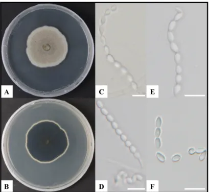
Colonies on PDA are flat, light gray, dense, with entire margins; reverse dark gray to black, with white margins. Aerial mycelia were smooth-walled, hyaline and 40~42 mm diameter after 6 days at 35°C. Conidia were hyaline, one-celled, ellipsoidal to fusiform, forming long chains with average length x width of 5.0±0.3 × 2.9±0.2 μm. Molecular analysis indicates that the internal transcribed spacer (ITS) region and partial beta-tubulin (tub2) gene sequence showed 100% and 99% similarities, respectively with Acrophialophora ellipsoidea CGMCC 3.15255 collected from China. Phylogenetic analysis by the neighbor-joining (NJ) method shows that the KNU-US-1802E was clustered with A. ellipsoidea CGMCC 3.15255 in a phylogenetic tree constructed using the concatenated sequences of ITS region and tub2 gene sequences with a high bootstrap value. Based on these findings, the isolate KNU-US-1802E was identified as Acrophialophora ellipsoidea, and this is the first report of this isolate in Korea.
Keywords: Acrophialophora ellipsoidea, Morphological characteristic, Phylogenetic analysis, Soil-inhabitant
INTRODUCTION
The genus Acrophialophora is monophyletic and belongs to the family Chaetomiaceae [1]. Edward (1959) established Acrophialophora genus with A. nainiana Edward as the type [1]. The genus Taifanglania was considered synonymous to Acrophialophora, and accordingly, all Taifanglania species were transferred to genus Acrophialophora [2]. The species of Acrophialophora are saprophytic, thermotolerant, and have characteristics that may play an essential role in the degradation of cellulose [3-5]. A. ellipsoidea is characterized by solitary phialides tapering into thin necks and long chains of ellipsoidal to fusiform conidia. A. ellipsoidea is found most frequently in soil, and it is widely distributed in temperate and tropical regions. It is also commonly isolated as a decomposer of compost and other self-heating substrates [6, 7].
During a survey, the strain KNU-US-1802E was isolated and identified as A. ellipsoidea, which until now
was an unreported fungal species in Korea. In this study, we describe morphological features and cultural characteristics of the A. ellipsoidea KNU-US-1802E strain and its phylogenetic relationship with allied species.
MATERIALS AND METHODS
Soil sample collection and fungal isolation
In 2018, the soil sample was collected from a field in Uiseong, Korea (N 36°25'12.8", E 128°45'33.6") at a depth of 15 to 30 cm. The sample was transferred to polyethylene zipper bags after air drying, and then stored at 4oC until use. KNU-US-1802E was isolated by a conventional dilution plating technique [8]. Briefly, 1 g of soil was suspended in 10 mL of sterile distilled water and gently vortexed. The suspension was serially diluted, and a defined volume spread on potato dextrose agar (PDA; Difco, Detroit, USA) plates.
Morphological characterization
The isolate, KNU-US-1802E, was cultured on PDA media and incubated at 35°C. Colony characteristics such as color, shape, and size were identified and recorded, and the morphological description was made from colonies on PDA media after 6 days in culture [2]. The model BX-50 light microscope (Olympus, Tokyo, Japan) was used to observe the morphological structures of the isolate.
Genomic DNA extraction, PCR amplification and sequencing
Using a HiGene Genomic DNA prep kit (Biofact, Daejeon, Korea) and following the instructions of the manufacturer, we extracted genomic DNA for molecular identification based on multiple genes. Polymerase chain reaction (PCR) amplification was performed to amplify two gene markers; ITS1F and ITS4 [9, 10] were used to amplify the internal transcribed spacer (ITS) regions, and Bt2a and Bt2b [11] were used to amplify a portion of the beta-tubulin (tub2) gene. Then, the amplified PCR products were purified with ExoSAP-IT (Thermo Fisher Scientific, Waltham, USA) and sequenced (Macrogen, Daejeon, Korea).
Phylogenetic analyses
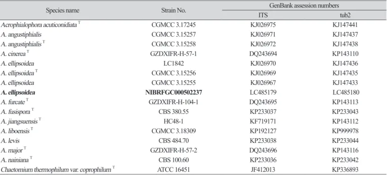
The DNA sequences obtained from our isolate were compared with reference sequences from the GenBank database of the National Center for Biotechnology Information (NCBI), using the basic local alignment search tool (BLAST). The isolates used to construct the phylogenetic tree are summarized in Table 1 with their strain and GenBank accession numbers. Based on Kimura�s neighbor-joining (NJ) algorithm, the evolutionary distance matrices were generated [12]. Phylogenetic analyses were performed using the program MEGA 7 [13] with bootstrap values based on 1,000 replications. The analyzed sequences were deposited at the collection facility of NCBI GenBank, with accession numbers of LC485179 and LC485180 for the ITS region and partial of tub2 gene sequences, respectively.
RESULTS AND DISCUSSION
Morphology of the KNU-US-1802E isolate
Colony diameters were 42 mm at 6 days of culture at 35°C on PDA. Colonies on PDA were flat, light gray, dense, with entire margins; reverse dark gray to black, with white margins (Fig. 1). Conidia were hyaline, one-celled, ellipsoidal to fusiform, forming long chains of more than ten conidia with an average length
× width of 5.0±0.3 × 2.9±0.2 μm (n=50). Although the isolate showed small differences in culture characteristics, most of morphological characteristics were like those previously reported for A. ellipsoidea CGMCC 3.15255 (Table 1). This result suggests that the fungal isolate KNU-US-1802E was closely related to A. ellipsoidea.
Table 1. Comparison of morphological characteristics of isolate KNU-US-1802E with the reference strain Acrophialophora ellipsoidea Characteristics Acrophialophora ellipsoidea KNU-US-1802Ea Acrophialophora ellipsoidea CGMCC 3.15255b
Colony Color and shape Flat, light gray with entire margins, has velvet like aerial-mycelia, reverse dark gray to black with white margin Flat, light gray, margin entire, velvet-like aerial mycelia, reverse dark gray to black, with no pigmentation in agar
Growth (Diam.) 42 mm after 6 days at 35°C on PDA 63 mm after 6 days at 35°C on PDA Conidia Conidial size 5.0 ± 0.3 × 2.9 ± 0.2 μm (n=50) 4.9 ± 0.7 × 2.8 ± 0.2 μm (n=50)
Shape and position Hyaline, one-celled, ellipsoidal to fusiform, forming long chains of more than 10 conidia One-celled, ellipsoidal or fusiform, hyaline, smooth-walled, forming chains of more than 10 conidia Phialide Shape Cylindrical, tapering at one end, solitary and lateral Solitary and lateral on somatic hyphae, Cylindrical or ellipsoidal
swollen bases, tapering into thin necks
a Fungal isolate studied in this paper.
b Source of description (Zhang et al., 2015)
Fig. 1. Cultural and morphological characteristics of Acrophialophora ellipsoidea KNU-US-1802E. A, B, Colony on potato dextrose agar (Front and reverse sides, respectively) after 6 days at 35°C; C, D and E, Chains of conidia on
Molecular phylogeny of the KNU-US-1802E isolate
In studying the phylogenetic relationship between isolate KNU-US-1802E and the previously reported A.
ellipsoidea, their ITS regions and a portion of their tub2 gene sequences were compared and analyzed. After the sequencing analysis, sequences of 507 bp and 488 bp were obtained from the ITS region and tub2 gene, respectively. BLAST search results indicated that the ITS region and the partial tub2 gene sequence showed 100% and 99% similarities, respectively, with A. ellipsoidea CGMCC 3.15255 collected from China. The concatenated sequences of the ITS region and tub2 gene were used to determine the molecular relationships between the sequences of A. ellipsoidea retrieved from GenBank (Table 2). A neighbor-joining method generated a phylogenetic tree showing that the strain KNU-US-1802E clustered in the same clade as other Acrophialophora strains, indicating that KNU-US-1802E is a strain of A. ellipsoidea (Fig. 2). Thus, fungal strain KNU-US-1802E was identified as A. ellipsoidea, and the fungal isolate KNU-US-1802E was deposited in the National Institute of Biological Resources (NIBRFGC000502237).
Genus Acrophialophora has been known to be a monophyletic and belongs to the family Chaetomiaceae [1].
Some species of these fungi can produce highly active laccase and cellulose as well as useful thermostable enzymes [14], while other species have been predicted to be emerging opportunistic pathogens in humans, associated with keratitis pulmonary colonization and infection [15] and devastating cerebral infections requiring intensive antifungal therapy [16]. According to previous reports, A. ellipsoidea is thermotolerant, having ideal growth temperatures of 37~40oC and maximum growth temperatures near 50oC, which are key to its acceptance into the Acrophialophora genus [6]. Therefore, the fungi can produce thermostable enzymes that help the food and paper industries and enhance agriculture, notwithstanding the fact that the species can cause infections in humans. Further studies are required to provide in-depth knowledge about this species. In this study, we report Acrophialophora ellipsoidea for the first time in Korea.
Table 2. List of the sequences used in this study
Species name Strain No. GenBank assession numbers
ITS tub2
Acrophialophora acuticonidiata T CGMCC 3.17245 KJ026975 KJ147441
A. angustiphialis CGMCC 3.15257 KJ026971 KJ147437
A. angustiphialis T CGMCC 3.15258 KJ026972 KJ147438
A. cinerea T GZDXIFR-H-57-1 DQ243694 KP143110
A. ellipsoidea LC1842 KJ026970 KJ147436
A. ellipsoidea T CGMCC 3.15256 KJ026969 KJ147435
A. ellipsoidea CGMCC 3.15255 KJ026967 KJ147433
A. ellipsoidea NIBRFGC000502237 LC485179 LC485180
A. furcate T GZDXIFR-H-104-1 DQ243695 KP143113
A. fusispora T CBS 380.55 KP233037 KP233043
A. jiangsuensis T HC48-1 KF719171 KP143112
A. liboensis T CGMCC 3.18309 KP192127 KP999978
A. levis CBS 484.70 KP233038 KP233044
A. major T GZDXIFR-H-57-2 DQ243696 KP143116
A. nainiana T CBS 100.60 KP233036 KP233042
Chaetomium thermophilum var. coprophilum T ATCC 16451 JF412013 KP336893
TType strain; bold letters: the isolates used in this study
ACKNOWLEDGEMENTS
This research was supported by the project on the survey and excavation of Korean indigenous species of the National Institute of Biological Resources (NIBR 201801105) under the Ministry of Environment, Republic of Korea.
REFERENCES
1. Edward JC. A new genus of the Moniliaceae. Mycologia 1959;51:781–6.
2. Zhang Y, Liu F, Wu W, Cai L. A phylogenetic assessment and taxonomic revision of the thermotolerant Hyphomycete genera Acrophialophora and Taifanglania. Mycologia 2015;107:768–79.
3. Liang ZQ, Han YF, Chu HL. A new thermotolerant Paecilomyces species that produces laccase and a biform sporogenous structure. Fungal Divers 2007;27:95–102.
4. Han YF, Liang JD, Dong X, Zou X, Du W, Liang ZQ. Research progress on Taifanglania. J Guizhou Agric Sci 2010;38:76–8.
5. Zhang YW, Wang Y, Zeng GP, Chen W, Xiao Z, Han YF, Qiu SY, Liang ZQ. A new species of Acrophialophora from Guizhou Province, China. Phytotaxa 2017;302:266–72.
6. Al-Mohsen IZ, Sutton DA, Sigler L, Almodovar E, Mahgoub N, Frayha H, Al-Hajjar S, Rinaldi MG, Walsh TJ. Acrophialophora fusispora brain abscess in a child with acute lymphoblastic leukemia: review of cases and taxonomy. J Clin Microbiol 2000;38:4569–76.
Fig. 2. Phylogenetic tree constructed with neighbor-joining (NJ) method, based on the concatenated ITS and tub2 gene sequences, shows the phylogenetic position of Acrophialophora ellipsoidea KNU-US-1802E among members of the genus Acrophialophora. The strain isolated in this study is shown in boldface. Bootstrap values (based on 1,000 replications) are shown at the branch points. Chaetomium thermophilum var. coprophilum T ATCC 16451 was used as an outgroup. Bar means 0.02 substitutions per nucleotide position.
7. Barros RR, Oliveira RA, Gottschalk LM, Bon EP. Production of cellulolytic enzymes by fungi Acrophialophora nainiana and Ceratocystis paradoxa using different carbon sources. Appl
Biochem Biotechnol 2010;161:448–54.
8. Park S, Ten L, Lee SY, Back CG, Lee JJ, Lee HB, Jung HY. New recorded species in three genera of the Sordariomycetes in Korea. Mycology 2017;45:64–72.
9. Gardes M, Bruns TD. ITS primers with enhanced specificity for basidiomycetes- application to the identification of mycorrhizae and rusts. Mol Ecol 1993;2:113–8.
10. White TJ, Bruns T, Lee S, Taylor J. Amplification and direct sequencing of fungal ribosomal RNA genes for phylogenetics. In: Innis MA, Gelfand DH, Sninsky JJ, editors. PCR protocols:
a guide to methods and applications. New York: Academic Press; 1990. p. 315–22.
11. Glass NL, Donaldson GC. Development of primer sets designed for use with the PCR to amplify conserved genes from filamentous ascomycetes. Appl Environ Microbiol 1995;61:1323–30.
12. Kimura M. A simple method for estimating evolutionary rates of base substitutions through comparative studies of nucleotide sequences. J Mol Evol 1980;16:111–20.
13. Kumar S, Stecher G, Tamura K. MEGA7: Molecular evolutionary genetics analysis version 7.0 for bigger datasets. Mol Biol Evol 2016;33:1870–4.
14. Yang SQ, Yan QJ, Jiang ZQ, Li LT, Tian HM, Wang YZ. High-level of xylanase production by the thermophilic Paecilomyces themophila J18 on wheat straw in solid-state fermentation.
Bioresour Technol 2006;97:1794–800.
15. Guarro J, Mendiratta DK, de Sequeira H, Rodríguez V, Thamke D, Gomes AM, Shukla AK, Menezes F, Narang P, Roldão Vieira J, Gené J. Acrophialophora fusispora: an emerging agent of human mycoses: a report of 3 new clinical cases. Diagn Microbiol Infect Dis 2007;59:85–8.
16. Li CW, Lee HC, Chang TC, Wan JY, Chen HM, Wu CJ, Lee NY, Chang CM, Lee CC, Ko WC. Acrophialophora fusispora brain abscess in a patient with acquired immune deficiency syndrome: a case report and review of the literature. Diagn Microbiol Infect Dis 2013;76:368–
71.