ISSN 2288-0356(Online) http://dx.doi.org/10.14518/crals.2013.31.4.002 Short Communications
Molecular Characterization of Fusarium proliferatum Causing Leaf Blight Symptoms on Chinese chive ( Allium tuberosum ) in Korea
Kyong-Han Kim, Seung-Yeol Lee, Chang-Gi Back, Hee-Young Jung
*School of Applied Biosciences, Kyungpook National University, Daegu 702-701, Republic of Korea
Abstract
In 2008, leaf blight symptoms were observed on several Chinese chive farms in Sangju. The Pathogenicity of the isolate was confirmed by artificial inoculation, where the pathogen exhibited a strong pathogenicity toward healthy plants. Morphological classification identified the isolate as from the Fusarium genus. For further analysis, PCR and phylogenetic classification were performed with ITS region and 28S rRNA gene which are commonly used for fungal identification. However, the results provided a poor resolution. To solve this problem, we analyzed translation elongation factor 1-alpha (TEF-1 α ) gene. The analyzed results using TEF-1 α gene indicated that the isolate was F. proliferatum . Therefore, it is assumed that TEF-1 α gene is important when Fusarium sp. was identified using molecular classification method.
Keywords:Chinese chive, Classification, Fusarium proliferatum , Leaf blight
Chinese chives ( Allium tuberosum ) is one of the vegetables being cultivated in Korea. Both young seedlings and adult plants of Chinese chive are high value products used for kimchi and soup. Chinese chive that cultivated continuously in fields throughout the year to fill market demand surpasses bulb onion.
However, during the summer of 2008, wilted plants were frequently found on several farms in the Chinese chive-growing areas of Sangju city. Initial symptoms were blight lesions on the tips of the leaves which eventually progressed to entire leaf necrosis and plant death. The affected plant had a brown discoloration. The symptoms clearly differed from those of Fusarium basal rot of Chinese chive caused by F. oxysporum f. sp. cepae .
1)To examine pathogenicity of microorganisms isolated from the affected Chinese chive, the Chinese chive plants infected by pathogen were received from several farms at Sangju city.
Diseased tissues were sectioned and sterilized in 70% ethanol for 30 sec followed by 0.5% (v/v) NaOCl for 30 sec, then rinsed with sterile distilled water 3 times and air-dried on sterile filter paper. Sectioned tissues were cut into 3~6mm pieces, and the small pieces were placed on potato dextrose medium (PDA) and incubated for 10~14 days at 25℃. After 2 or 3 days, Fusarium -like colonies were observed, and the hyphal tips of the colonies were transferred to a PDA medium. To test the pathogenicity of the isolate, spore suspension after filtering with
sterile gauze cloth was inoculated on healthy plants by three methods; pin-prickling, spray, and paper disk-covering. The isolate cultures were aseptically filtered through sterile gauze cloth. The spore concentration was estimated using a haemocytometer and adjusted to 1.6 x 10
2conidia/ml concentrations.
For the control, sterile distilled water was used for inoculation.
Thereafter, the pots were separated according to each treatment group to avoid cross-contamination with humidity for 24 hr.
Treated plants were watered as required to maintain normal growth.
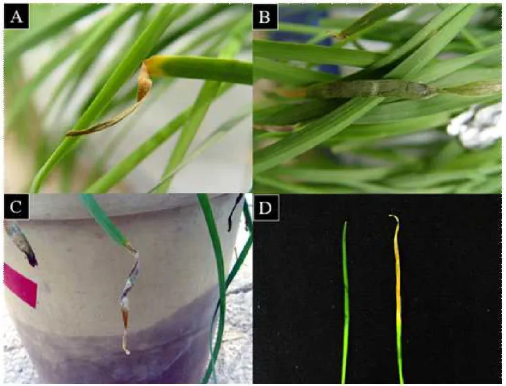
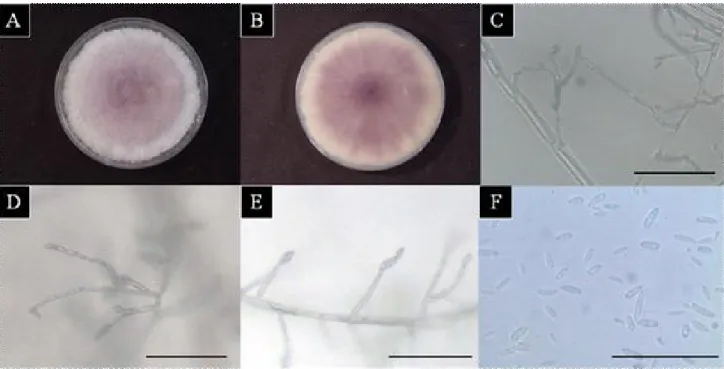
After 7 days, signs of leaf blight appeared on the leaf tips, which turned yellow and began to die back (Figure 1), whereas none of the control plants showed any symptoms during experiments. For the morphological analysis, the macroscopic and microscopic characteristics of the pure cultures were observed after 10 days. The mycelia were initially white and then developed a dark violet by time (Figure 2 A-B). Microconidia were produced from monophialides and polyphialides in long chains (Figure 2 C-E) in the aerial mycelium. In every case, the isolates of these species produced abundant microconidia that were monocellular and oval or elliptical in shape. The isolate was identified as from the Fusarium genus based on the characteristics of the microconidia (4.0-)7~10(-13)x 2.5~3.5㎛
that formed chains (Figure 2F) and the absence of chlamydospores.
These results were similar to a previous report (Shin and Kim
Received: December 11 2013 / Revised: December 16 2013 / Accept: December 31 2013
*
Corresponding Author: Hee-Young Jung, Tel. 82-53-950-5760, Fax. 82-53-950-6758, Email. [email protected]
©2012 College of Agricultural and Life Science, Kyungpook National University
Fusarium proliferatum causing leaf blight symptoms on Chinese chive 246
2001), it was hard to verify that the isolate was Fusarium sp., because macroconidia were absent.
2)To differentiate Fusarium sp. in detail, molecular biological analyses including PCR methods and Phylogenetic classification were performed. For PCR analysis, the isolate was grown in a PDA medium and incubated for 3 days at 25℃ for DNA extraction. The culture was then cut using a sterilized knife, and the harvested mycelium placed in a 1.5 ml Eppendorf tube.
To extract genomic DNA, a method described by Liu et al.
(2000) was applied after minor modification. PCR was performed with a thermal cycler (Astec, Japan) in a reaction solution including primers sets (Table 1), polymerase (Bioneer, Korea), 10 mM of dNTP (Bioneer, Korea), 2 μl of 10× reaction buffer (20 mM Tris-HCl, 400 mM KCl, 15 mM MgCl
2, pH 9.0, Bioneer, Korea) and 2 μl of the template DNA. Deionized
Figure 1. Symptoms of leaf blight of Allium tuberosum .
Pin-prickling inoculation (A), paper disk-covering inoculation (B), spray inoculation(C), and comparison of between healthy plant (Left) and inoculated plant (Right) (D).
distilled water as a negative control was included in every assay.
Amplicons were separated by electrophoresis on 0.7% agarose gels and visualized by staining with ethidium bromide. Under a UV illuminator, amplified and separated PCR products were purified by using ExoSAP-IT (USB co., USA) and prepared for DNA sequencing using Solgent (Daejeon, Korea).
Sequencing of each amplicon of the band, followed by BLAST
analysis (Altschul et al., 1997) and multiple alignments of ITS
region, 28S rRNA gene and TEF-1 α gene was done using the
SeqMan II software (DNASTAR, USA). We used sequence
analysis tools available at DDBJ. Alignment was made with
the program CLUSTALW (DDBJ version) and then automatically
converted to tree files. The trees were calculated with 100
bootstrap replications (Kimura 1980), and the results were
visualized using the TREEVIEW (win32). By BLAST analysis,
Figure 2. Mycological characteristics of the isolate from Allium tuberosum .
Photographs (A)-(B) show colonies of isolate on PDA medium after nine days using petri dishes 9cm in diameter; (C)-(E) show that chain of microconidia chains were produced on PDA medium after 3 days cultivation in the dark; (F) shows microconidia after 3 days. Scale bar represents 50㎛ for (C)-(F).
Table 1. PCR primer pairs used in this study Amplified
region Primer name Sequence(5´-3´) Resource
ITS region ITS1F
ITS4 CTTGGTCATTTAGAGGAAGTAA
TCCTCCGCTTATTGATATGC (http://www.biology.duke.edu/fungi/mycolab/primers.htm) 28s rRNA gene NL1
NL4 GCATATCAATAAGCGGAGGAAAAG
GGTCCGTGTTTCAAGACGG Sugita et al. (2003)
TEF-1αgene EF1
EF2 ATGGGTAAGGA(A/G)GACAAGAC
GGA(G/A)GTACCAGT(G/C)ATCATGTT O’Donnell et al. (1998)
the isolate can be included in Fusarium and phylogenetic analyses were proceeded to identify to its species level. For the partial 28S rRNA gene, 8 sequences were downloaded from Genbank (Figure 3) and the isolate’s ITS sequence was compared with species of other Fusarium deposited at Genbank (Figure 4). In line with the 28S rRNA gene analysis, the isolate can be included in F. fujikuroi complex, however further analyses were still needed to identify it to species level. For the partial ITS region, 13 sequences were downloaded from GenBank. We conducted phylogenetic distance tree which was constructed by the neighbor-joining method. We conducted phylogenetic analyses of 28S rRNA gene and ITS region data sets provided the results which showed the poor resolution.
To overcome this problem, other faster evolving parts of the genome must be analyzed. TEF-1 α gene-based analysis may be highly informative for phylogenetic classification Fusarium , because the gene encodes an essential part of the protein translation machinery and non-orthologous copies of the gene have not been detected in the genus (O’Donnell et al. 1998).
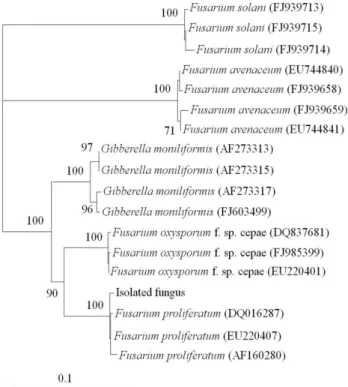
We used TEF-1 α gene and conduct its phylogenetic distance tree (Figure 5).
3)Molecular phylogenetic analyses demonstrated that the isolated fungus forms a monophyletic group with F.
proliferatum which confirmed us to accommodate the isolated fungus as F. proliferatum (Figure 5). We found that F.
proliferatum can infect Chinese chive plants and induce leaf
blight symptom. Recently, F. proliferatum was reported in Japan
Fusarium proliferatum causing leaf blight symptoms on Chinese chive 248
Figure 3. Phylogenetic distance tree was constructed using neighbor-joining method to compare the 28S rRNA genes of isolated fungus with those of other Fusarium sp. that cause Allium diseases from Genbank.
F. solani AY097317 and AY097318 were used as outgroups. Accession numbers are shown in parentheses. Numbers on branches are confidence values obtained for 100 replicates (only values above 70% are shown). Bar represents phylogenetic distance of 1%.
4)