- 49 -
서 론
소엽성 모세관 혈관종은 과거 화농성 육아종으로 불리 던 질환으로 피부, 비강, 입술, 혀, 구강 점막 등에서 빠르 게 성장하며, 쉽게 출혈하는 경향을 가진 양성 종양이 다.1) 후두에 발생하는 경우는 매우 드물며, 기관 삽관 및 인후두 역류증 등이 원인인자로 알려져 있다.2) 모든 연령층에서 발생 가능하지만 30대에서 흔하며, 특히 가 임기 여성에서 호발한다.3) 병리학적으로 내피세포의 과 분열과 증식에 의해 발생하는 것으로, 국소적인 외상에
대한 염증 반응으로 알려져 있으며, 가임기 여성에서 호 발하는 것으로 보아, 호르몬의 변화도 일부 관여한다고 한다.4) 후두에 발생시 흔한 증상은 발성장애, 애성, 호흡 곤란 및 기침 등의 증상이 나타날 수 있으며, 보존적인 치료에 반응이 없거나, 호흡곤란 등의 증상이 있는 경우 에는 수술의 적응이 된다.5)
저자들은 비소세포성 폐암으로 Gefitinib (Iressa®)치료 를 받은 병력이 있는 74세 남환의 거대 성문부 종물을 제거하여, 소엽성 모세관 혈관종으로 진단된 드문 증례 를 치험하였기에 문헌 고찰과 함께 보고 하고자 한다.
증 례
74세 남자 환자가 내원 1개월 전부터 서서히 심해지는 호흡곤란과 애성을 주소로 내원하였다. 인후두 역류증 의 전형적인 증상은 호소하지 않았다. 경성 후두내시경 검사상 성문의 중간부위와 후방부에 위치하며, 성문 입 Received
Re v i se d Accepted
: May 1, 2016 : May 20, 2016 : May 24, 2016
+Corresponding author: Seung Woo Kim, MD, Department of Otolaryngology-Head and Neck Surgery, Veterans Health Service Medical Center, 53, Jinhwangdo-ro 61-gil, Dunchon-dong, Gangdong-gu, Seoul, Korea. 05368 Tel: (02) 2225-1384 Tel: (02) 2225-1385 E-mail: [email protected]
대한두경부종양학회지, 제32권 제1호, 2016. pp.49-52 Korean Journal of Head & Neck Oncology, Vol.32, No.1
http://dx.doi.org/10.21593/kjhno/2016.32.1.49 ISSN 1229-5183(Print)
기도 폐색을 유발한 성문부 거대 소엽성 모세관 혈관종 1예
최전하1·임성환1·이미지2·김승우1+
중앙보훈병원 이비인후과1, 병리과2
A Case of Giant Lobular Capillary Hemangioma in Glottis Causing Airway Obstruction
Jeon Ha Choi, MD1, Sung Hwan Lim, MD1, Mi Ji Lee, MD2, Seung Woo Kim, MD1+
Department of Otolaryngology-Head and Neck surgery1 and Pathology,2 Veterans Health Service Medical Center, Seoul, Korea
= Abstract =
The lobular capillary hemangioma (LCH) was previously known to pyogenic granuloma and is benign vascular lesion which grows rapidly on skin and mucosa. It arises from whole body, but oral and nasal cavities are most predilection sites in the head and neck area. The laryngeal LCH looks like a granulomatous lesion of posterior glottis and its common etiology are tracheal intubation and laryngopharyngeal reflux disease etc. The LCH in larynx can cause blood tinged sputum and lump sense. The lesions refractory to medical therapy or causing dyspnea may require surgical excision. A 74-year-old man who presented gradually aggravated dyspnea, lump sensation and hoarseness of one month came to our hospital. The stroboscopic examination revealed large well-margined glottic mass. It was excised with CO2 laser and finally diagnosed as LCH. We present a rare unique case of glottic LCH with a review of literatures.
KEY WORDS:Lobular capillary hemangiomaㆍGlottis.
- 50 - 구의 약 80% 정도를 차지하고 있는 경계가 명확한 종물 이 관찰되었다. 종물은 분홍빛이었고, 우측 경계부에 소 량의 출혈소견이 관찰되었으며, 발성 및 호흡 시 약간의 가동성이 관찰되었다(Fig. 1A). 환자는 4년 전 비소세포 성 폐암으로 엽절제술을 시행 받았고, 수술 3년 후 재발 하여 표피성장인자 수용체 억제제인 Gefitinib (Iressa®)로 12회의 구제 항암치료를 받았다.
경부컴퓨터단층촬영에서 비교적 경계가 명확하며, 성 문부를 채우고 있는 약 1.3 × 1.1 cm 크기의 균일한 조영 증강을 보이는 종물이 관찰되었으나, 주변 림프절증대 의 소견은 관찰되지 않았다(Fig. 2A and B). 술 전 음성검
사에서 GRBAS척도는 전체적인 애성도에서 2, 조조성 (Rough, R)과 기식성(Breathy, B)은 2, 노력성(Strained, S) 은 1로 평가되었다. 최대 발성지속시간은 6.8초로 감소 되었고, 기본 주파수는 163.8 Hz, 주파수 변동률(jiitter)은 1.626 (참고값, <1.1), 진폭 변동률(shimmer)은 7.442 (참 고값, < 3.8), 잡음 대 배음비는 0.190 (참고값, <0.2)로 조조성 및 기식성 애성의 소견이었다.
성문부에 발생한 육아종을 우선적으로 고려하고, 증상 이 심해서 보존적 치료 없이 레이져를 이용한 후두미세 수술을 계획하였다. 마취는 기관절개술과 고빈도 환기 법 등을 준비한 상태에서, 5.5F 크기의 튜브로 기관 삽관 에 성공하였다. 종물의 위치는 우측 진성대의 중간부위 에서 후방부와 피열간극의 점막에 기저부를 두고 있었으 며, 겸자로 종물을 잡았을 때 쉽게 출혈하는 양상이었다.
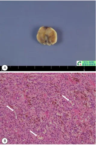
2.5watt 의 강도로 CO2 레이져를 이용하여, 피막의 파열 없이 조심스럽게 종물을 절제하였다. 최종 병리조직검 사에서 소엽성 모세관 혈관종으로 진단되었다(Fig. 3A and B). 환자는 술 후 2일째 특별한 합병증 없이 퇴원하였 고, 주요 증상은 소실되었다(Fig. 1B). 술 후 6주에 실시한 음성학적 검사에서 주요 음성학적 지표가 술 전에 비하
A
B
C
Fig. 1. Rigid laryngoscopic findings. It shows about 1.3×1.1 cm sized mass around the posterior glottis at first examina- tion (white arrow). The sessile granuloma on the posterior larynx is causing airway obstruction(A). It shows black am- munition formed by CO2 laser at the right posterior true vo- cal cord at the first day after operation (white arrow)(B). The fibrotic change and tiny granulation is observed on the same site after six months (white arrow)(C).
A
B
Fig. 2. Neck computed tomography (CT) scans. Preoperative non-enhanced axial CT shows 1.3 x 1.1cm sized soft tissue density mass in posterior glottis area (arrowhead)(A).
Preoperative enhanced axial CT shows 1.3 x 1.1cm sized homogeneously enhanced soft tissue mass in posterior glot- tic area (arrowhead)(B).
- 51 - 여 호전되었다. 술 후 6개월째 후두 내시경 소견에서 우 측 성대 돌기 주변의 미세한 육아 조직 이외에는 특이 소견소견 없이 외래 추적 관찰 중이다(Fig. 1C).
고 찰
소엽성 모세관 혈관종은 피부, 구강 및 비강 점막 등의 표면에서 종종 발견되며 빠르게 성장하며 쉽게 출혈하는 특성을 지니고 있는, 용종형의 모세관 혈관종이다.6) 30 대에서 잘 발생하며, 가임기 여성에서 호발한다.3,7) 비강, 혀, 결막, 외음부, 대장에 발생한 보고는 비교적 흔하게 있지만, 후두에서 발견되는 경우는 약 1~2% 이하로 매우 드물다.8)
성문부의 소엽성 모세관 혈관종의 흔한 증상은 애성, 호흡곤란, 기침 등이며, 그 기전은 궤양 및 출혈 등을 일으켜서, 성대 점막의 진동을 방해하여 발성장애를 유 발하고, 크기가 큰 경우는 상기도 폐쇄의 원인이 될 수 있다.8) 성문부 육아종의 호발 부위는 삽관 육아종의 경 우는 주로 후방성대에 발생하지만, 다른 육아종의 발생 부위는 비교적 다양하다.9) 육아종 형성의 기여인자로는
기도 삽관과 같은 외부 자극이 가장 흔하고, 위식도 역류 증, 음성의 오남용, 진균, 결핵균, 바이러스 등에 의한 감염 등이다.10)
본 증례의 환자는 평소 위식도 역류증상이 없었고, 증 상 발생 1년 이내에 기도 삽관의 이력도 없었다. 환자는 비소세포성 폐암의 재발로 증상 발생 약 1년 전부터 Gefitinib (Iressa®)으로 항암치료를 약 12회 시행 받았다.
Gefitinib은 표피성장인자 수용체(EGFR, epithelial growth factor receptor)인 티로신 인산화효소 억제제(tyrosine kin- ase inhibitor)로 비소세포성 폐암을 포함하여 EGFR 돌연 변이와 관련된 다양한 암치료에 사용되고 있으며, 이물 질의 주요 부작용으로는 여드름양 발진, 피부건조증 그 리고 손톱주위 염증 등이 보고되고 있으며 그 기전으로는 아직 더 많은 연구가 요구되지만, 티로신 인산화효소 억 제제가 EGFR에 공유 결합하여 비가역적으로 EGFR 신호 체계를 억제하기 때문으로 생각된다.11) 현재까지 표피성 장인자 저해제로 인한 상기도 점막에 소엽성 모세관 혈관 종이 발생했다는 보고는 없었지만, Gefitinib, Afatinib 등 으로 치료를 받은 후 조갑 주변 피부에 화농성 육아종이 발생했고, 그 치료를 중단 한 후 화농성 육아종이 완전히 사라졌다는 일련의 보고가 있었으며, 병변 주위 괴사조 직을 제거해주거나 레이져 치료가 치료속도를 더 증가시 켜 준다고 한다.11,12) 현재까지 이런 병변의 발생 기전은 명확히 알려져 있지 않다. 보다 체계적인 다기관 공동 연구가 필요하겠지만, 피부 및 상기도에 발생하는 소엽 성 모세관 혈관종의 발생 기전과 호발 부위와의 유사성을 고려하면, 본 증례의 경우 Gefitinib에 의한 육아종의 발생 가능성을 조심스럽게 추론해 볼 수 있다.
영상학적 소견은 전산화 컴퓨터 단층촬영에서는 균질 또는 국소적으로 조영증강 되지만, 특징적인 소견은 없 다.13) 확진은 병리학적 소견이 중요하며, 약한 염증성 변 화와 섬유성 기질에 의해 나뉘어진 소엽들 내부에 수많은 모세혈관의 배열이 관찰되었을 때 진단할 수 있다.13) 치 료는 음성치료, 생활 습관의 개선, 병변 내 스테로이드 주입술, 인후두 역류 치료 등을 시행 할 수 있으며,10) 보존 적 치료에 반응이 없는 경우는 악성 병변, 결핵, 진균, Wegener 육아종증 등을 감별해야 한다.10,14) 보존적 치료 에도 실패한 경우, 호흡곤란 및 기도폐쇄가 있는 경우 등은 수술의 적응이 되며, 마취 시 구강을 통한 삽관이 불가능 할 때는 기관절개술, 고빈도 환기법 등이 필요할 수 있다.10) 술 후에는 재발을 방지하기 위해 양성자 펌프 억제재나 항생제 등을 사용할 수 있으며, 술 후 재발이 빈번하므로 지속적으로 경과 관찰을 해야 한다.15)
저자들은 성문부의 대부분을 차지할 정도인 거대 소
A
B
Fig. 3. Pathologic findings. The cut surface of resected mass shows irregular shaped grayish white soft tissue(A). It shows numerous capillaries arranged in a lobular pattern (H&E, X200)(white arrows)(B).
- 52 - 엽성 모세관 혈관종을 CO2 레이져를 이용한 후두미세수 술로 치험하여 보고하고자 한다. 향후 표피성장인자 억 제제 사용과호흡기계 점막에서 소엽성 모세관 혈관종의 발생의 연관성에 관한 추가 연구가 필요할 것으로 사료 된다.
중심 단어:소엽성 모세관 혈관종ㆍ성문.
References
1) El-Sayed Y, Al-Serhani A. Lobular capillary hemangioma (pyogenic granuloma) of the nose. J Lanryngol otol 1997;
111:941-945.
2) Matthew M, Garett BA, Walter T. Obstructive pyogenic gran- uloma as a result of blunt laryngeal trauma. Otolaryngology- Head and Neck surgery 2007;136:89-90.
3) Leyden JJ, Master GH. Oral cavity pyogenic granuloma. Arch Dermatol 1997;108:226-228.
4) Puxeddu R, Berlucchi M, Ledda GP, Parodo G, Farina D, Nicolai P. Lobular capillary hemangioma of the nasal cavity:
a retrospective study on 40 patients. Am J Rhinol 2006;20(4):
480-484.
5) Kenneth O. Devancy, Alessandra R, Alfio F. Vocal process granuloma of the larynx-recognition, differential diagnosis and treatment. Oral oncology 2005;41(7):666-669.
6) Mills SE, Cooper PH, Fechner RE. Lobular capillary he- mangioma: the underlying lesion of pyogenic granuloma. A study of 73 cases from the oral and nasal mucous membranes.
Am J Surg Pathol. 1980;4:470- 479.
7) Walner DL, Parker NP, Kim OS, Angeles RM, Stich DD.
Lobular capillary hemangioma of the neonatal larynx. Arch Otolaryngol Head Neck Surg. 2008;134:272-277.
8) Matthew M, Garett BA, Walter T. Obstructive pyogenic gran- uloma as a result of blunt laryngeal trauma. Otolaryngol Head Neck Surg 2007;136:489-490.
9) Wang SG, Chon KM, Yoon IK, Kim DK, Lee SH, Park JW, et al. The effect of topical inhalant steroid (Budesonide, Pulmocort) in treatment of intubation granuloma. Korean J Otolaryngol 1992;35:183-190.
10) Ko JS, Park HW, Kim JP, Woo SH. One case report of ob- structing pyogenic granuloma. J Korean Soc Laryngol Phoniatr Logoped 2010;21(2):139-141.
11) Ehmann LM, Ruzicka T, Wollenberg A. Cutaneous side-ef- fects of EGFR inhibitors and their management. Skin Therapy Lett. 2011;16:1–3.
12) Whitney A. High. Gefitinib: A Cause of Pyogenic Granuloma like Lesions of the Nail.Arch Dermatol 2006;142(7):927-947.
13) Xu Q, Yin X, Sutedjo J, Sun J, Jiang L, Lu L. Lobular Capillary Hemangioma of the Trachea. Arch Iran Med. 2015;
18(2):127–129.
14) Kwak SG, Kim CD, Kim EJ, Kim SW. A case of coexistent squamous cell cancer with granuloma in posterior glottis.
Korean J Otorhinolaryngol-Head Neck Surg 2016;59(3):
246-249.
15) David L, Noah P, Parkerm BA, Oliver S, Ronald M, Duane D.
Lobular Capillary Hemangioma of the Neonatal Larynx.
Arch otolaryngology, Head & Neck surgery 2008;134(3):
272-277.