Cervical spondylolysis is a rare condition, and fewer than 100 cases have been reported in the English litera- ture since Perlman and Hawes first described the condi- tion in 1951 (1). Cervical spondylolysis is a bilateral de- fect in the posterior element of a cervical vertebra, and is condition usually diagnosed in patients after minor trauma or as an incidental finding on routine radi- ographs. However, the etiology of cervical spondylolysis is unknown. We report a case of cervical spondylolysis involving four levels in a child, and include radiographs and CT images.
Case Report
A 9-year-old boy visited to our hospital complaining of posterior neck pain of two days duration. The physical
examination of the cervical spine conducted at the time, was unremarkable.
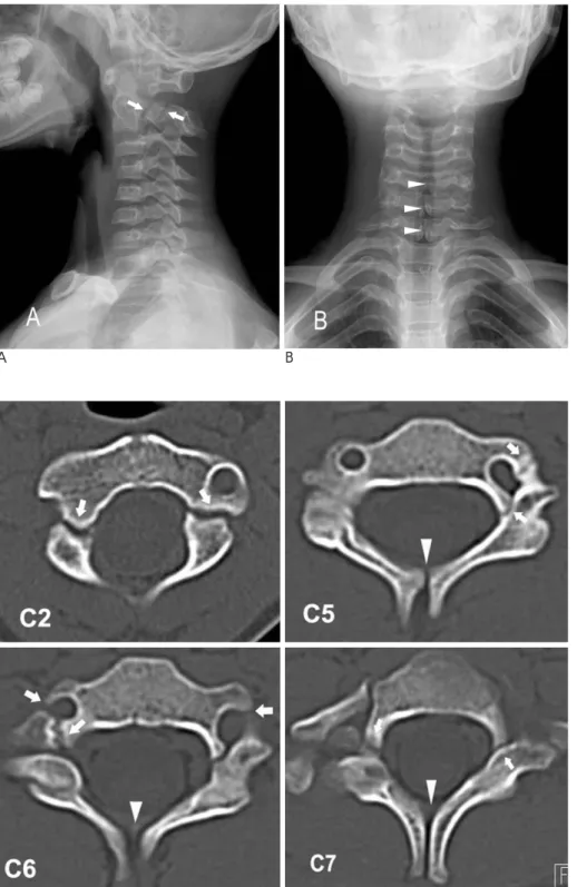
However, a plain lateral radiographic view of the cer- vical spine revealed radiolucent defects in the pedicle- neural arch regions at C2 (Fig. 1A), representing spondy- lolysis. Additionally, 2-mm of anterolisthesis of the C2 vertebral body upon C3 was observed, but there was no evidence of hypermobility or instability of the cervical spine. Associated spina bifida occulta was also observed to involve the C5, C6 and C7 levels (Fig. 1B).
Computerized tomography (CT, Somatom Sensation 16, Siemens, Erlangen, Germany) demonstrated bilater- al neural arch defects at the C2, C6 and C7 levels, with a unilateral defect at the C5 level on the left (Fig. 2 & 3). In addition, spina bifida occulta and dysplastic changes were evident at three abnormal cervical levels (C5-C7).
The patient was treated conservatively, and no manip- ulation was performed over the levels affected by spondylolysis. The presenting clinical symptom re- solved spontaneously.
Discussion
Cervical spondylolysis is defined as a corticated cleft
J Korean Radiol Soc 2006;55:619-622
─ 619 ─
Cervical Spondylolysis in Child with Four Levels of Simultaneous Involvement: A Case Report1
Gang Deuk Kim, M.D., Hye Won Kim, M.D., Sung Jo Jang, M.D.2, Jung Taek Oh, M.D.3
1Department of Diagnostic Radiology, Wonkwang University School of Medicine
2Department of Neurosurgery, Wonkwang University School of Medicine
3Department of General Surgery, Wonkwang University School of Medicine
* 이 논문은 2004년도 원광대학교 교비지원에 의해서 시행됨.
Received December 28, 2005 ; Accepted October 11, 2006
Address reprint requests to : Gang Deuk Kim, M.D., Department of Diagnostic Radiology, Gunsan Medical Center, 29-1 Jigok dong, Gunsan, Jeonbuk 573-713, Korea
Tel. 82-63-472-5343 Fax. 82-63-851-4749 E-mail: [email protected]
Cervical spondylolysis is a rare condition, and less than 100 cases have been report- ed in the world literature. Cervical spondylolysis is defined as a well corticated defect in the posterior element of a cervical vertebra. Although the etiology of cervical spondylolysis is unknown, its association with dysplastic changes and spina bifida oc- culta suggest that the lesion is congenital. Here, we describe the radiographs and CT images of cervical spondylolysis involving four levels in a 9 year old boy.
Index words :Spondylolysis Neck, abnormalities Neck, CT
between the superior and inferior articular facets of the articular pillar; the cervical equivalent of pars interartic- ularis in the lumbar spine (2). Patients experience markedly varied symptoms, ranging from asympto- matic to a mild nonspecific neck pain, neck stiffness, and radiculopathy, and the majority present after an episode of minor trauma or with chronic neck and shoulder pain. Pateint ages range from 20 to 81 years at diagnosis, and the C6 level and the left side of the neck
are most often affected. The fulcrum of motion in the cervical spine in children is at the C2-C3 level, whereas in the adult cervical spine it is located at the C5-C6 level (3, 4). Cervical spine injuries in children usually occur in the upper cervical spine from the occiput to C3.
The principal imaging modalities used to diagnose cer- vical spondylolysis are radiography and CT. Reported imaging findings include a well-corticated cleft between facets, a triangular configuration of pillar fragments on
Gang Deuk Kim, et al : Cervical Spondylolysis in Child with Four Levels of Simultaneous Involvement
─ 620 ─
A B
Fig. 1. Plain radiographs show spondy- lolytic defects (arrows) and associated spina bifida occulta (arrowhead).
Fig. 2. Axial CT images showing the well-corticated, smoothly marginated spondylolytic defects (arrow) and spina bifida occulta (arrowhead).
either side of spondylolytic defects, posterior displace- ment of the dorsal triangular pillar fragment, hypoplasia of the ipsilateral pedicle, spina bifida at the involved lev- el, compensatory hyper- or hypoplasia of the ipsilateral articular pillars at the levels above and/or below a de- fect, and spondylolisthesis (2, 3, 5, 6, 7).
The exact etiology of cervical spondylolysis remains unknown, although several theories have been pro- posed (8, 9). Failure of chondrification and ossification centers to unite is one such theory, and may account for the spectrum of observed posterior arch defects, which include cervical spondylolysis and an absent pedicle.
However, the presence of associated anomalies favors a congenital cause, but autopsy studies of newborns have never mentioned spondylolysis (8). It may be that a dys- plastic spine is simply more predisposed to the develop- ment of spondylolytic defects from whatever cause, and although repetitive microtrauma or post-traumatic nonunion have been suggested, no direct evidence sup- ports these theories.
Numerous conditions must be considered in the dif- ferential diagnosis of defects affecting the neural arch.
These include spinal cord tumors, bone tumors, trauma, abnormalities of the vertebral artery, and surgical laminectomy (5). Since the mainstay treatment is con- servative, it is most important to differentiate cervical spondylolysis from an acute fracture, which may re- quire acute surgical intervention (2, 6). Surgery should be considered when instability is present or when con- servative therapy fails. Acute fractures are not smoothly corticated, and the presence of soft tissue swelling or neurologic symptoms favor a diagnosis of acute frac- ture. Differentiation from a chronic fracture can be diffi-
cult, but corticated margins and associated congenital anomalies favor spondylolysis.
In summary, cervical spondylolysis is a rare anomaly, but its radiologic features are distinctive, and diagnosis often can be strongly suspected from plain films.
Moreover, CT can help confirm a diagnosis in equivocal cases. Well-corticated spondylolysis and spina bifida fa- vor a congenital rather than acute fracture based etiolo- gy. An awareness of the radiologic features of cervical spondylolysis reduces the potential for misdiagnosis and inappropriate therapy.
References
1. Perlman R, Hawes LE. Cervical spondylolisthesis. J Bone Joint Surg Am 1951;33-A:1012-1013
2. Forsberg DA, Martinez S, Vogler JB 3rd, Wiener MD. Cervical spondylolysis: imaging findings in 12 patients. AJR Am J Roentgenol 1990;154:751-755
3. Lustrin ES, Karakas SP, Ortiz AO, cinnamon J, Castillo M, Vaheesan K, et al. Pediatric cervical spine: normal anatomy, vari- ants, and trauma. Radiographics 2003;23:539-560
4. Roche C, Carty H. Spinal trauma in children. Pediatr Radiol 2001;31:677-700
5. Yochum TR, Carton JT, Barry MS. Cervical spondylolysis: three levels of simultaneous involvement. J Manipulative Physiol Ther 1995;18:411-415
6. Poggi JJ, Martinez S, Hardaker WT, Richardson WJ. Cervical spondylolysis. J Spinal Disord 1992;5:349-356.
7. Schwartz JM. Case 36: Bilateral cervical spondylolysis of C6.
Radiology 2001;220:191-194
8. Charlton OP, Gehweiler JA, Morgan CL, Martinez S, Daffner RH.
Spondylolysis and spondylolisthesis of the cervical spine. Skeletal Radiol 1978;3:79-84
9. Schwartz AM, Wechsler RJ, Landy MD, Wetzner SM, Goldstein SA. Posterior arch defects of the cervical spine. Skeletal Radiol 1982;8:135-139
J Korean Radiol Soc 2006;55:619-622
─ 621 ─
A B
Fig. 3. (A) Left paramedian sagittal multiplanar reformation and (B) three- dimensional images show the spondy- lolytic defects (arrow).
Gang Deuk Kim, et al : Cervical Spondylolysis in Child with Four Levels of Simultaneous Involvement
─ 622 ─
대한영상의학회지 2006;55:619-622
다발성 소아 경추 척추분리증: 증례 보고1
1원광대학교 의과대학 영상의학과
2원광대학교 의과대학 신경외과
3원광대학교 의과대학 일반외과
김강득・김혜원・장성조2・오정택3
경추 척추분리증은 전 세계적으로 100예 이하로 보고된 드문 질환으로 피질화가 잘된 결손이 척추 후방의 신경 궁에 있는 경우를 말한다. 원인은 아직 잘 모르나 동반된 이형성증이나 척추 이분증 등이 있어 선천성으로 알려져 있다. 저자들은 9세 소아에서 네 개의 경추에 발생한 척추분리증의 일반촬영과 CT 소견을 보고 한다.