- 65 -
R eceived R e v i s e d A ccepted
: July 31, 2018 : August 17, 2018 : September 7, 2018
+Corresponding author: Seung Woo Kim, MD
Department of Otolaryngology-Head and Neck Surgery, Veterans Health Service Medical Center, 53, Jinhwangdo-ro 61-gil, Gangdong-gu, Seoul, Korea. 05368
Tel: +82-2-2225-1384, Fax: +82-2-2225-1385 E-mail: [email protected]
대한두경부종양학회지, 제34권 제2호, 2018. pp.65-67 Korean Journal of Head & Neck Oncology, Vol.34, No.2
https://doi.org/10.21593/kjhno/2018.34.2.65 ISSN 1229-5183(Print) / ISSN 2586-2553(Online)
상측경부에 발생한 악하선 외 다형선종 1예
홍석정1⋅이미지2⋅김승우1+
중앙보훈병원 이비인후과1, 병리과2
A Case of Extra-Submandibular Gland Pleomorphic Adenoma in Upper Lateral Neck
Seok Jung Hong, MD1, Mi Ji Lee, MD2, Seung Woo Kim, MD1+
Department of Otolaryngology-Head and Neck Surgery1 and Pathology,2 Veterans Health Service Medical Center, Seoul, Korea
= Abstract =
Pleomorphic adenoma (PA) is a benign tumor which usually originates from major and minor salivary glands.
This tumor arising outside submandibular gland (SMG) is extremely rare. To author’s knowledge, only four cases have been reported so far in English literature. Its pathogenesis is still unclear, but it can be explained by embryo- logic theory of major salivary gland. A 68-year-old man with an incidental mass on left upper lateral neck visited to our clinic. The radiologic findings showed well-margined round mass outside left SMG. The excisional biopsy revealed a pleomorphic adenoma. We report the rare and unique case with a brief literature review.
Key W ords : Extra-submandibular gland, Pleomorphic adenoma
서 론
타액선 종양은 이하선에 가장 호발하며, 다양한 위치에 발생하는 소 타액선 기원 종양도 있다.1)소 타액선 종양이 아니면서 주 타액선 외에 발생하는 종물은 상경부에 발생 하는 왈틴 종양이 가장 흔하다.2,3)주 타액선 외 다형선종 은 같은 양상의 왈틴 종양보다 매우 드물며, 특히 악하선 외에서 발생한 경우는 이제까지 영문 검색에서 4예만 보 고되었다.4,5)저자들은 상측경부 종물을 주소로 내원한 68 세 남자 환자에서 악하선 외 다형선종으로 진단된 매우 드문 증례를 경험하여 문헌고찰과 함께 보고한다.
증 례
68세 남자 환자가 경동맥 초음파 시행 중에 우연히 발 견된 좌측 상측경부 종물을 주소로 본과에 의뢰되었다.
과거력에서 당뇨, 고혈압 등이 있었고, 음주력은 주 1회 소주 1병이었고, 흡연력은 없었다. 신체검사 소견에서 좌측 악하선과 인접하여, 직후방으로 약 2 X 2.4cm 크기 의 경계가 명확한 원형의 비교적 단단한 종물이 촉지되 었다(Fig. 1). 경부의 다른 곳에 비정상적으로 촉지되는 종물은 없었다. 신경학적 이상 소견은 없었으며, 후두 내시경 소견에서도 특이 소견 없었다. 경부 초음파 검사 에서 좌측 II 구역에 1.8 X 2.2cm 크기의 저 에코 음영을 보이는 종물이 악하선과 이하선 미부 사이에서 관찰되었 다(Fig. 2A). 전산화 단층 촬영에서 좌측 악하선의 직후 방, 이하선 및 후안면 정맥보다 내측에 위치하는 1.6 X 2.3cm 크기의 경계가 명확하고, 비균질하게 조영 증강되 는 종물이 관찰되었다(Fig. 2B and C). 초음파 유도 하 세침흡인검사에서는 유두상 배열과 비정형성을 가지는 상피성 종양 소견이었다. 확진 및 치료 등을 위하여 국소
- 66 -
A B C
Fig. 2. Radiologic findings. Transverse scan of neck ultrasonography shows that the 1.8 X 2.2cm sized hypoechoic mass with lobulated margin on left level II (asterisks) is located between left submandibular gland (arrow) and parotid tail (arrowhead)(A).
The axial enhanced neck CT scan reveals 1.6 X 2.3cm sized well-margined mass (long arrow) in medial to left parotid gland (arrowheads) and posterior facial vein (B). The heterogeneously enhanced mass (long arrow) is obviously dissociated from just posterior to left submandibular gland (short arrow)(C).
A B C
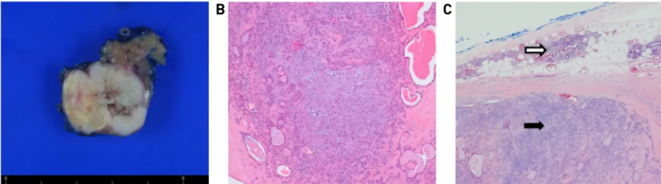
Fig. 3. Pathological findings. The surgical specimen shows 3.3 X 2 X 1.6cm sized grayish-white colored multi-lobulated mass (A).
The tumor is composed of variable epithelial and myoepithelial stromal components (H&E, X100)(B). Around the well-delineated tumor with fibrous capsule (black arrow), a normal salivary gland tissue is observed (white arrow)(H&E, X200)(C).
Fig. 1. External photograph shows a 2 X 2.4cm sized rubbery hard mass at left upper lateral neck (arrowheads).
마취 하에 절제 생검을 시행하였다. 종물의 상방에 절개 선을 가하고, 주변 구조물과 박리하여 종물을 노출시켰 다. 종물은 악하선과는 확연히 구분되어, 악하선의 직후 방에 위치하였고, 주변 구조물과의 유착이 관찰되었다.
종물의 외측면과 심부로 안면 동맥과 정맥의 분지가 주 행하여 이들을 결찰하고, 수술을 종료하였다.
병리 육안 소견에서 종물의 절단면은 3.3 X 2 X 1.6cm
크기의 경계가 명확한 구형의 여러 개 결절로 이루어져 있었다(Fig. 3A). 현미경 소견에선 종양 세포는 근상피 세포로 둘러싸인 관상구조에 점액물질을 함유하고 있고 주변에 정상 타액선 조직들이 일부 관찰되었다(Fig. 3B and C). 이상의 소견 등을 종합하여 악하선 외 다형선종 으로 최종 진단되었고, 특별한 합병증 없이 퇴원하였다.
술 후 9개월이 지난 현재까지 특이 소견 없이 추적 관찰 중이다.
고 찰
다형선종은 상피세포성분과 중배엽성 조직의 이중 기 원 때문에, 혼합종으로도 불린다.1) 가장 흔한 양성 타액선 종양으로, 주 타액선에서 호발하며 구순부, 협부, 설부, 구인두 및 비강 등의 소 타액선에서도 드물게 발생한다.1,2) 다형선종의 발생에 대한 가설은 예비세포 이론(reserve cell theory)이 가장 인정받고 있다. 이는 이하선 개재관 (intercalated duct)의 예비 세포들이 다형선종의 조직생성 전구물질로 작용하여, 점액양, 섬유양, 연골양 및 유골의 형태가 단독 혹은 복합적으로 나타난다는 이론이다.6)
주 타액선 외 양성 종양의 기원은 불명확하지만 타액
- 67 - 선의 발생과 관련되며, 이는 소 타액선 종양과는 다른 기전에 의한 것으로 추정된다.4)이하선은 악하선 및 설 하선 등에 비하여 조기에 발생하지만 피막 형성은 발생 14주로 상대적으로 가장 늦다.3)또한, 림프계의 발생은 이하선에 피막이 형성되기 이전, 악하선 및 설하선 등에 피막이 형성된 이후에 주로 일어나기 때문에 이하선 주 변에 타액선 상피조직이 잔존 할 수 있다.7)이런 발생학 적 배경 때문에 이소성 타액선 조직이 발생할 수 있으며, 주로 이하선 하방 및 상경부 림프절 등에서 발견되며, 악하선 주변에서는 극히 드물다.5)또한 이소성 타액선 조직이 존재하더라도, 종양이 형성되는 경우는 매우 드 물다고 알려져 있다.8)이렇게 발생한 주 타액선 외 종양 의 조직 소견은 타액선 내 종양과 유사하며, 약 80%는 양성으로 이하선 외 왈틴 종양이 가장 흔하고, 다형선종, 유두낭샘종, 호산성과립세포종 및 점액표피양 암종 등 의 보고도 있다.5,8,9)
소 타액선 기원이 아니면서 주 타액선 외에 발생한 다 형선종은 현재까지 20예 정도가 보고되었고, 가장 호발 하는 곳은 흉쇄유돌근 전연의 상부 및 경정맥-이복근 림 프절 등이다.4,5,10,11)이하선 외 종양과 악하선 외 종양의 감별은 병리 소견으로는 불가능하고, 이학적, 영상학적 및 수술 소견 등으로 구분하게 된다.4,5,8)이런 방법으로도 구분이 어려운 경우는 “주 타액선 외 종양”으로 명명하 기도 한다.5,8) 또한, 상경부에 발생한 이하선 외 왈틴 종양 및 다형선종 등은 일반적으로 본 증례보다 외측에 발견 되고, 종종 다발성 양상을 보인다.10)
타액선 외에 발생하는 다형선종은 서서히 커지는 무통 성 및 유동성 종물의 양상을 보인다.5)대부분 우연히 발 견되며, 세침흡인검사 및 영상 검사 등이 진단에 도움이 된다. 타액선 외 다형선종의 세침흡인검사 결과는 진단 적 가치가 높지만, 저 악성도의 점액표피양 암종, 선양낭 성 암종 등으로 오인될 수 있다.12)또한, 다형선종의 전산 화 단층 소견은 진단이 비특이적이다.11)대부분 최종 진 단을 위해서는 절제 생검이 필요하다.
다형선종의 예후는 양호하지만, 악성 변화 및 국소 재 발을 예방하기 위해 충분한 경계를 확보한 수술적 절제 가 필요하다.10)주 타액선 외 다형선종의 재발은 타액선 내 다형선종과 비슷하지만, 악성화의 위험성은 더 높다 고 알려져 있어 보다 세심한 초음파 추적관찰이 필요하 다.13)저자들은 본 증례를 통해서 경부 I, II 구역 및 악하 공간 주변에 종물이 관찰될 때 감별진단에 드물지만 이
소성 타액선 조직에서 발생한 종물의 가능성도 염두에 두어야 한다는 교훈을 얻었다.
References
1) Lee NH, Choi HJ, Yeo JO, Lee SH. A case of minor salivary gland pleomorphic adenoma arising from larynx. Korean J Otolaryngol- Head Neck Surg. 2008;51:1147-1150.
2) Choi KM, Yang SC, Kim SW. Two cases of extraparotid Warthin’s tumor in lateral cervical region. Korean J Head Neck Oncol.
2010;26:232-235.
3) Hwang JY, Kim SW, Yang SC, Kim CD. Extraparotid Warthin tumor in upper cervical lymph node accompanied by primary cervical tuberculosis. Otolaryngol Head Neck Surg. 2011;144:
646-647.
4) Kubota Y, Nitta S, Takenoshita Y, Shimizu M, Shirasuna K.
Pleomorphic adenoma originating from submandibular hetero- topic salivary gland tissue: A case report and review of the literature. Oral Oncology Extra. 2005;41:93-96.
5) Luksić I, Suton P, Manojlović S, Macan D, Dediol E. Pleomorphic adenoma in ectopic salivary gland tissue in the neck. Coll Antropol. 2012;36 Suppl 2:133-136.
6) Angelov A, Dikranian K, Trosheva M. Immunomorphological characteristics of pleomorphic adenoma of salivary glands. Bull Group Int Rech Sci Stomatol Odontol. 1996;39:67-75.
7) Kim JY, Kim JP, Lee EJ, Woo SH. A case of multifocal multisite Warthin’s tumor. Korean J Otolaryngol- Head Neck Surg. 2010;
53:778-780.
8) Daniel E, McGuirt WF Sr. Neck masses secondary to heterotopic salivary gland tissue: A 25-year experience. Am J Otolaryngol.
2005;26:96-100.
9) Sáenz-Santamaría J, Catalina-Fernández I, Fernández-Mera JJ, Villarreal-Renedo P. Low grade mucoepidermoid carcinoma arising in cervical lymph node. A report of two cases with fine needle aspiration findings. Acta Cytol. 2003;47:470-474.
10) Kim JM, Kim YJ, Kim CH, Kim SW. A case of multiple ex- traparotid pleomorphic adenomas in subcutaneous peri-auric- ular area. Korean J Otolaryngol- Head Neck Surg. 2013;56:
314-316.
11) Kim HJ, Hwang EG, Kim JH. Pleomorphic adenoma arising from heterotopic salivary gland tissue in the neck: A case report.
J Korean Radiol Soc. 1997;37:1021-1023.
12) Gudmundsson JK, Ajan A, Abtahi J. The accuracy of fine-needle aspiration cytology for diagnosis of parotid gland masses: A clin- icopathological study of 114 patients. J Appl Oral Sci. 2016;
24:561-567.
13) Cotelingam JD, Gerberi MP. Parotid heteropia with pleomorphic adenoma. Arch Otolaryngol. 1983;109:563-565.