■Received: 2013. 6. 8. ■ Revised: 2013. 10. 12.
■Accepted: 2014. 3. 1.
■Address reprint requests to Yu Cheol Kim, MD
Department of Ophthalmology, Keimyung University Dongsan Medical Center, #56 Dalseong-ro, Jung-gu, Daegu 700-712, Korea
Tel: 82-53-250-8026, Fax: 82-53-250-7705 E-mail: [email protected]
* This study was presented as a poster at the 109th Annual Meeting of the Korean Ophthalmological Society 2013.
Purpose: To report a case of lipemia retinalis in a patient with diabetes.
Case summary: A 27-year-old female with type 2 diabetes visited our clinic with visual disturbance in her left eye while being fol- lowed up from a pars plana vitrectomy in her right eye for proliferative diabetic retinopathy. On fundus examination of both eyes, the retinal vessels were creamy white and the retinal veins were undistinguishable from the retinal arteries. The serum trigly- ceride level was 2,676 mg/dL. The patient was asymptomatic except for visual impairment due to vitreous hemorrhage in her left eye. The patient was diagnosed with lipemia retinalis and chylomicronemia syndrome. After controlling the triglyceride level, fun- duscopic findings in the both eyes were improved. However, the visual acuity in her right eye remained unchanged.
Conclusions: Lipemia retinalis can be a sign of a systemic condition although it may not affect visual acuity. Fundus examination may be a useful tool in the early diagnosis of hyperlipidemia.
J Korean Ophthalmol Soc 2014;55(4):623-627
Key Words: Chylomicronemia syndrome, Lipemia retinalis, Triglyceride
ⓒ2014 The Korean Ophthalmological Society
This is an Open Access article distributed under the terms of the Creative Commons Attribution Non-Commercial License (http://creativecommons.org/licenses/by-nc/3.0/) which permits unrestricted non-commercial use, distribution, and reproduction in any medium, provided the original work is properly cited.
망막지혈증은 혈중 중성지방(트리글라세라이드) 수치가 2,000 mg/dL 이상이 될 때 나타나는 드문 안저소견이다. 혈 장의 중성지방 수치가 증가되면 혈액이 우유빛을 나타내게 되어 큰 망막혈관은 오렌지색을 나타내며 동맥과 정맥의 구 분이 어려워진다. 안저검사에서 이러한 망막혈관의 변화와 더불어 망막은 연어색(salmon color)을 나타내며 심할 경우 크림(creamy appearance)처럼 보이기도 한다. Brunzell and
Bierman1의 연구에 따르면 안저소견은 중성지방의 혈중 농 도에 따라 변화를 보이며 중성지방 수치의 증가로 인하여 망막지혈증의 소견을 보이더라도 중성지방 수치가 회복이 되면 안저소견 역시 원상태로 회복이 된다고 알려졌다. Lipid Research Program prevalence study에 따르면 고중성지 방혈증은 인구 10,000명당 1.79명의 유병률을 나타내며2 인 슐린 저항성 당뇨와 같은 대사장애 질환의 경우에 더 빈번 하게 발생한다고 알려졌다.3 저자들은 당뇨 환자에서 혈중 중성지방 수치 증가로 인한 망막지혈증 증례가 있어 이를 보고하고자 한다.
증례보고
증식당뇨망막병증으로 경과관찰 중인 27세 여자 환자가 좌안의 시력 저하를 주소로 내원하였다. 환자는 20세에 2형 당뇨로 진단받았고 5년 후 본원에서 증식당뇨망막병증으로 진단받았으며 우안은 황반부를 포함한 망막앞출혈이 있어
Figure 1. (A) Fundus photograph shows preretinal hemorrhage involving the macula. (B) Fundus photograph after pars plana vitrectomy.
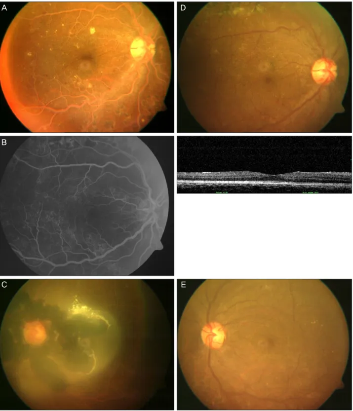
유리체절제술을 시행 받았다(Fig. 1). 내원 시 좌안 시력은 0.1, 안압은 18 mmHg로 3개월 전 검사 시의 0.5보다 저하되 어 있었고 우안 시력은 0.6, 안압은 16 mmHg로 차이가 없었 다. 안저검사에서 양안의 망막 혈관은 오렌지 색을 띠고 있었고, 좌안에는 지질(lipid)로 추정되는 노란색의 삼출물 이 포함된 유리체출혈 및 망막전출혈이 동반되어 있었다 (Fig. 2A, C). 빛간섭단층촬영(Spectral OCT/SLO; OTI, Ophthalmic Technology, Toronto, Ontario, Canada)에서 황반 부종이나 심한 경성 삼출물의 소견은 관찰되지 않았고 형광 안저혈관조영검사(FF450 plus fundus camera, Carl Zeiss Inc., Germany)에서는 변화된 혈관이 확장(dilation)되어있고 사행(tortuosity)이 관찰되었으나 누출이나 충만지연 등의 소 견은 확인되지 않았다(Fig. 2B). 환자의 과거력에서 당 조절 이 잘 이루어지지 않았던 것과 망막 혈관의 이상 소견으로 보아 망막지혈증을 의심하고 혈액검사를 시행하였다. 혈액 검사 결과, 공복 혈당수치는 313 mg/dL, 혈중 중성지방은 2,676 mg/dL, 당화혈색소는 13.9%, HDL-콜레스테롤 68.5 mg/dL, LDL-콜레스테롤 250.6 mg/dL로 측정되었고, 원심 분리 후의 혈장은 혼탁한 흰색을 띠고 있었다(Fig. 3A). 증 식당뇨망막병증과 망막지혈증으로 진단하고 좌안 유리체 절제술을 시행하고 혈중 중성지방 수치가 조절이 되지 않 을 경우 급성 췌장염이나 혈액 점성의 증가로 인한 심혈관 계 질환도 발생할 수 있기 때문에 식이조절과 내과치료를 병행하였다. 7일간의 입원 치료 후 공복 혈당수치는 113 mg/dL, 혈중 중성지방 수치는 425.1 mg/dL으로 호전되었고 원심분리 후의 혈장도 과거에 비해 맑아진 소견을 보였다 (Fig. 3B). 망막소견도 치료 전과 비교하여 망막 혈관의 동, 정맥의 구별이 가능할 정도로 호전을 보였으나(Fig. 2D), 우 안의 시력은 0.6으로 안저소견의 호전에 따른 시력 변화는
없었고 좌안은 수술 후 출혈이 제거되어 0.2로 시력이 호 전되었다(Fig. 2E).
고 찰
혈중의 중성지방 수치는 200 mg/dL 이하가 정상이지만 조절되지 않는 당뇨 환자와 비만, 알코올 중독, 신부전, 간 기능저하, 갑상선기능저하와 같은 대사 장애가 있는 경우 고중성지방혈증의 발생 빈도는 증가하며 에스트로젠과 같 은 호르몬제나 베타 차단제 같은 약물에 의해서도 유발이 가능하다.4 이 같은 경우는 이차적으로 혈중의 중성지방 수 치를 올리는 원인들로 알려졌으며 가족성 고중성지방혈증 같은 유전적인 원인으로도 가능하다.5 망막지혈증은 혈중의 중성지방 수치가 2,000-2,500 mg/dL 이상으로 증가 시 발현 될 수 있는 망막혈관의 이상 소견으로 망막혈관이 우유빛 (creamy-white)으로 변하는 것을 관찰할 수 있다.6 망막과 망 막혈관의 색 변화는 적혈구들이 얇게 분포되어 있는 주변 부 망막에서부터 먼저 관찰되고 혈중 중성지방 수치가 증 가될수록 후극부까지 변화가 미치게 되지만 직접적으로 시 력에는 영향을 미치지는 않으며7 혈중 중성지방 수치가 정 상화되면 망막 소견도 회복된다고 알려졌다.Lu et al8의 연 구에 따르면 고중성지방혈증에서 망막지혈증 소견을 보일 때 시력에는 영향을 미치지 않지만 전기생리학적 검사를 시행한 결과, 간상세포와 원추세포의 a-, b- 파장의 진폭이 정상치보다 감소되어 있는 것을 확인할 수 있었고 중성지 방 수치의 회복 시에는 전기생리학적 검사에서도 회복을 보인다고 하였다. 망막지혈증이 동반된 고중성지방혈증을 가진 환자의 치료는 혈중의 중성지방 수치 정도에 따라 시 행되며 내과적 약물과 식이조절 및 금식을 병행하여 혈중
A B
Figure 2. Fundus photography, fluorescein angiogram and optical coherence tomography before (A, B, C) and after (D, E) reso- lution of elevated triglyceride. (A) Retinal vessels are orange-colored and arterioles and venules are indistinguishable. (B) Fluorescein angiogram (left) shows tortuous and dilated large retinal vessels without leakage. Optical coherence tomography (right) reveals normal retinal thickness with foveal depression. (C) Vitreous hemorrhage and preretinal hemorrhage with suspected lipid materials were seen. (D, E) In both eyes retinal vessels have recovered their own colors.
중성지방 수치를 500 mg/dL 이하로 낮추는 것을 목표로 한 다.9 환자의 경우 당화혈색소는 13.9%, 공복 혈당 수치 313
mg/dL로 조절되지 않는 당뇨병을 가진 환자로 망막 혈관의 이상 소견을 보여 망막지혈증 의심하에 혈액검사를 시행하 B
C E
Figure 3. (A) A blood specimen was lipemic with a triglyceride concentration of 2,676 mg/dL. (B) The blood specimen with a trigly- ceride concentration of 425.1 mg/dL was less lipemic than (A).
여 혈중 중성지방 수치가 2,676 mg/dL으로 측정되면서 유 미지립혈증 증후군에 의한 망막지혈증으로 진단할 수 있었 다. 우안은 중성 지방 수치의 변화에 따른 안저소견의 변화 는 있었지만 시력은 0.6으로 유지되며 변화는 없었던 것으 로 보아 망막지혈증이 시력에는 영향을 미치지 않는다는 것을 확인할 수 있었고 빛간섭단층촬영과 형광안저혈관조 영검사에서도 혈관의 확장과 뒤틀림(tortuosity) 외에 일반적 인 당뇨망막병증의 소견과 크게 다르지 않아 안저소견에서 혈관의 변화 외의 다른 안과검사의 영상에는 영향이 없음 을 보여주었다. 좌안은 기존의 망막지혈증 증례에서 볼 수 없었던 유리체출혈이 있었던 경우로 망막지혈증에서 유리 체강 내에 출혈이 있을 경우 지질이 출혈과 함께 관찰될 수 있음을 알 수 있었다.
저자들은 조절되지 않는 당뇨병 환자에서 안저소견의 변 화가 고중성지방혈증을 진단할 수 있는 좋은 진단적 방법 이 될 수 있기에 이를 보고하는 바이다.
REFERENCES
1) Brunzell JD, Bierman EL. Chylomicronemia syndrome. Interaction of genetic and acquired hypertriglyceridemia. Med Clin North Am 1982;66:455-68.
2) Citkowitz E. Hypertriglyceridemia. Emedicine Online. Updated November 2013. http://emedicine.medscape.com/article/126568- overview.
3) Mostaza JM, Vega GL, Snell P, Grundy SM. Abnormal metabolism of free fatty acids in hypertriglyceridaemic men: apparent insulin resistance of adipose tissue. J Intern Med 1998;243:265-74.
4) Yuan G, Al-Shali KZ, Hegele RA. Hypertriglyceridemia: its etiol- ogy, effects and treatment. CMAJ 2007;176:1113-20.
5) Fredrickson DS, Levy RI, Lees RS. Fat transport in lipoprotein- sdan integrated approach to mechanisms and disorders. N Engl J Med 1967;276:34-44.
6) Vinger PF, Sachs BA. Ocular manifestations of hyperlipopro- teinemia. Am J Ophthalmol 1970;70:563-73.
7) Gopal L, Sunder KS, Rao SK, et al. Hyperlipidemia in a poorlycon- trolled diabetic presenting with lipemic aqueous and lipemia retinalis. Retina 2004;24:312-5.
8) Lu CK, Chen SJ, Niu DM, et al. Electrophysiological Changes in lipaemia retinalis. Am J Ophthalmol 2005;139:1142-5.
9) Leaf DA. Chylomicronemia and the chylomicronemia syndrome: a practical approach to management. Am J Med 2008;121:10-2.
A B
결론: 안저검사에서 보이는 망막지혈증은 직접적으로 시력을 저하시키지는 않지만, 고중성지방혈증을 의심할 수 있는 좋은 진단적 소견으로 생각된다.
<대한안과학회지 2014;55(4):623-627>