Case Report J Clin Med Res • 2011;4(1):61-63
Elmer ress
Articles © The authors | Journal compilation © J Clin Med Res and Elmer Press™ | www.jocmr.org
Subcutaneous Sacrococcygeal Myxopapillary Ependymoma in Asian Female:A Case Report
Kyung-Jae Lee a, b , Byung-Woo Min a , Hyuk-Jun Seo a , Chul-Hyun Cho a
Abstract
Subcutaneous sacrococcygeal myxopapillary ependymoma is ex- tremely rare tumor that has a tendency to develop in children and adolescents. There have been several case reports and sporadic re- ports in the literature. However, no case has been reported in an Asian patient, to the best of our knowledge. We describe a 25-year- old Asian female patient with a subcutaneous sacrococcygeal myxopapillary ependymoma that had been clinically diagnosed as a pilonidal cyst. The tumor was treated successfully by surgical ex- cision and the patient is doing well without evidence of local recur- rence or distant metastasis at 2 years after surgery.
Keywords: Myxopapillary ependymoma; Subcutaneous; Sacro- coccygeal
Introduction
Ependymomas are slowly growing glial cancers of the central nervous system that account for over 60% of spinal tumors of glial origin [1]. However, they rarely occur outside of the central nervous system known as extraspinal ependymomas.
The majority of the extraspinal ependymomas occur in the sacrococcygeal subcutaneous tissue or the presacral regions [2-7]. Although the incidences of ependymoma do not differ with respect to race [8], no case of subcutaneous sacrococ- cygeal myxopapillary ependymoma has been reported in an Asian patient. We present a case of an ependymoma arising from the sacrococcygeal subcutaneous tissue in a 25-year- old Asian female.
Case Report
A previously healthy 25-year-old-Asian woman was referred for evaluation of an enlarging, painless, subcutaneous mass located intergluteal fold. It was clinically diagnosed as a pi- lonidal cyst by previous examiner. She could remember that a mass had been present in this region for 2 years. During the 2-year period the mass had slowly increased in size from less than 0.5 cm to over 2.5 cm in diameter. During the past few months, the mass had become tender and sometimes making it uncomfortable to sit down. There was no history of urinary or faecal problems. Physical examination revealed a 2 × 3 cm sized, solid, mildly tender, mobile mass over the coccyx.
It was well circumscribed proximally but not in distally on palpation. Neurological examination was normal.
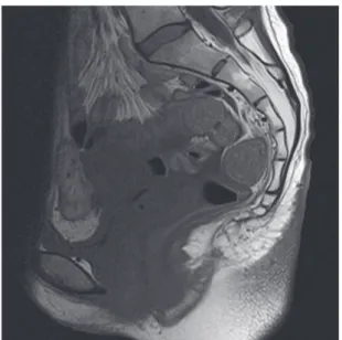
Magnetic resonance imaging (MRI) revealed a 6 ×3 cm well-circumscribed subcutaneous mass located over the coc- cyx. This tumor was comprised of two ovoid mass and there was no direct invasion to the coccyx (Fig. 1). Ultrasonogra- phy (USG)-guided needle biopsy was performed and micro- scopic examination showed a myxopapillary ependymoma.
Computed tomography (CT) scanning of the thorax and ab- domen demonstrated no evidence of lung or liver metasta- ses. Isotope bone scanning revealed no evidence of skeletal metastases.
The mass was completely excised. Intraoperatively, the lesion was seen near the tip of the coccyx and well-cir- cumscribed. It was composed of two ovoid but contiguous masses, the larger 3.0 × 2.5 × 1.5 cm, and the smaller 1.2
×1.0 ×0.8 cm (Fig. 2). The histology of the specimens again confirmed a myxopapillary ependymoma (Fig. 3). Her post- operative course was uneventful and no adjuvant therapy
Manuscript accepted for publication December 5, 2011
a
Department of Orthopaedic Surgery, College of Medicine, Keimyung University, Daegu, Korea
b