• 검색 결과가 없습니다.

Sudden Cardiac Arrest with Acute Myocardial Infarction Induced by Left Subclavian Artery Occlusion in a Patient with Prior Coronary Artery Bypass Surgery

N/A
N/A
Protected

Academic year: 2022

Share "Sudden Cardiac Arrest with Acute Myocardial Infarction Induced by Left Subclavian Artery Occlusion in a Patient with Prior Coronary Artery Bypass Surgery"

Copied!
3
0
0

로드 중.... (전체 텍스트 보기)

전체 글

(1)

866 Copyright © 2012 The Korean Society of Cardiology Korean Circulation Journal

Introduction

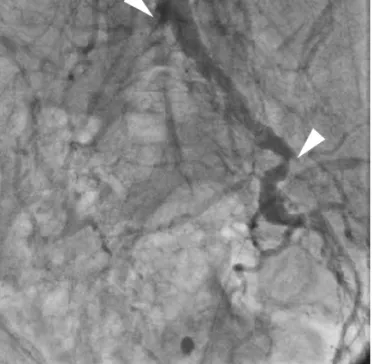
Coronary artery disease (CAD) is a significant factor in sudden car- diac arrest (SCA), and multivascular therapeutic approaches are nec- essary in CAD patients because atherosclerosis has a common sys- temic pathogenesis and simultaneously affects multiple circulation.

We report a rare case of SCA with acute myocardial infarction induc- ed by the total occlusion of left subclavian artery (LSCA) in CAD pa- tient with a history of coronary artery bypass surgery (CABG).

Case

A 70-year-old male presented at the emergency room with SCA

Case Report

http://dx.doi.org/10.4070/kcj.2012.42.12.866 Print ISSN 1738-5520 • On-line ISSN 1738-5555

Sudden Cardiac Arrest with Acute Myocardial Infarction Induced by Left Subclavian Artery Occlusion in a Patient with Prior

Coronary Artery Bypass Surgery

Ki-Bum Won, MD, Yun-Hyeong Cho, MD, and Deok-Kyu Cho, MD

Department of Cardiology, Myongji Hospital Cardiovascular Center, Kwandong University College of Medicine, Goyang, Korea

Multivascular preventive and therapeutic approaches are necessary in patients with coronary artery disease because atherosclerosis has a common systemic pathogenesis. We present a rare case of sudden cardiac arrest with acute myocardial infarction induced by the total occlusion of left subclavian artery (LSCA) in a patient with a history of previous coronary artery bypass surgery using the left internal mam- mary artery. We initially performed blind-puncture of left brachial artery, attempting percutaneous coronary intervention because pulses were absent in both upper and lower extremities. However, the cause of sudden cardiac arrest was atherosclerotic total occlusion of LSCA.

The patient was stabilized after successful revascularization of LSCA by percutaneous transluminal angioplasty with stent insertion.

(Korean Circ J 2012;42:866-868)

KEY WORDS: Sudden cardiac arrest; Coronary artery bypass surgery; Atherosclerosis.

Received: April 24, 2012 Revision Received: May 29, 2012 Accepted: June 1, 2012

Correspondence: Deok-Kyu Cho, MD, Department of Cardiology, Myongji Hospital Cardiovascular Center, Kwandong University College of Medicine, 697-24 Hwajeong-dong, Deogyang-gu, Goyang 412-270, Korea

Tel: 82-31-810-6776, Fax: 82-31-810-6778 E-mail: [email protected]

• The authors have no financial conflicts of interest.

This is an Open Access article distributed under the terms of the Creative Commons Attribution Non-Commercial License (http://creativecommons.

org/licenses/by-nc/3.0) which permits unrestricted non-commercial use, distribution, and reproduction in any medium, provided the original work is properly cited.

after complaining of typical chest pain in December 2011. We had only limited information that he had coronary risk factors including hypertension, diabetes, and smoking, and he underwent CABG 4 ye- ars ago in another hospital. After cardiopulmonary resuscitation for 15 minutes, the electrocardiogram showed ST elevation in the ante- rior leads. We promptly referred the patient to the cathroom for pri- mary percutaneous coronary intervention.

Initiall physical examination revealed absent pulses in both upper and lower extremities. We perforce performed blind-puncture at left brachial artery, and attempted coronary angiography. However, the 035’’ hydrophilic soft terumo wire (Terumo, Tokyo, Japan) did not pass the LSCA. Transbrachial peripheral angiography showed total occlusion of LSCA, but the graft from left internal mammary artery (LIMA) to mid left anterior descending artery (LAD) was patent (Fig.

1A). After 014’’ guide wire (Pilot, Abbott, Santa, Clara, CA, USA) pass-

ed the lesion, the LSCA was progressively dilated by a 2.0×20 bal-

loon (Lacrosse, Goodman Medical Ireland Ltd., Galway, Ireland) inflat-

ed at 16 atm, and a 4.0×20 mm balloon (Lacrosse, Goodman Me-

dical Ireland Ltd., Galway, Ireland) at 18 atm (Fig. 1B). A 7.0×24 mm

stent (Coroflex Blue, Braun Vascular Systems, Berlin, Germany) was

deployed at the LSCA (Fig. 1C). Transbrachial coronary angiography

showed 3 vessels with CAD presenting significant atherosclerotic

stenosis of proximal LAD, big obtuse marginal artery, and mid right

coronary artery (Fig. 2). Peripheral angiography showed total occlu-

sion of right common iliac artery and left external iliac artery (Fig. 3).

(2)

867 Ki-Bum Won, et al.

http://dx.doi.org/10.4070/kcj.2012.42.12.866 www.e-kcj.org

Because the patient’s vital signs were unstable, we attempted to insert the intra-aortic balloon pump (IABP) via left brachial artery. The IABP successfully passed the deployed stent of LSCA, and was opti- mally located in descending thoracic aorta. However, the IABP was not operating properly, and we finally removed the IABP. The patient was stabilized with medical treatment after percutaneous translu- minal angioplasty (PTA), but unfortunately had sequela of ischemic brain damage.

Discussion

Coronary artery disease is known to be one of the most common causes of SCA. As newer treatments of CAD continues to reduce mortality and morbidity, cardiovascular physicians increasingly con- front the problem of concomitant peripheral arterial disease. There is

a need to encourage new therapeutic approaches for the treatment of multivascular atherosclerotic disease in CAD patients.

Left internal mammary artery is the conduit of choice for CABG.

Preoperative assessment of LIMA before CABG should be carefully performed because most subclavian atherosclerotic stenoses exist in the absence of symptoms. The comparison of blood pressure mea- surements in both arms is important to exclude significant subcla- vian stenoses that may interfere with successful cardiac circulation of the LIMA graft.

1)

Selective left subclavian angiography with seg- mental pressure measurement is indicated for patients with sus- pected subclavian disease who need CABG, and pseudolesions ca- used by respirophasic kinking of the subclavian arteries should be considered. In our case, unfortunately, we could not know whether or not preoperative evaluation of subclavian artery was precisely per- formed because this was our patient’s first visit with SCA to our car-

A   B   C  

Fig. 1. Transbrachial peripheral angiography demonstrates (A) total occlusion of left subclavian artery (LSCA) (arrow) with patent graft from left internal mammary artery to mid left anterior descending artery, (B) balloon angioplasty of LSCA (arrow), (C) successful revascularization with stent insertion at LSCA (arrow).

A   B  

Fig. 2. Transbrachial coronary angiography demonstrates (A) significant atherosclerotic stenoses of proximal left anterior descending artery (arrow), big

obtuse marginal artery (arrowhead), and (B) mid right coronary artery (arrow).

(3)

868 Left Subclavian Artery Occlusion

http://dx.doi.org/10.4070/kcj.2012.42.12.866 www.e-kcj.org

diovascular center.

Risk factors for atherosclerotic disease of the upper extremity are identical to those for atherosclerosis in coronary or lower extremity arteries with the exception of other etiologies specific to the upper extremity.

2-6)

The incidence of severe obstruction in the subclavian arteries is known to be lower than that in the iliofemoral systems, and ischemic symptoms are also less frequent because of the low- er metabolic work and the propensity for collaterals formation ar- ound shoulder and scapula. PTA of atherosclerotic obstruction of the subclavian arteries should be considered for patients with reprodu- cible arm claudication, distal hand atheroembolism, subclavian steal syndrome, and impaired inflow into internal mammary grafts, axillo- femoral bypasses, and dialysis access fistulas.

7)

Although experi- ences with subclavian stenting are limited to small series, it is known

that stenting is superior to balloon dilatation alone.

8)9)

In this case, we performed primary PTA with stent insertion at the ostium of LSCA because total occlusion of LSCA interfere with cardiac circulation of the LIMA graft, and the lesion was the culprit of SCA in this patient.

Multivascular preventive and therapeutic approaches for patients with diffuse atherosclerotic disease are certainly needed. Cardiovas- cular surgeons and interventionists should take care of a more pro- active clinical role, along with their vascular medicine colleagues.

They should also encourage optimal treatment of arterial disease af- fecting multiple vascular beds guided by an appreciation of the di- verse natural histories associated with peripheral arterial disease.

References

1. Shadman R, Criqui MH, Bundens WP, et al. Subclavian artery stenosis:

prevalence, risk factors, and association with cardiovascular diseases.

J Am Coll Cardiol 2004;44:618-23.

2. Cherry KJ. Atherosclerotic occlusive disease of brachiocephalic arteries.

In: Rutherford RB, editor. Vascular Surgery. 5th ed. Philadephia: W.B.

Saunders Company;2000. p.1140-1.

3. Arend WP, Michel BA, Bloch DA, et al. The American College of Rheuma- tology 1990 criteria for the classification of Takayasu arteritis. Arthritis Rheum 1990;33:1129-34.

4. Singer AJ, Hollander JE. Blood pressure. Assessment of interarm differ- ences. Arch Intern Med 1996;156:2005-8.

5. Fairbairn JF, Juergens JL, Spittell JA. Peripheral Vascular Diseases. 4th ed.

Philadelphia: W.B. Saunders Company;1972.

6. Kappert A. Lehrbuch und Atlas der Angiologie. 12th ed. Bern: Huber;

1987.

7. Azzarone M, Cento M, Mazzei M, Tecchio T, Ugolotti U. Symptomatic subtotal occlusion of the innominate artery treated with balloon an- gioplasty and stenting. J Endovasc Ther 2000;7:161-4.

8. Rodriguez-Lopez JA, Werner A, Martinez R, Torruella LJ, Ray LI, Dieth- rich EB. Stenting for atherosclerotic occlusive disease of the subclavian artery. Ann Vasc Surg 1999;13:254-60.

9. Hadjipetrou P, Cox S, Piemonte T, Eisenhauer A. Percutaneous revas- cularization of atherosclerotic obstruction of aortic arch vessels. J Am Coll Cardiol 1999;33:1238-45.

Fig. 3. Transbrachial peripheral angiography demonstrates total occlusion of

right common iliac artery (arrow) and left external iliac artery (arrowhead).

수치

Fig. 1. Transbrachial peripheral angiography demonstrates (A) total occlusion of left subclavian artery (LSCA) (arrow) with patent graft from left internal  mammary artery to mid left anterior descending artery, (B) balloon angioplasty of LSCA (arrow), (C)
Fig. 3. Transbrachial peripheral angiography demonstrates total occlusion of  right common iliac artery (arrow) and left external iliac artery (arrowhead).

참조

관련 문서

Cerebral Cerebral Cerebral angiography Cerebral angiography angiography angiography shows shows shows shows feeding feeding feeding artery feeding artery

In 4-week group, the group filled with bone graft with decortication revealed larger new bone formation area than shown in the group that had a defect area

The locations of aneurysms were middle cerebral artery in 15 patients, cerebral artery in 15 patients, cerebral artery in 15 patients, cerebral artery in

82 year-old female patient with traffic accident, (A,B,C) Initial x-rays show open fracture of left distal femur(AO 33-C3, Gustilo-Anderson II) with patella

Pilot Confirm runway 09 left for landing. ATC gives you further instructions to turn right heading 310. On the radar display, there was an area to avoid which requires a

activation(electroencephalolgram: left alpha wave, right alpha wave, left sensory motor rhythm, right sensory motor rhythm, right mid-beta wave, left attention concentration

Sizes of left and right pupils Intensities of left and right pupils Ellipse ratios of left and right

 Although van der Waals equation is still less accurate at high