• 검색 결과가 없습니다.

A Case of Three Consecutive Events of Acute Myocardial Infarctions in Three Different Vessels

N/A
N/A
Protected

Academic year: 2022

Share "A Case of Three Consecutive Events of Acute Myocardial Infarctions in Three Different Vessels"

Copied!
5
0
0

로드 중.... (전체 텍스트 보기)

전체 글

(1)

Korean Circulation Journal

Introduction

Multivessel myocardial infarction (MI) is a rare case. Most cases of multivessel MI are simultaneous events and consecutive events of MI usually involving the same vessel. A case with consecutive events of acute MI in different vessels is rare. In addition, patients of consecutive events of acute MI have multiple risk factors or spe- cific underlying diseases. There have been no reports on 3 consec- utive events of acute MI in each 3 vessels during a long-term inter- val. Herein, we report the first case with 3 consecutive events of acute MI in 3 different vessels during a long-term interval.

Case

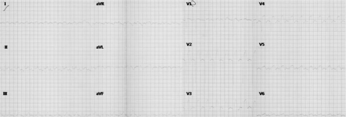
On September 25, 2012, a 51-year-old man visited the emergency

Print ISSN 1738-5520 • On-line ISSN 1738-5555

A Case of Three Consecutive Events of Acute Myocardial Infarctions in Three Different Vessels

Hyun Yang, MD 1 , Sung-Ho Her, MD 1 , Mahn Won Park, MD 1 , Jung Sun Cho, MD 1 ,

Chan Joon Kim, MD 1 , Jong-Bum Kwon, MD 2 , Sang Mi Ro, MD 1 , and Yun Kyung Park, MD 1

1

Division of Cardiology, Department of Internal Medicine and

2

Division of Thoracic and Cardiovascular Surgery, Daejeon St. Mary’s Hospital, College of Medicine, The Catholic University of Korea, Daejeon, Korea

A 51-year-old man was being admitted to the emergency department with chest pains. He had a history of acute myocardial infarction (MI) on two prior occasions and was successfully treated with drug eluting stents. He was diagnosed with 3 consecutive events of acute MI in 3 different vessels. The consecutive events of acute MI in different vessels are a very rare case. He did not have risk factors, such as coagulation abnormality, clopidogrel resistance, patient’s compliance and vessel abnormality, except for his cigarette smoking. We reported the first case with 3 consecutive events of acute MI in each 3 vessels during a long-term interval. (Korean Circ J 2013;43:694-698) KEY WORDS: Myocardial infarction; Coronary vessels; Thrombosis.

Received: December 6, 2012 Revision Received: March 19, 2013 Accepted: May 16, 2013

Correspondence: Sung-Ho Her, MD, Division of Cardiology, Department of Internal Medicine, Daejeon St. Mary’s Hospital, College of Medicine, The Ca- tholic University of Korea, 64 Daeheung-ro, Jung-gu, Daejeon 301-723, Korea Tel: 82-42-220-9504, Fax: 82-42-226-9686

E-mail: [email protected]

• The authors have no financial conflicts of interest.

This is an Open Access article distributed under the terms of the Creative Commons Attribution Non-Commercial License (http://creativecommons.

org/licenses/by-nc/3.0) which permits unrestricted non-commercial use, distribution, and reproduction in any medium, provided the original work is properly cited.

room for acute retrosternal squeezing chest pains for the past tw- elve hours. He had no history of diabetes mellitus, hyperlipidemia, and nor a familial history of coronary artery disease. However, he was a current smoker (30 pack years). He had hypertension and his- tory of two consecutive acute MI.

The first acute MI was on February 21, 2000. The electrocardiogram (ECG) showed ST-segment elevations in the inferior leads (II and aVF) and the anterior (V 2-4) leads (Fig. 1). The peak levels of creatine kinase-MB (CK-MB) and troponin T were 31.71 and 2.51 ng/mL. The coronary angiography was performed. The coronary angiography showed a total occlusion with thrombus and Thrombolysis in Myo- cardial Infarction (TIMI) 0 distal flow in the proximal portion of the left anterior descending from the coronary artery (LAD), which was successfully treated with a bare metal stent (3.0×15 mm, NIR Roy- al® stent, Boston Scientific Corporation, Natick, MA, USA). In addi- tion, he had taken aspirin 100 mg, atenolol 25 mg, captopril 37.5 mg and simvastatin 10 mg once daily and cilostazol 100 mg twice daily, on a regular basis after the first MI event.

The second acute MI was on December 30, 2007. The ECG show- ed ST-segment elevation in the inferior (II, III, and aVF) leads (Fig. 2).

The peak levels of CK-MB and troponin T were 164.7 and 4.01 ng/

mL. Emergent coronary angiography was performed. The coronary

angiography showed total occlusion with thrombus, with TIMI 0 dis-

tal flow in the mid portion of the right coronary artery (RCA), which

was successfully treated with a paclitaxel eluting stent (4.0×24 mm,

TAXUS®, Boston Scientific Corporation, Natick, MA, USA) (Fig. 3-1),

and a 95% restenosis in a stent with thrombus, and TIMI 0 distal

(2)

flow in the proximal portion of the LAD, which was successfully treated with a zotarolimus eluting stent (2.75×30 mm, ENDEAV- OR®, Medtronic Inc., Minneapolis, MN, USA) and overlapping a siro- limus eluting stent (3.0×23 mm, CYPHER®, Cordis Corporation, War- ren, NJ, USA) (Fig. 3-2). There were no other stents for appropriate lesion lengths and sizes, except ENDEAVOR and CYPHER stents at

that time.

He had taken aspirin 100 mg, clopidogrel 75 mg, ramipril 2.5 mg and simvastatin 10 mg once daily and molsidomine 2 mg twice dai- ly, on a regular basis after a second MI event.

At the third acute MI, physical examination showed that his blood pressure was 90/60 mm Hg, the heart rate was 64/min, and respi-

Fig. 1. ECG on February 21, 2000. The ECG shows ST-segment elevation in the inferior leads (II and aVF) and the anterior (V 2-4) leads. ECG: electrocardio- gram.

Fig. 2. ECG on December 30, 2007. The ECG shows ST-segment elevation in the inferior (II, III, and aVF) leads. ECG: electrocardiogram.

Fig. 3-1. Right coronary angiography on December 30, 2007. A: the initial coronary angiography shows a total occlusion with thrombus and TIMI 0 distal flow in the mid portion of the RCA (white arrow). B: the post stent coronary angiography shows TIMI 3 distal flow in the RCA, which is successfully treated with a paclitaxel eluting stent (white arrow). TIMI: Thrombolysis in Myocardial Infarction, RCA: right coronary artery.

A B

(3)

ratory rate was 20/min. He had a regular heart beat and clear heart sounds without murmurs.

Electrocardiogram showed Q-wave and ST elevation in the V 1-5 and T-inversion in V 4-6 (Fig. 4). The peak levels of CK-MB and tro- ponin T were 17.84 and 0.741 ng/mL.

Echocardiographic evaluation revealed aneurysmal changes at the apicoseptal wall in the left ventricle, and ischemic insult in the LAD and the RCA territory. An ejection fraction of 40% was observed.

Emergent coronary angiography showed a total occlusion with thrombus and TIMI 0 distal flow in the proximal portion of the left circumflex coronary artery and collateral flow from RCA, which was successfully treated with a zotarolimus eluting stent (3.5×22 mm,

RESOLUTE INTEGRITY®, Medtronic Inc., Minneapolis, MN, USA) (Fig.

5). After primary percutaneous interventions, the patient became stable without chest pains and was discharged on the fourth hos- pital day.

Hematological analyses were performed and showed the follow- ing results: D dimer 0.38 ug/mL (<0.5 ug/mL), antithrombin III 107%

(80-120%), protein C activity 131% (70-130), and protein S activity 90% (70-130). In addition, immunological analyses were perform- ed and indicated the following results: lupus anticoagulant titer 1.01 (0.8-1.2), anti-beta2-glycoprotein1 immunoglobulin G/M titer negative/negative, anti-cardiolipin antibody immunoglobulin G/M titer negative/negative and homocystein 12.33 umol/L (5.08-15.4

Fig. 4. ECG on September 25, 2012. The ECG shows Q-wave and ST elevation in V 1-5 and T-inversion in V 4-6. ECG: electrocardiogram.

Fig. 3-2. Coronary angiography on December 30, 2007. A right anterior oblique view. A: the initial coronary angiography shows a 95% restenosis in stent with thrombus and TIMI 0 distal flow in the proximal portion of the LAD (white arrow). B: the post stent coronary angiography shows TIMI 3 distal flow in the LAD, which is successfully treated with a zotarolimus eluting stent (black arrow) and overlapping a sirolimus eluting stent (white arrow). TIMI: Throm- bolysis in Myocardial Infarction, LAD: left anterior descending coronary artery.

A B

(4)

umol/L). Further, the platelet drug response assay was also per- formed and displayed the following results [The results of the as- say are reported as absolute P2Y12-Reaction-Units (PRU), as well as percent inhibitions (% inhibition), the latter calculated as 100- (TEST/BASE×100) or {(BASE-TEST)/BASE}×100]: PRU 215 (0-240),

% inhibition 31% (20-80).

1)

He was discharged after uneventful re- covery with triple anti-platelet therapy using aspirin 100 mg and clopidogrel 75 mg once daily and cilostazol 100 mg twice daily. He had also been treated with ramipril 2.5 mg, bisoprolol 2.5 mg and rosuvastatin 10 mg once daily and molsidomine 2 mg twice daily.

The patient has been followed-up at the outpatient department without further symptoms.

Discussion

Coronary artery thrombosis is the pathogenic mechanisms of MI.

The formation of an intraluminal clot is a result from the loss of in- tegrity of a protective covering over an atherosclerotic plaque. The loss of integrity from the protective covering induces plaque rup- tures or erosions. This disruption allows blood to come in contact with the highly thrombogenic contents of the necrotic core of the plaque and luminal thrombosis to occur.

2)3)

There are many risk factors of acute MI, such as cigarettes smok- ing, hypertension, dyslipidemia, diabetes mellitus, family history of coronary heart diseases, old age, obesity, physical inactivity, and ath- erogenic diets. Other rare risk factors of acute MI are hyperhomo- cysteinemia, dysregulated coagulation and fibrinolysis. However, he had no conventional risk factor, except for smoking. There was also no abnormal values for homocystein, coagulation factors, and in-

flammatory factor.

The second MI event was de novo lesion and stent thrombosis.

Several factors have been associated with stent thrombosis, includ- ing older age, black race, diabetes mellitus, bifurcation lesion, in- stent restenosis lesion, and other procedure-related factors, such as stent malposition, greater stent length, post-procedure acute renal failure, non-compliance to anti-platelet agent and anti-platelet re- sistance.

4)

Clopidogrel resistance has been defined as incomplete blockade of the platelet membrane P2Y12 receptor, as measured by various laboratory tests specifically to clopidogrel’s mechanism of platelet inhibition according to a patient compliant with clopidogrel therapy.

5-7)

We checked the patient’s platelet drug response assay, but there was no abnormal values. Furthermore, we also checked the patient’s compliance of medication; he had good compliances.

We reviewed other cases of acute MI, including consecutive events or simultaneous events, as well as for same vessels or differ- ent vessels. Some cases were reported simultaneously with multi- vessels coronary artery thrombosis.

8-10)

Several possible underlying conditions for coronary thromboses have been suggested, such as cocaine use, hypercoagulable state and essential thrombocytosis.

In addition, most patients had multiple risk factors for coronary ar- tery diseases. Acute multiple coronary thromboses may be associ- ated with a systemic prothrombotic condition. Another mechanism is that the first event, causing impairments of other vessel flows, can lead to acute secondary thrombosis. The other mechanism is due to aortic or mitral valve endocarditis. Some cases on consecutive ev- ents of acute MI were also reported. The reasons of consecutive acute MI are clopidogrel resistance,

11)

anatomical abnormality (pa- pillary fibroelastoma

12)

), hematologic abnormality (antithrombin III Fig. 5. Coronary angiography on September 25, 2012. A left anterior oblique view. A: the initial coronary angiography shows a total occlusion with throm- bus and TIMI 0 distal flow in the proximal portion of the LCX (white arrow). B: the post stent coronary angiography shows TIMI 3 distal flow in the LCX, which is successfully treated with a zotarolimus eluting stent (white arrow). TIMI: Thrombolysis in Myocardial Infarction, LCX: left circumflex coronary ar- tery.

A B

(5)

deficiency

13)

), or other specific underlying diseases. One case was re- ported with two consecutive events of acute MI in two different vessels during a four-year interval. The patient in the above men- tioned case has the Budd-Chiari syndrome and Behcet’s disease.

14)

There was no reported case for three consecutive events of acute MI in three different vessels during a long-term interval. The evalu- ation of the causes for consecutive MI attack is important in order to prevent further coronary vascular events which could happen in such manner. However, he did not have risk factors, such as coagul- ation abnormality, clopidogrel resistance, patient’s compliance and vessel abnormality, except for smoking. He has already stopped smoking, thus, there was no correctable factor. So we treated him with triple antiplatelet therapy and planned short term interval fol- low-ups.

References

1. Godino C, Mendolicchio L, Figini F, et al. Comparison of VerifyNow- P2Y12 test and Flow Cytometry for monitoring individual platelet re- sponse to clopidogrel. What is the cut-off value for identifying pa- tients who are low responders to clopidogrel therapy? Thromb J 2009;

7:4.

2. Virmani R, Kolodgie FD, Burke AP, Farb A, Schwartz SM. Lessons from sudden coronary death: a comprehensive morphological classifica- tion scheme for atherosclerotic lesions. Arterioscler Thromb Vasc Biol 2000;20:1262-75.

3. Burke AP, Farb A, Malcom GT, Liang Y, Smialek JE, Virmani R. Plaque rupture and sudden death related to exertion in men with coronary ar-

tery disease. JAMA 1999;281:921-6.

4. Holmes DR Jr, Kereiakes DJ, Garg S, et al. Stent thrombosis. J Am Coll Cardiol 2010;56:1357-65.

5. Wang TH, Bhatt DL, Topol EJ. Aspirin and clopidogrel resistance: an emerging clinical entity. Eur Heart J 2006;27:647-54.

6. Michos ED, Ardehali R, Blumenthal RS, Lange RA, Ardehali H. Aspirin and clopidogrel resistance. Mayo Clin Proc 2006;81:518-26.

7. Sweeny JM, Gorog DA, Fuster V. Antiplatelet drug ‘resistance’. Part 1:

mechanisms and clinical measurements. Nat Rev Cardiol 2009;6:273- 82.

8. Kanei Y, Janardhanan R, Fox JT, Gowda RM. Multivessel coronary artery thrombosis. J Invasive Cardiol 2009;21:66-8.

9. Falk E. Multiple culprits in acute coronary syndromes: systemic disease calling for systemic treatment. Ital Heart J 2000;1:835-8.

10. Gan F, Hu D, Dai T. Acute multivessel coronary artery occlusion: a case report. BMC Res Notes 2012;5:523.

11. Kim SS, Jeong MH, Kim HK, et al. Acute and subacute stent thrombosis in a patient with clopidogrel resistance: a case report. Korean Circ J 2009;39:434-8.

12. Brito JD, Almeida MS, Ribeiras R, Melo JQ, Almeida RH, Silva JA. Recur- rent myocardial infarction in a patient with papillary fibroelastoma.

Arq Bras Cardiol 2012;98:e7-10.

13. Peovska I, Maksimovic J, Kalpak O, Pejkov H, Bosevski M. Recurrent myocardial infarction in a young football player with antithrombin III deficiency. Cardiol J 2008;15:463-6.

14. Chang MH, Bang SY, Kim TH, et al. Recurrent acute myocardial infarc-

tions and Budd-Chiari syndrome in young woman with Behcet’s dis-

ease. J Korean Rheum Assoc 2007;14:96-100.

수치

Fig. 1. ECG on February 21, 2000. The ECG shows ST-segment elevation in the inferior leads (II and aVF) and the anterior (V 2-4) leads
Fig. 4. ECG on September 25, 2012. The ECG shows Q-wave and ST elevation in V 1-5 and T-inversion in V 4-6

참조

관련 문서

Conventional angiography revealed com- plete occlusion of the M1 segment of MCA (TIMI 0 flow) in all 10 patients with contrast filling the distal branches in a retrograde

Although acute diastolic and systolic dysfunction after AMI may improve with the successful restoration of coronary blood flow by means of a percutaneous coronary intervention, some

Marijuana has various effects, such as vaso- constriction or vasodilation on different vascular beds and, may also cause coronary vasospasm or slow coronary flow (SCF) by its

Even after restoration of thrombolysis in myocardial in- farction (TIMI) 3 flow, the symptoms from the ruptured plaque persisted after therapy. IVUS was performed and