Effect of Proapoptotic Bcl-2 on Naringenin-induced Apoptosis in Human Leukemia U937 Cells
Cheol Park1, Cheng-Yun Jin2, Tae Hyun Choi3, Su Hyun Hong4and Yung Hyun Choi4,5*
1Department of Molecular Biology, College of Natural Sciences, Dongeui University, Busan 614-714, Korea
2School of Pharmaceutical Science, Zhengzhou University, Henan 450001, China
3Department of Ophthalmic Optics, Daegu Technical University, Daegu 704-742, Korea
4Department of Biochemistry, Dongeui University College of Oriental Medicine, Busan 614-052, Korea
5Anti-Aging Research Center & Blue-Bio Industry RIC, Dongeui University, Busan 614-714, Korea Received August 15, 2013 /Revised August 16, 2013 /Accepted September 17, 2013
Naringenin, a naturally occurring citrus flavonone, is a potentially valuable candidate for cancer chemotherapy. However, the cellular and molecular mechanisms responsible for its anticancer activity are largely unknown. In the present study, we attempted to elucidate the mechanisms responsible for naringenin-induced apoptosis in human leukemic U937 cells. We found that naringenin markedly in- hibited the growth of U937 cells by decreasing cell proliferation and inducing apoptosis, which was associated with the activation of caspases. A pan-caspase inhibitor, z-VAD-fmk, significantly inhibited naringenin-induced U937 cell apoptosis, indicating that caspases are key regulators of apoptosis in re- sponse to naringenin in U937 cells. Although the levels of antiapoptotic Bcl-2 and proapoptotic Bax proteins remained unchanged in naringenin-treated U937 cells, Bcl-2 overexpression attenuated nar- ingenin-induced apoptosis. Furthermore, combined treatment with naringenin and HA14-1, a small-molecule Bcl-2 inhibitor, effectively increased the apoptosis through enhancement of XIAP down-regulation, Bid cleavage, and caspase activation, suggesting that the synergistic effect was at least partially mediated through the death receptor-mediated apoptosis pathway.
Key words : Naringenin, U937 cells, apoptosis, Bcl-2, caspase
*Corresponding author
*Tel : +82-51-850-7413, Fax : +82-51-853-4036
*E-mail : [email protected]
This is an Open-Access article distributed under the terms of the Creative Commons Attribution Non-Commercial License (http://creativecommons.org/licenses/by-nc/3.0) which permits unrestricted non-commercial use, distribution, and reproduction in any medium, provided the original work is properly cited.
Journal of Life Science 2013 Vol. 23. No. 9. 1118~1125 DOI : http://dx.doi.org/10.5352/JLS.2013.23.9.1118
서 론
Apoptosis는 프로그램된 세포 죽음의 한 기전으로서 개체 의 발생단계나 DNA 손상, 바이러스 감염 등에 따른 유전적 조절 하에서 일어나는 개체보존 수준에서 손상된 세포들의 제거를 위한 중요한 방어기전이다[15, 30]. 특히 종양의 자연 치유나 항암제에 의한 암세포의 죽음은 apoptosis 유발과 밀 접한 관련이 있다. Apoptosis 과정은 mitochondria 비의존적 인 extrinsic pathway와 mitochondria 의존적인 과정인 in- trinsic pathway로 크게 구별되어지며, 이들 두 가지 pathway 는 각각 별개로 작용하기도 하지만 Bid라는 단백질에 의하여 연결되기도 한다[1, 28, 29]. Apoptosis의 조절에는 많은 유전 자들이 관여하고 있으며, 그 중 caspase라고 이름 붙여진 ICE/CED-like protease family는 apoptosis 유발에 가장 중요 한 중심 조절자로서 알려져 있다. 이들은 pro-enzyme 형태로
존재하다가 apoptosis 유도를 활성화시키는 신호에 의해 활성 화된 cysteine-related protease로 전환되어 직ㆍ간접적으로 세포 내에 존재하는 많은 표적 단백질의 분해에 관여하게 된 다[7, 10]. 또한 apoptosis 유발에는 mitochondria의 기능 이상 이 중요한 작용을 하는 것으로 알려져 있으며, 여러 가지 외부 자극에 의하여 mitochondrial membrane permeability (MMP, Δψm)의 변화가 유발되어 mitochondria 내에 존재하 는 apoptotic molecule들이 세포질로 방출됨으로서 apoptosis 가 시작된다[20, 27]. Mitochondria 기능 이상에 관여하는 MMP 변화를 유발하는 데 있어서 Bcl-2 family가 핵심적인 역할을 하는 것으로 알려져 있는데, 특히 apoptosis 유발을 억제하는 것으로 알려진 anti-apoptotic 유전자인 Bcl-2는 대부 분의 암세포에서 과발현되어 있으며 항암제 내성의 원인이 된다고 알려져 있다[5, 16]. 따라서 Bcl-2의 발현을 조절하는 것은 암치료의 효율을 높이는 한 방법으로 유용하게 사용될 수 있다.
Flavone을 기본 구조로 가지는 flavonoid는 식물에 많이 함 유되어 있는 화합물로서 항산화작용을 할 뿐만 아니라 여러 가지 인체 질병을 예방하는 효과가 있는 것으로 알려져 있다 [3, 6]. Naringenin은 자몽이나 감귤류 과일에 많이 함유되어 있는 flavonoid계 화합물로서 CYP3A4 activity 억제, anti-oxi- dant, anti-estrogenic, anti-inflammation, anti-proliferative 및
anti-metastatic 효능을 가지는 것으로 알려져 있다[8, 9, 14, 17, 24]. 최근 연구에 따르면 naringenin은 hepatitis C virus 감염을 억제하며, carbon tetrachloride에 의하여 유도된 급성 nephrotoxicity를 억제하는 것으로 보고된 바 있다[12, 22]. 또 한 정상세포에는 독성을 나타내지 않으며, 인체 혈구암세포인 HL-60 세포에서 caspase-3/CPP32 활성 증가를 통하여 apop- tosis를 유발하고, aflatoxin B1에 의하여 유도된 carcino- genesis를 억제하는 등 여러 종류의 인체 암세포에서 apopto- sis 유발을 통하여 암세포의 증식을 억제하는 것으로 확인된 바 있다[3, 11, 13, 25]. 그러나 항암제 내성의 가장 큰 원인 중의 하나로서 많은 종양조직에서 과발현을 보이는 Bcl-2 유전 자 연관성에 관한 정보는 매우 희박하다.
따라서 본 연구에서는 naringenin에 의한 apoptosis 유발 과정에서 Bcl-2 유전자의 역할을 확인하기 위하여 인체 혈구암 세포인 U937 세포 및 Bcl-2가 과발현된 U937/Bcl-2 세포에 미치는 apoptosis 유발 정도 및 유전자 발현을 비교 분석하여 유의적인 결과를 얻었기에 이를 보고하는 바이다.
재료 및 방법 세포배양
인체혈구암 U937 세포는 American Type Culture Collec- tion (ATCC, Manassas, VA, USA)에서 분양 받았으며 90%의 RPMI-1640 배지(Gibco-BRL, Grand Island, NY, USA), 10%
fetal bovine serum (FBS)에 2 mM glutamine, 100 U/ml pen- icillin 및 100 μg/ml streptomycin (Gibco-BRL)이 포함된 배지 를 사용하여 5% CO2, 37°C의 조건하에서 배양하였다. Bcl-2가 과발현된 U937/Bcl-2 세포는 계명대학교 의과대학 권택규 교 수 연구실에서 분양받았으며, 0.7 μg/ml geneticin (G418 sul- fate, Calbiochem, Cambridge, MA, USA)을 첨가하여 배양하 였다.
A trypan blue exclusion assay
Naringenin 처리에 의한 세포의 생존율을 측정하기 위하여 세포 배양용 6 well plate에 U937 세포를 1X10⁵개/ml의 개수 로 well 당 2 ml씩 분주한 다음 naringenin을 적정농도로 처리 하여 37°C, 5% CO2조건 하에서 배양하였다. 24시간 후 배지 를 제거하고 phosphate-buffered saline (PBS)를 각 well 당 1 ml을 첨가하여 세포를 부유시킨 다음 0.5% trypan blue sol- ution (Gibco-BRL)을 동량으로 첨가하여 2분간 처리하였다.
처리된 sample을 hemocytometer에 적용한 후 도립 현미경 (Carl Zeiss, Germany)을 이용하여 살아있는 세포의 수를 비 교하였다.
Apoptosis 유발의 정량적 분석
U937 및 U937/Bcl-2 세포에서 naringenin이 유발하는
apoptosis 정도를 정량적으로 분석하기 위하여 정상 및 nar- ingenin이 24시간 동안 처리된 세포들을 annexin V binding buffer (10 mM HEPES/NaOH, pH 7.4, 140 mM NaCl and 2.5 mM CaCl2)에 부유시킨 후 annexin V fluorescein iso- thiocyanate (FITC, Sigma-Aldrich, St. Louis, MO, USA) 및 propidium iodide (PI, Sigma-Aldrich)를 첨가하여 암실에서 15분 동안 반응을 시켰다. 반응시킨 세포를 35-mm mesh를 이용하여 단일세포로 분리한 후 FACSCalibur (Becton Dickinson, San Jose, CA, USA)를 적용시켜 apoptosis가 유발 된 세포(V+/PI-)의 빈도를 측정하였다.
DNA fragmentation
Apoptosis 유발의 또 다른 증거인 DNA 단편화 현상의 분 석을 위하여 준비된 세포에 850 μl의 lysis buffer [5 mM Tris-HCl (pH 7.5), 5 mM EDTA, 0.5% Triton X-100]를 첨가하 여 상온에서 1시간 동안 lysis 시켰다. 회수된 상층액에 protei- nase K solution (Sigma-Aldrich)을 0.5 mg/ml의 농도로 처리 하여 50°C에서 3시간 동안 반응시킨 후 phenol : chloroform : isoamyl alcohol 혼합 용액(25 : 24 : 1, Sigma-Aldrich)을 첨가 하고 30분간 회전교반 시킨 다음 14,000 rpm에서 10분간 원심 분리하였다. 여기서 얻어진 상층액에 isopropanol (Sigma- Aldrich)과 5 M의 NaCl를 첨가하여 4°C에서 24시간 동안 반 응시킨 후, 14,000 rpm, 4°C에서 30분간 원심 분리시켜 상층액 을 제거하고 DNA pellet을 추출하였다. DNA pellet에 RNase A가 적당량 들어있는 TE buffer를 이용하여 녹인 후, 6X gel loading dye (Bioneer, Daejeon, Korea)를 섞어 주었다. 마지막 으로 1.5% agarose gel을 만들어서 1시간 동안 50 V로 전기영 동 시킨 후 ethidium bromide (EtBr, Sigma-Aldrich)로 염색하 여 DNA 단편화 현상을 확인하였다.
Western blot analysis
준비된 세포에 적당량의 lysis buffer [25 mM Tris-Cl (pH 7.5), 250 mM NaCl, 5 mM EDTA, 1% NP-40, 1 mM phenyme- thylsulfonyl fluoride (PMSF), 5 mM dithiothreitol (DTT)]를 첨가하여 4°C에서 1시간 동안 반응시킨 후, 14,000 rpm으로 30분간 원심 분리하여 상층액에 있는 총 단백질을 분리하였 다. 상층액의 단백질 농도는 Bio-Rad 단백질 정량 시약 (Bio-Rad, Hercules, CA, USA)과 그 사용방법에 따라 정량 한 다음 동량의 Laemmli sample buffer (Bio-Rad)를 섞어서 sam- ple을 만들었다. 동량의 단백질을 sodium dodecyl sulphate (SDS)-polyacrylamide gel을 이용하여 전기영동으로 분리한 후, nitrocellulose membrane (Schleicher and Schuell, Keene, NH, USA)으로 전이시켰다. 단백질이 전이된 membrane을 5% skim milk를 처리하여 비특이적인 단백질들에 대한 blocking을 실시하고 1차 항체를 처리하여 상온에서 2시간 이 상 또는 4°C에서 over night 시킨 다음 PBS-T로 세척하고 처리
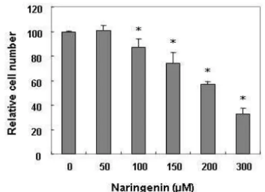
Fig. 1. Inhibition of cell proliferation by naringenin in human leukemic U937 cells. Cells were seeded at a density of 2x10⁵cells/ml and then treated with the indicated con- centrations of naringenin. After treatment for 24 hr, the proliferation of U937 cells was evaluated with a trypan blue exclusion assay. The results are expressed as the mean ± SD of three independent experiments. The stat- istical significance of the results was analyzed by a stu- dent’s t-test (* p<0.05 vs. untreated control).
A B Fig. 2. Induction of apoptotic cell death by naringenin in U937
cells. (A) After treatment for 24 hr, the cells were stained with FITC-conjugated Annexin-V and PI for flow cytometry analysis. The apoptotic cells were de- termined by counting the percentage of Annexin V+/PI-cells and the percentage of Annexin V+/PI-cells.
The data is the mean of two different experiments. (B) For the DNA fragmentation analysis, genomic DNA was extracted, separated by 1.5% agarose gel electro- phoresis, and visualized under UV light after staining with EtBr. The marker indicates a size marker of the DNA ladder.
된 1차 항체에 맞는 2차 항체를 사용하여 상온에서 1시간 정도 반응시켰다. 반응이 끝난 후 암실에서 enhanced chem- iluminoesence (ECL) slution (Amersham Life Science Co., Arlington Heights, IL, USA)을 적용시킨 다음 X-ray film에 감광시켜 특정단백질의 발현 양을 분석하였다. 본 실험에 사 용된 1차 항체들(Caspase-3, -8, Bcl-2, Bax, XIAP 및 Bid)은 Santa Cruz Biotechnology Inc. (Santa Cruz, CA, USA) 및 Calbiochem (PARP 및 actin)에서 구입하였으며, 2차 항체들은 Amersham Life Science Corp.에서 구입하였다.
DAPI 염색에 의한 핵의 변화 관찰
Apoptosis가 유발되었을 경우 특이적으로 나타나는 핵의 형태적 변화를 관찰하기 위하여 준비된 세포를 37% form- aldehyde 용액과 PBS를 1 : 9의 비율로 섞은 fixing solution을 이용하여 고정하였다. 고정된 세포를 cytospin하여 slide glass 에 부착하고 slide glass를 PBS로 2∼3회 정도 세척하고 0.2%
의 Triton X-100 (Amresco, Solon, OH, USA)을 첨가하여 상온
에서 10분간 고정한 후 2.5 μg/ml 농도의 4',6-diamidino-2- phenylindole (DAPI, Sigma-Aldrich) 용액을 처리하여 상온 에서 15분간 염색하였다. 염색이 끝난 후 DAPI 용액을 충분하 게 세척하고 mounting solution을 처리한 후 형광 현미경(Carl Zeiss)을 이용하여 400배의 배율로 각 농도에 따른 암세포의 핵의 형태 변화를 관찰하였다.
통계분석
모든 실험 결과는 SPSS ver. 18.0 (SPSS Inc., Chicago, IL, USA) 통계 프로그램을 이용하여 평균(mean)±표준편차(SD) 로 나타냈다. 각 실험군의 분석 항목별 통계의 유의성 검증은 분산분석(Analysis of Vatiance, ANOVA)을 한 후, Student t-test와 Duncan's multiple range test를 이용하여p<0.05 수 준에서 검증하였다.
결 과
Naringenin에 의한 U937 세포의 증식 억제 및 apoptosis 유발
U937 세포의 증식에 미치는 naringenin의 영향을 조사한 결과, Fig. 1에 나타난 바와 같이 50 μM 처리군에서는 큰 변화 가 나타나지 않았지만 100 μM 처리군에서부터 증식억제 현상 이 나타나기 시작하여 200 μM 및 300 μM 처리군에서 약 40%
및 65%의 증식억제 효과가 있는 것으로 나타났다. Naringen- in 처리에 의해 유발되는 증식억제가 apoptosis 유발과 직접 연관이 있는지를 확인하기 위하여 flow cytometry를 이용한 annexin V positive 세포(V+/PI-)의 빈도를 측정한 결과는 Fig.
2A에 나타낸 바와 같다. 결과에서 알 수 있듯이 정상 배지에서 자란 암세포에서의 자연적 apoptosis 유발 빈도는 약 3.57%로 매우 낮았으나 naringenin 처리농도가 증가할수록 apoptosis 가 유발된 세포의 빈도가 증가하여 300 μM 처리군에서는 약 39.87%에 해당하는 세포에서 apoptosis가 유발된 것으로 관찰 되었다. 다음으로 apoptosis 유발의 직접적인 증거에 해당하 는 DNA 단편화 여부를 agarose gel 전기영동으로 조사한 결 과, Fig. 2B에 나타난 바와 같이 naringenin을 처리하지 않았을
Fig. 3. Caspase-dependent apoptosis by naringenin in U937 cells. (A) Cells were incubated with the indicated concentrations of naringenin for 24 hr. Equal amounts of cell lysates were then resolved by SDS-polyacrylamide gels and transferred to nitrocellulose membranes. The proteins were visualized using the indicated antibodies and an ECL detection system.
Actin was used as an internal control. (B) The cells grown under the same conditions as (A) were stained with FITC- conjugated Annexin-V and PI for flow cytometry analysis. The data is the mean of two different experiments.
경우에는 DNA 단편화에 의한 DNA 단편화 현상을 관찰할 수 없었지만 naringenin 처리농도 증가에 따라 DNA 단편화 현상이 점차 증가되었음을 알 수 있었다. 이상의 결과는 nar- ingenin에 의하여 유발되는 U937 세포의 증식억제는 apopto- sis 유발과 밀접한 연관이 있음을 의미한다.
Naringenin에 의한 U937 세포의 caspase 의존적 apoptosis 유발
Apoptosis 유발에는 여러 종류의 유전자들이 관여하는 것 으로 알려져 있는데 이들 중 특히 caspases가 중요한 유전자로 밝혀져 있다. 따라서 naringenin 처리에 의한 apoptosis 유발 에 있어서 이들 유전자들이 어떠한 영향을 미치는 지를 Western blotting을 통하여 확인하였다. Fig. 3A에서 나타난 바와 같이 extrinsic pathway의 활성화를 통한 apoptosis 유발 에 관여하는 caspase-8의 경우에는 활성형 단백질의 발현이 관찰되지 않았지만 불활성형 단백질의 발현이 감소하는 것으 로 나타났으며, caspase cascade 중 가장 마지막에 활성화되어 apoptosis 유발에 직접적으로 관여하는 caspase-3의 경우에는 불활성형 단백질의 발현감소와 함께 활성형 단백질의 발현증 가가 관찰되었다. 또한 caspase-3의 활성화에 의하여 단편화되 는 표적단백질인 poly(ADP-ribose) polymerases(PARP)의 경 우에는 주단백질의 발현 감소와 더불어 단편화된 단백질의 발현 증가가 관찰되었다. 다음으로 caspases의 활성을 억제하 였을 경우에 apoptosis가 억제되는지를 확인하기 위하여 an- nexin V positive 세포(V+/PI-)를 측정한 결과는 Fig. 3B에 나타 낸 바와 같다. Naringenin이 처리된 배지에서 배양된 U937 세포의 경우는 apoptosis 유발 빈도가 약 38.64%로 나타났지 만 pan-caspase inhibitor인 z-VAD-fmk를 1시간 선처리하여 caspases의 활성을 억제한 다음 naringenin을 처리하였을 경 우에는 apoptosis 유발 빈도가 약 8.63%로서 naringenin 단독 처리군에 비교하여 현저하게 억제되는 것으로 나타났다.
Naringenin에 의한 U937 세포의 apoptosis 유발에 미 치는 Bcl-2 과발현의 영향
다음은 U937 세포에서 naringenin에 의한 apoptosis 유발 에 있어서 anti-apoptotic 유전자인 Bcl-2의 과발현이 미치는 영향을 비교하였다. 먼저 Fig. 4A에 나타낸 바와 같이 U937 세포에 naringenin을 처리하였을 경우에는 Bcl-2 및 Bax의 발 현에는 아무런 변화가 나타나지 않았으며, U937 및 U937/
Bcl-2 세포에서 Bcl-2의 과발현이 naringenin에 의한 apoptosis 유발에 어떠한 영향을 미치는 지를 조사한 결과는 Fig. 4B 및 C에 나타낸 바와 같이 U937 세포에서 naringenin에 의하여 유발된 apoptosis가 U937/Bcl-2 세포에서는 현저하게 억제되 어 Bcl-2의 고하발현은 naringenin에 의하여 유발되는 mi- tochondria의 기능 손상을 억제함으로서 apoptosis를 막아주 는 것으로 생각된다.
Naringenin에 의한 U937 세포의 apoptosis 유발에 미 치는 Bcl-2 inhibitor의 영향
U937 세포에서 naringenin에 의하여 유발되었던 apoptosis 가 Bcl-2의 과발현에 의하여 억제되는 것으로 관찰되었으므로 이러한 현상을 재확인하기 위하여 Bcl-2 억제제인 HA14-1을 이용하여 U937/Bcl-2 세포에서 Bcl-2의 기능을 억제하였을 경 우 naringenin에 의한 apoptosis가 다시 유발되는 지를 확인하 였다. 먼저 U937/Bcl-2 세포에서 naringenin 및 HA14-1 처리 에 의하여 유발되는 apoptosis 관련 단백질들의 변화를 조사 한 결과는 Fig. 5A에 나타난 바와 같다. 결과에서 알 수 있듯이 U937/Bcl-2 세포에서는 naringenin 처리에 의하여 apoptosis 관련 단백질들의 발현 변화가 나타나지 않았지만 HA14-1을 선처리하여 Bcl-2의 발현을 다시 억제하였을 경우에는 cas- pases의 활성을 억제하는 단백질인 XIAP의 발현이 감소하였 으며, caspase-8의 활성화에 의하여 단편화되어 mitochondria 의 기능 손상을 유발하는 단백질로 알려진 Bid의 단편화가
Fig. 4. Effects of Bcl-2 overexpression on naringenin-induced apoptosis in U937 cells. (A) Cells were incubated with the indicated concentrations of naringenin for 24 hr. Equal amounts of cell lysates were then resolved by SDS-polyacrylamide gels and transferred to nitrocellulose membranes. The proteins were visualized using anti-Bcl-2 and anti-Bax antibodies and an ECL detection system. Actin was used as an internal control. (B and C) U937/vector or U937/Bcl-2 cells were treated with 300 μM naringenin for 24 hr, after which they were stained with FITC-conjugated Annexin-V and PI for flow cytometry analysis.
The data is the mean of two different experiments.
Fig. 5. Bcl-2 inhibitor, HA14-1, enhanced naringenin-induced apoptosis in U937/Bcl-2 cells. (A) Cells were treated with naringenin (150 μM) alone or in combination with HA14-1 (10 μM) for 24 hr, after which equal amounts of cell lysates were resolved by SDS-polyacrylamide gels, transferred to nitrocellulose, and then probed with the indicated antibodies. The proteins were visualized using the indicated antibodies and an ECL detection system. Actin was used as an internal control. (B) The cells grown under the same conditions as (A) were fixed and stained with DAPI. The stained nuclei were then observed under a fluorescent microscope using a blue filter (Magnification, ×400).
유발되었을 뿐만 아니라 caspase-3 활성형 단백질도 증가하는 것으로 나타났다. 또한 U937/Bcl-2 세포를 대상으로 apopto- sis 유발 시 특이하게 관찰되는 핵의 형태변화를 확인한 결과, Fig. 5B에 나타낸 바와 같이 naringenin을 단독 처리하였을 경우에는 아무런 변화가 나타나지 않았지만 HA14-1을 선처리 한 후 naringenin을 처리하였을 경우에는 전체적인 핵의 밀도 감소와 더불어 apoptosis가 일어난 세포에서 전형적으로 관찰 되는 염색질 응축에 의한 apoptotic body가 관찰되었다.
고 찰
본 연구에서는 다양한 약리학적 활성을 가지는 것으로 알려 진 naringenin의 항암효능을 알아보기 위하여 인체 혈구암세
포인 U937 세포의 증식에 미치는 영향과 이와 연관된 apopto- sis 유발 여부와 함께 대부분의 인체 암세포에 과발현되어 있 으며 여러 가지 항암제에 의한 암치료에 있어서 저항성을 가 지는 원인 중 하나인 Bcl-2를 과발현시켰을 경우 naringenin에 의하여 유발되는 apoptosis가 억제되는 지를 조사하였다. 먼 저 naringenin의 처리에 따른 증식억제 정도를 조사한 결과, naringenin 처리 농도의존적으로 증식억제 현상이 나타났으 며, 이러한 증식억제 현상은 apoptosis 유발과 밀접한 관련이 있었다.
Apoptosis 유발에는 여러 가지 유전자들이 관여하지만 특 히 caspases가 중요한 역할을 하는 것으로 알려져 있다.
Caspases는 cystein-containing aspartate-specific protease family로서 세포가 정상적으로 성장 및 생존할 경우에는 핵과
mitochondria 외막에 불활성 상태인 pro-enzyme 형태로 존재 하고 있지만 세포 내외부의 다양한 자극에 의하여 활성화되어 여러 가지 표적 단백질들의 분해를 통하여 apoptosis를 유발 하는 것으로 알려져 있다[2, 23]. Caspase cascade 중에서 가장 하위단계에 존재하는 caspase-3에 의하여 분해되는 대표적인 표적 단백질로인 PARP는 DNA의 결함을 인지하는 F1 및 F2 라고 하는 두 개의 zinc finger motif를 가지고 있고 molecular nick sensor 기능을 하는 N-terminal DNA binding domain과 NAD+가 결합하는 C-terminal catalytic domain으로 구성되어 있으며, DNA repair에 관여하는 것으로 알려져 있다[21]. 따라 서 본 연구에서는 apoptosis 유발에 직접적으로 관여하는 것 으로 알려진 caspase-8 및 -3와 caspase-3의 표적 단백질인 PARP의 발현에 미치는 naringenin의 영향에 대해서 조사하 였다. 결과에서 볼 수 있듯이 naringenin은 caspase-8의 불활 성형 단백질의 발현 감소와 caspase-3의 활성형 단백질의 발현 증가와 함께 PARP의 단편화가 유발되는 것으로 나타났다. 다 음으로 caspases의 활성화가 apoptosis 유발에 직접적으로 관 여하는 지를 다시 한번 확인하기 위하여 pan-caspase in- hibitor인 z-VED-fmk를 이용하여 caspases의 활성을 억제하 였을 경우에 naringenin에 의한 apoptosis가 억제되는 것으로 나타났으므로 caspases가 중요한 조절자라는 것을 알 수 있었 다.
Bcl-2 family는 mitochondria 기능 이상을 통한 apoptosis 유발에 관여하며, Bcl-2 family 중 anti-apoptotic 유전자인 Bcl-2의 과발현은 apoptosis를 억제한다고 알려져 있다. 특히 Bcl-2 과발현에 의한 apoptosis 억제는 caspase-3 활성 억제를 통한 PARP 단편화 억제와 함께 DNA 단편화 억제를 통하여 유발된다고 보고되고 있다[18]. 본 연구에서는 이를 바탕으로 하여 대부분의 인체 암세포에 과발현되어 있으며 여러 가지 항암제에 의한 암치료에 있어서 저항성을 가지는 원인 중 하 나인 Bcl-2를 과발현시켰을 경우 naringenin에 의하여 유발되 는 apoptosis가 억제되는 지를 확인하였다. 이를 위하여 U937 및 U937/Bcl-2 세포에서 naringenin에 의한 apoptosis 유발 정도를 비교한 결과, U937/Bcl-2 세포에서 apoptosis가 현저 하게 억제되는 것으로 관찰되었다. U937/Bcl-2 세포에서의 apoptosis 억제가 Bcl-2의 과발현과 직접적인 연관이 있는 지 를 재확인하기 위하여 Bcl-2와 결합함으로서 Bcl-2의 기능을 억제하여 apoptosis 유발을 막는 것으로 알려진 강력한 Bcl-2 inhibitor인 HA14-1을 이용하였다[19, 26]. 본 연구의 결과에서 와 같이 U937/Bcl-2 세포에 naringenin을 처리하였을 경우에 는 몇 가지 유전자들의 발현 변화와 핵의 형태변화가 나타나 지 않았지만 HA14-1을 선처리하여 Bcl-2의 발현을 억제하였 을 경우에는 apoptosis 유발 관련 단백질들 중 XIAP 발현 감 소, Bid의 단편화 및 caspase-3 활성형 단백질의 발현 증가가 나타났으며, apoptosis 유발의 증거인 apoptotic body의 증가 도 관찰되었다. 따라서 Bcl-2의 과발현으로 항암제 저항성을
지닐 수 있는 암세포에서 Bcl-2 활성 억제제의 동시 처리는 항암제 내성을 극복할 수 있는 새로운 방법이 될 수 있음을 알 수 있었다.
이상의 결과들을 살펴볼 때 naringenin은 인체 혈구암세포 인 U937 세포에서 caspases의 활성을 증가시킴으로서 apop- tosis를 유발하지만 Bcl-2 과발현에 의하여 naringenin에 의한 apoptosis가 억제된다는 것을 알 수 있었다. 또한 Bcl-2 과발현 에 따른 apoptosis 저항성 극복을 위하여 Bcl-2 저해제의 혼용 처리가 중요하게 작용하므로 Bcl-2 발현의 조절을 통하여 암세 포의 약재 저항성을 극복할 수 있음을 시사하여 준다.
감사의 글
본 논문은 2013년도 동의대학교 교내연구비(2013AA106) 지원으로 이루어졌음.
References
1. Ashkenazi, A. 2002. Targeting death and decoy receptors of the tumour-necrosis factor superfamily.Nat Rev Cancer 2, 420-430.
2. Chai, F., Truong-Tran, A. Q., Ho, L. H. and Zalewski, P.
D. 1999. Regulation of caspase activation and apoptosis by cellular zinc fluxes and zinc deprivation: A review.Immunol Cell Biol 77, 272-278.
3. Chen, D., Chen, M. S., Cui, Q. C., Yang, H. and Dou, Q.
P. Structure-proteasome-inhibitory activity relationships of dietary flavonoids in human cancer cells. Front Biosci12, 1935-1945.
4. Chen, Y. C., Shen, S. C. and Lin, H. Y. 2003. Rutinoside at C7 attenuates the apoptosis-inducing activity of flavonoids. Biochem Pharmacol66, 1139-1150.
5. Cory, S., Huang, D. C. and Adams, J. M. 2003. The Bcl-2 family: roles in cell survival and oncogenesis.Oncogene22, 8590-8607.
6. Dixon, R. A. and Steele, C. L. 1999. Flavonoids and iso- flavonoids - a gold mine for metabolic engineering.Trends Plant Sci 4, 394-400.
7. Fiandalo, M. V. and Kyprianou, N. 2012. Caspase control:
protagonists of cancer cell apoptosis.Exp Oncol34, 165-175.
8. Galati, G., Moridani, M. Y., Chan, T. S. and O'Brien, P. J.
2001. Peroxidative metabolism of apigenin and naringenin versus luteolin and quercetin: glutathione oxidation and conjugation. Free Radic Biol Med30, 370-382.
9. Galluzzo, P., Ascenzi, P., Bulzomi, P. and Marino, M. 2008.
The nutritional flavanone naringenin triggers antiestrogenic effects by regulating estrogen receptor alpha-palmitoylation.
Endocrinology149, 2567-2575.
10. Ghavami, S., Hashemi, M., Ande, S. R., Yeganeh, B., Xiao, W., Eshraghi, M., Bus, C. J., Kadkhoda, K., Wiechec, E., Halayko, A. J. and Los, M. 2009. Apoptosis and cancer: mu- tations within caspase genes. J Med Genet 46, 497-510.
11. Guengerich, F. P. and Kim, D. H. 1990. In vitro inhibition of dihydropyridine oxidation and aflatoxin B1 activation in human liver microsomes by naringenin and other flavonoids.Carcinogenesis 11, 2275-2279.
12. Hermenean, A., Ardelean, A., Stan, M., Herman, H., Mihali, C. V., Costache, M. and Dinischiotu, A. 2013. Protective ef- fects of naringenin on carbon tetrachloride-induced acute nephrotoxicity in mouse kidney. Chem Biol Interact 205, 138-147.
13. Jin, C. Y., Park, C., Hwang, H. J., Kim, G. Y., Choi, B. T., Kim, W. J. and Choi, Y. H. 2011. Naringenin up-regulates the expression of death receptor 5 and enhances TRAIL-in- duced apoptosis in human lung cancer A549 cells.Mol Nutr Food Res 55, 300-309.
14. Kawaii, S., Tomono, Y., Katase, E., Ogawa, K. and Yano, M. 1999. HL-60 differentiating activity and flavonoid con- tent of the readily extractable fraction prepared from citrus juices. J Agric Food Chem47, 128-135.
15. Kerr, J. F., Wyllie, A. H. and Currie, A. R. 1972. Apoptosis:
a basic biological phenomenon with wide-ranging im- plications in tissue kinetics. Br J Cancer26, 239-257.
16. Kuwana, T. and Newmeyer, D. D. 2003. Bcl-2-family pro- teins and the role of mitochondria in apoptosis.Curr Opin Cell Biol 15, 691-699.
17. Lentini, A., Forni, C., Provenzano, B. and Beninati, S. 2007.
Enhancement of transglutaminase activity and polyamine depletion in B16-F10 melanoma cells by flavonoids nar- ingenin and hesperitin correlate to reduction of thein vivo metastatic potential. Amino Acids 32, 95-100.
18. Liu, X., Zou, H., Slaughter, C. and Wang, X. 1997. DFF, a heterodimeric protein that functions downstream of cas- pase-3 to trigger DNA fragmentation during apoptosis.Cell 89, 175-184.
19. Manero, F., Gautier, F., Gallenne, T., Cauquil, N., Grée, D., Cartron, P. F., Geneste, O., Grée, R., Vallette, F. M. and Juin, P. 2006. The small organic compound HA14-1 prevents Bcl-2 interaction with Bax to sensitize malignant glioma cells to induction of cell death. Cancer Res 66, 2757-2764.
20. Martinou, J. C. and Youle, R. J. 2011. Mitochondria in apop- tosis: Bcl-2 family members and mitochondrial dynamics.
Dev Cell21, 92-101.
21. Muller, S., Briand, J. P., Barakat, S., Lagueux, J., Poirier, G.
G., De Murcia, G. and Isenberg, D. A. 1994. Autoantibodies
reacting with poly(ADP-ribose) and with a zinc-finger func- tional domain of poly(ADP-ribose) polymerase involved in the recognition of damaged DNA. Clin Immunol Immunopathol 73, 187-196.
22. Nahmias, Y., Goldwasser, J., Casali, M., van Poll, D., Wakita, T., Chung, R. T. and Yarmush, M. L. 2008. Apolipoprotein B-dependent hepatitis C virus secretion is inhibited by the grapefruit flavonoid naringenin. Hepatology47, 1437-1445.
23. Oliver, F. J., de la Rubia, G., Rolli, V., Ruiz-Ruiz, M. C., de Murcia, G. and Murcia, J. M. 1998. Importance of poly(ADP-ribose) polymerase and its cleavage in apoptosis.
Lesson from an uncleavable mutant. J Biol Chem 273, 33533-33539.
24. Park, H. Y., Kim, G. Y. and Choi, Y. H. 2012. Naringenin attenuates the release of pro-inflammatory mediators from lipopolysaccharide-stimulated BV2 microglia by inactivat- ing nuclear factor-κB and inhibiting mitogen-activated pro- tein kinases. Int J Mol Med30, 204-210.
25. Park, J. H., Jin, C. Y., Lee, B. K., Kim, G. Y., Choi, Y. H.
and Jeong, Y. K. 2008. Naringenin induces apoptosis through downregulation of Akt and caspase-3 activation in human leukemia THP-1 cells.Food Chem Toxicol 46, 3684- 3690.
26. Pei, X. Y., Dai, Y. and Grant, S. 2004. The small-molecule Bcl-2 inhibitor HA14-1 interacts synergistically with fla- vopiridol to induce mitochondrial injury and apoptosis in human myeloma cells through a free radical-dependent and Jun NH2-terminal kinase-dependent mechanism. Mol Cancer Ther 3, 1513-1524.
27. Scatena, R. 2012. Mitochondria and cancer: a growing role in apoptosis, cancer cell metabolism and dedifferentiation.
Adv Exp Med Biol 942, 287-308.
28. Waterhouse, N. J., Sedelies, K. A. and Trapani, J. A. 2006.
Role of Bid-induced mitochondrial outer membrane per- meabilization in granzyme B-induced apoptosis. Immunol Cell Biol 84, 72-78.
29. Zamzami, N., Susin, S. A., Marchetti, P., Hirsch, T., Gómez-Monterrey, I., Castedo, M. and Kroemer, G. 1996.
Mitochondrial control of nuclear apoptosis.J Exp Med183, 1533-1544.
30. Zimmermann, K. C., Bonzon, C. and Green, D. R. 2001. The machinery of programmed cell death. Pharmacol Ther 92, 57-70.
초록:Naringenin에 의한 인체혈구암세포의 apoptosis 유발에 미치는 pro-apoptotic Bcl-2의 영향 박 철1․Cheng-Yun Jin2․최태현3․홍수현4․최영현4,5*
(1동의대학교 자연과학대학 분자생물학과,2중국 정주대학교 약학대학,3대구공업대학 안경광학과,4동의대학 교 한의과대학 생화학교실, 5동의대학교 항노화연구소 및 Blue-Bio 소재개발센터
감귤류에 많이 함유되어 있는 naringenin은 항암화학요법제로서 중요한 가능성을 가지고 있으나 항암활성에 대한 분자생물학적 기전에 대해서는 명확히 밝혀져 있지 않다. 본 연구에서는 인체 혈구암세포인 U937 세포에서 naringenin이 유발하는 항암효과 및 항암기전을 조사하였다. Naringenin 처리에 의한 U937 세포의 증식억제는 apoptosis 유발과 연관성이 있었으며, 이러한 현상은 caspases 활성화와 밀접한 관련이 있었다. 그러나 pan-cas- pase inhibitor인 z-VAD- fmk의 선처리에 의하여 U937 세포에서 naringenin이 유발하는 apoptosis가 억제되는 것으로 나타났으므로 caspases가 apoptosis 유발의 중요한 조절자라는 것을 알 수 있었다. 또한 U937 세포에 nar- ingenin을 처리하였을 경우 pro-apoptotic Bcl-2 및 anti-apoptotic Bax의 발현에는 아무런 변화가 나타나지는 않았 지만 Bcl-2가 과발현된 U937/Bcl-2 세포에서 naringenin에 의한 apoptosis가 억제되었다. 하지만 small-molecule Bcl-2 inhibitor인 HA14-1 및 naringenin을 같이 처리하였을 경우에는 XIAP 발현감소, Bid 단편화 및 caspase-3 활성화를 통하여 다시 apoptosis가 유발되었다. 따라서 HA14-1 및 naringenin에 의한 apoptosis 상승효과는 death receptor-mediated apoptosis pathway를 경유한다는 것을 제시하는 결과이다.