Copyrightⓒ 2009, The Korean Academy of Oral Biology
7
Journal of Oral Biology
Mechanism underlying NO-induced apoptosis in human gingival fibroblasts
In-Nam Hwang, Yeon-Jin Jeong, Ji-Yeon Jung, Jin-Ha Lee, Kang-Moon Kim, and Won-Jae Kim*
Dental Science Research Institute, Brain Korea 21 Project, School of Dentistry Chonnam National University, Gwangju 500-757, Korea
(received February 2, 2009 ; revised March 6, 2009 ; accepted March 13,2009)
Nitric oxide (NO) acts as an intracellular messenger at the physiological level but can be cytotoxic at high concentrations. The cells within periodontal tissues, such as gingival and periodontal fibroblasts, contain nitric oxide syntheses and produce high concentrations of NO when exposed to bacterial lipopolysaccharides and cytokines.
However, the cellular mechanisms underlying NO-induced cytotoxicity in periodontal tissues are unclear at present. In our current study, we examined the NO-induced cytotoxic mechanisms in human gingival fibroblasts (HGF). Cell viability and the levels of reactive oxygen species (ROS) were determined using a MTT assay and a fluorescent spectrometer, respectively. The morphological changes in the cells were examined by Diff-Quick staining. Expression of the Bcl-2 family and Fas was determined by RT-PCR or western blotting. The activity of caspase-3, -8 and -9 was assessed using a spectrophotometer. Sodium nitroprusside (SNP), a NO donor, decreased the cell viability of the HGF cells in a dose- and time-dependent manner. SNP enhanced the production of ROS, which was ameliorated by NAC, a free radical scavenger. ODQ, a soluble guanylate cyclase inhibitor, did not block the SNP-induced decrease in cell viability. SNP also caused apoptotic morphological changes, including cell shrinkage, chromatin condensation, and DNA fragmentation. The expression of Bax, a member of the pro- apoptotic Bcl-2 family, was upregulated in the SNP-treated HGF cells, whereas the expression of Bcl-2, a member of the anti-apoptotic Bcl-2 family, was downregulated. SNP
augmented the release of cytochrome c from the mitochondria into the cytosol and enhanced the activity of caspase-8, -9, and -3. SNP also upregulated Fas, a component of the death receptor assembly. These results suggest that NO induces apoptosis in human gingival fibroblast via ROS and the Bcl-2 family through both mitochondrial- and death receptor-mediated pathways.
Our data also indicate that the cyclic GMP pathway is not involved in NO-induced apoptosis.
Key words: Nitric Oxide, Human gingival fibroblast, Apoptosis, Cytochrome c, Caspases
Introduction
NO is a short lived, highly reactive free radical gas that is synthesized from L-arginine via a reaction catalyzed by nitric oxide synthase (NOS), of which there are three isoforms. Periodontal tissues, such as gingival fibroblasts and periodontal fibroblasts, express at least one of these isoforms; inducible NOS (iNOS), which is activated by bacterial lipopolysaccharide or cytokines (Daghigh et al., 2002; Kendall et al., 2000).
NO generally acts as an intracellular messenger at the physiological level, whereas it can be cytotoxic at high concentrations resulting in cell death, such as necrosis and apoptosis (Gross and Wolin, 1995; Dawson et al., 1996).
However, NO-induced cytotoxicity and its underlying mechanisms in periodontal tissues are not completely understood.
Necrosis is often characterized by cell swelling followed by a rupture of the plasma membrane. Apoptosis is characterized by the early condensation of nuclear
*Corresponding author: Won-Jae Kim, Department of Oral Physiology, School of Dentistry Chonnam National University, Gwangju 500-757, Korea. Tel.: +82-62-530-4881, Fax.: +82-62- 530-4885, E-mail: [email protected]
chromatin, cell shrinkage and DNA fragmentation.
Apoptosis is driven by the activation of a family of cysteine proteases, called caspases, which then cleave a critical set of cellular proteins to initiate apoptotic cell death (Roth et al., 2000). This family is expressed as proenzymes and activated by upstream stimuli. Among mammalian caspases, of which there are at least 14 known members, those involved in apoptosis can be further subdivided into the initiator caspases (2, 8, 9, 10) and effector caspases (3, 6, 7) (Adams and Cory, 1998; Tsujimoto and Shimizu, 2000).
Two main pathways that activate caspases are the death receptor-mediated and mitochondria-mediated mechanisms.
Both pathways involve the activation of caspase-3 as an executioner caspase, which activates caspase-activated DNase, causing apoptotic DNA fragmentation. The death receptor pathway is stimulated by cell surface death receptors, such as the tumor necrosis factor (TNF) receptor and Fas (Beer et al., 2000). The receptors activated by ligands lead to caspase-8 activation, with the subsequent activation of caspase-3. The mitochondrial pathway is initiated by the release of cytochrome c from the mitochondria into the cytosol. This results in caspase-9 activation, which causes the activation of caspase-3.
In addition to caspase, members of the Bcl-2 protein family are also essential for regulating apoptosis. The Bcl-2 family controls the release of mitochondrial cytochrome c by regulating the permeability of the outer mitochondrial membrane. The Bcl-2 family members are divided functionally into anti-apoptotic molecules (Bcl-2, Bcl-X
L, Bcl-W, Mcl-1, A1) and pro-apoptotic molecules (Bax, Bcl- 1s, Bid, Bad, Bim, Bik) (Adams and Cory, 1998; Tsujimoto and Shimizu, 2000). Among the Bcl-2 protein family, Bcl-2 and Bcl-X
Lare prominent members of the anti-apoptotic family, whereas Bax, Bid and Bak are prominent members of the pro-apoptotic family (Cheng et al., 1997). There is increasing evidence that NO may be cytotoxic to human gingival fibroblastic cells because it is mass produced by bacterial lipopolysaccharide and cytokines in periodontal tissues (Susilowati, et al., 2002; Daghigh et al., 2002).
Nevertheless, NO-induced cytotoxicity in periodontal tissue is not completely understood.
This study examined the roles of the molecules associated with the mitochondria- and death receptor-mediated apoptotic pathways during NO-induced apoptosis of human gingival fibroblasts.
Materials and methods
Cell culture and cell viability assay
HGF cells were obtained from the healthy gingival tissue of patients in Chonnam National Hospital. The HGF cells were maintained in DMEM medium supplemented with 10% fetal bovine serum (Gibco, USA) under 5% CO
2at
37
oC. Sodium nitroprusside (SNP) (Sigma, USA) was dissolved in distilled DMEM and sterilized through a 0.2 µm filter. The viable cells were detected using a 3-(4,5- dimethylthiazol-2-yl)-2,5-diphenyltetra zolium bromide (MTT) assay (Sigma, USA).
Nuclear staining with Diff-Quick
The morphological changes of apoptotic cells were investigated by Diff-Quick staining. The cells were plated on an 8-well chamber slide at a density of 1 × 10
5and incubated for 18 h. They were then treated with 5 mM SNP for 12 h. The cells were washed with 1 × PBS and fixed with acetone and methanol (1:1). After incubating for 20 min at - 20
oC, the cells were stained with a solution containing 10 µg/ml Diff-Quick in PBS and observed by fluorescence microscopy (Olympus, JAPAN).
Detection of ROS production and caspase activity The production of reactive oxygen species (ROS) production was monitored using a fluorescence spectrometer (Hitachi F-4500, JAPAN) with a 2', 7'- dichlorofluor- bescin diacetate (DCF-DA) stain. The cells were plated on a 96-well plate and treated with N-acetyl- cysteine (NAC) (Sigma, USA) and SNP. DCF-DA (25 µM) was then added to the media and the cells were incubated for a further 10 min at 37 C. The emission was measured at 530 nm. The caspase activities were assayed using a caspase-3, -9 activity assay kit (Calbiochem, CA) and caspase-8 activity Kit (Santa Cruz, USA) on a spectrometer according to the manufacturer's instructions.
Isolation of total RNA and reverse transcription polymerase chain reaction (RT-PCR)
The total RNA was extracted by homogenizing the cells using a polytron homogenizer in Trizol reagent (Gibco- BRL, USA). The RNA samples were quantified by spectrophotometry at 260 nm. For the synthesis of cDNA, 2 µg of the total RNA and 2 ㎕ of Oligo-dT (10 pmoles) were mixed with 50 ㎕ RNase-free water, and then incubated at 42
oC for 1 h and 94
oC for 5 min. The PCR products were generated in a PCR buffer containing 10 pmoles of each primer using a PCR-premix kit (Bioneer, Korea). A GeneAmp PCR system (Perkin-Elmer 2400) was used for RT-PCR. After the first denaturation step (5 min at 95
oC), samples were subjected to 30 cycles consisting of the following: 40 sec at 95
oC, 40 sec at 55
oC, and 1 min 30 sec at 72
oC. A final extension step of 10 min was performed.
The following primer pairs were used: for Bax, 5'- GTTTCATCCAGGATCGAGCAG-3' (senseprimer) and 5'- CATCTTCTTC CAGATGGTGA-3' (antisense primer) ; for Bcl-2, 5'-CCTGTGGATGAC TGGTACC-3' (sense primer), 5'-GAGACAGCCAGGAGAAATCA-3' (antisense primer) ; for Fas, 5'-CAAGGGACTGATAGCATCTTT- GAGG-3' (sense primer), 5'-TCCAGATTCAGGGTCA- CAGGTTG-3' (antisense primer). The amplified products
o–
were analyzed on 1.5% agarose gels containing ethidium bromide and visualized using a UVP Transilluminator/
Polaroid camera System (UVP Laboratories, CA). RT-PCR was performed with the primers for GAPDH, a housekeeping gene, as the control. The following primer pairs for GAPDH were used: 5'-TGC ATCCTGCACCA- CCAACT-3' (sense primer) and 5'-CGCCTGCTTCACCA CCTTC-3' (antisense primer). The intensities of the bands were determined using NIH Scion Image Software.
Western blotting
The cells were washed twice with PBS and the proteins were solubilized in a lysis buffer (500 mM Tris-HCl, pH 7.4, 150 mM NaCl, 5 mM EDTA, 1 mM Benzamiden, 1 µg/
ml Trypsin inhibitor) containing a cocktail of protease inhibitors (Complete, Germany). To determinate the level of cytosolic cytochrome c, a pellet was resuspended in an extraction buffer containing 220 mM mannitol, 68 mM sucrose, 50 mM PIPES-NaOH (pH 7.4), 50 mM Kcl, 5 mM EGTA, 2 mM MgCl
2, and 1 mM DTT. The lysates were incubated for 30 min at 4
oC, centrifuged at 11000 ×g for 20 min and the protein concentrations were determined using a BCA protein assay (Pierce, IL). The protein extracts (100~500 µg) were boiled for 5 min with the SDS-sample buffer and subjected to electrophoresis on 12%
polyacrylamide gel. The proteins were electroblotted onto a nitrocellulose membrane (Amersham Pharmacia Biotech, UK) and blocked with 5% skim milk (Becton Dickinson, USA) in Tris-buffered saline-0.1%Tween 20 (TBS-T). The primary antibodies used were a Rat monoclonal anti- cytochrome c (Pharmingen, CA), and Bid (Santa Cruz, USA). The blots were then washed three times in TBS-T for 5 min and incubated with the specific peroxidase-coupled secondary antibodies (Sigma, USA). The bound antibodies were visualized using an enhanced chemiluminescent detection system (Amersham Pharmacia Biotech, UK).
Results
NO induces ROS production and apoptosis in HGF cells
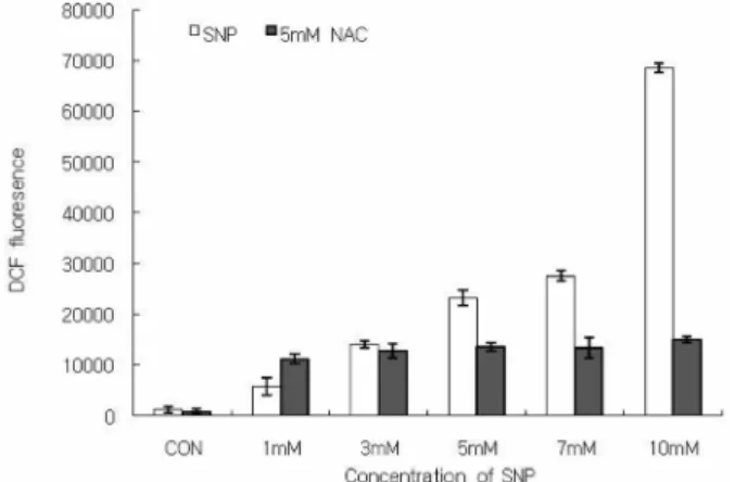
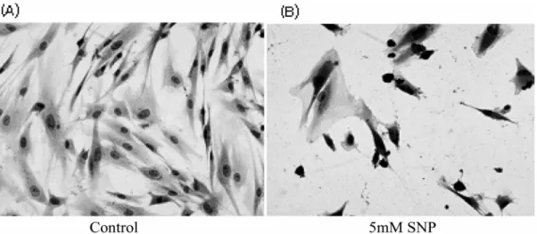
To determine the involvement of ROS in NO-induced cell death of HGF cells, the level of ROS production was measured using DCF-DA. Fig.1 shows that SNP enhanced the production of ROS in the HGF cells in a dose- dependent manner. Pretreatment of the cells with 5 mM NAC inhibited the SNP-induced increase in ROS. The cell viability was determined using a MTT assay. As shown in Fig. 2, the cell viability decreased gradually in a dose- and time-dependent manner when the HGF cells were exposed to SNP (Fig. 2A, 2B). The cell survival was < 80% when the cells were treated with 5 mM SNP for 24 h. In the presence of 5 mM SNP for 12 h, Diff-Quick staining revealed apoptotic morphological changes, including chromatin condensation
and nuclear fragmentation (Fig. 3B). Because NO usually targets soluble guanylate cyclase, the effects of a soluble gyanylate cyclase inhibitor on NO-induced cell death were
Fig. 1. ROS production was enhanced in SNP-treated HGF cells.The HGF cells loaded with DCF were incubated for 12 h with SNP alone or with 5 mM N-acetyl-L-cysteine (NAC) for 1 h. The intra- cellular levels of ROS were detected by measuring the DCF-DA fluorescence. The data is reported as the mean± SD of 5 indepen- dent experiments. SNP augmented the production of ROS in a dose-dependent manner, and NAC, a free radical scavenger, ame- liorated the increase in ROS produced by SNP.
Fig. 2. NO induced cell death in HGF cells. The cell viability was determined using a MTT assay, as described in materials and meth- ods. The HGF cells were incubated 5 mM SNP for the indicated times (A) and with SNP at the indicated concentrations (B). The viability of the cells without the SNP treatment was defined as 100%. The viability of SNP-treated HGF cells decreased in a dose- and time-dependent manner. The data is reported as the mean± SD of 5 independent experiments.
examined. ODQ (10
-4M) did not recover the cell viability reduced by SNP (Fig. 4), indicating that NO-induced apoptosis does not occur through cyclic GMP.
NO-induced apoptosis is mediated with mitochondria in HGF cells
In order to determine if the mitochondria are involved in the NO-induced apoptosis of HGF cells, the amount of cytochrome c released from the mitochondria into the cytosol was determined using the cytosolic fractions, as previously reported (Boulares et al., 2002). The cytosolic level of cytochrome c was assessed from the amount of cytochrome c released from the mitochondria into the cytoplasm. The HGF cells were incubated with varying concentrations of SNP for different periods, and subjected to Western blotting. Cytosolic cytochrome c was enhanced in a dose-dependent manner in response to SNP exposure (Fig. 5A). Cytochrome c reached a peak value after 4 h
incubation with SNP and remained higher than control values, even at 24 h (Fig. 5B). These results show that cytochrome c is released from the mitochondria into the cytoplasm during NO-induced apoptosis of HGF cells.
Bax is upregulated and Bcl-2 is downregulated during NO-induced apoptosis
Generally, the Bax to Bcl-2 expression ratio is significant for determining apoptosis because a high ratio denotes a low apoptotic threshold, while a low ratio indicates a higher apoptotic threshold. After treating the HGF cells with 10 mM SNP for 12 h, the changes in the mRNA level of Bax and Bcl-2 in the HGF cells was determined using RT-PCR.
Fig. 6 shows that SNP upregulated the expression of Bcl-2 and downregulated the expression of Bax in a dose dependent manner.
Fig. 3. NOinduced apoptotic morphologic changes in HGF cells. The HGF cells were treated with 5 mMSNP for 12 h, fixed with ethanol and stained with Diff-Quick. Cell shrinkage, chromatin condensation, and DNA fragmentation were observed in the SNP-treated HGF cells (B).
Fig. 4. Cyclic GMP is not involved in the NO-induced cell death of HGF cells. ODQ did not recover the cell viability decreased by SNP. The results are reported as the mean± SD of 5 independent experiments.
Fig. 5. Enhancement of cytochrome c released from the mitochon- dria into the cytosol in the SNP-treated HGF cells. Cytosolic cyto- chrome c was analyzed by immunoblotting with the antibody against cytochrome c. SNP increased the amount of cytochrome c released from the mitochondria into the cytosol in dose-dependent manner (A) and at 4 h as the peak time (B).
Caspases are involved in the NO-induced apoptosis of HGF cells
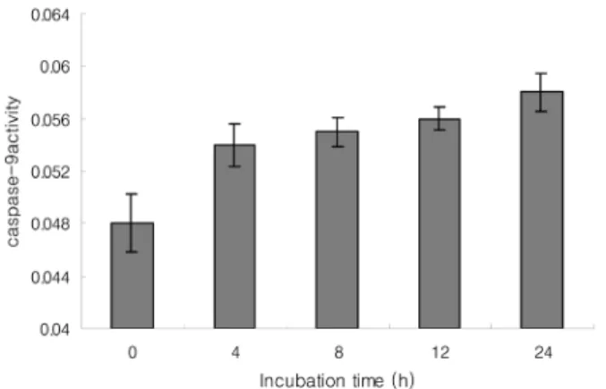
It is important to identify the intracellular apoptotic pathways induced by NO in HGF cells. Therefore, the caspases activities were measured based on the fact that active caspases consequently cleave their substrate at a specific site. LEHD-pNA (200 µM), IETD-pNA (200 µM), and DEVD-pNA (200 µM) were used as substrates for caspase-9, -8, and -3, respectively. 5 mM SNP enhanced the activities of caspase-8, -9 and -3 in a time-dependent manner. Therefore, it is believed that both the mitochondria and death receptor-dependent apoptotic pathway are involved in the NO-induced apoptosis of HGF cells (Fig.
7, 8, 9).
NO upregulates Fas expression and cleaves Bid in HGF cells
The mRNA levels of Fas, a death receptor assembly, were
measured using RT-PCR to determine if the death receptor- mediated apoptosis pathway is activated in HGF cells.
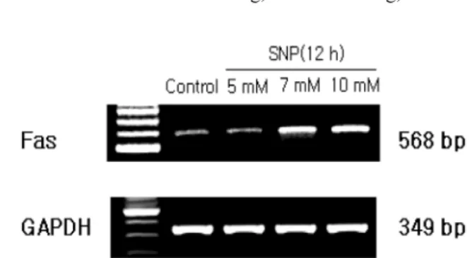
5 mM SNP upregulated the expression of Fas in a dose dependent manner (Fig. 10). Bid expression was determined because activated caspase-8 is known to cleave proform Bid (pro-Bid) into truncated Bid (tBid). The Pro-Bid protein was decreased by SNP in a dose- and time-dependent manner (Fig. 11). These results suggest that the death receptor mediated pathway plays a key role in the NO-induced apoptosis of HGF cells.
Discussion
Periodontal tissues, such as gingival and periodontal fibroblasts, contain NOS and mass produce NO at high
Fig. 6. Altered expression of Bax and Bcl-2 in the SNP-treatedHGF cells. After incubating the HGF cells with SNP for 12 h, RT- PCR was performed forBax and Bcl-2 expression. SNP upregu- lated the expression of Bax and downregulated the expression of Bcl-2 in a dose-dependent manner.
Fig. 7. Caspase-9 was activated in SNP-treated HGF cells. The absorbance for caspase-9 activity was measured at 405 nm using a ELISA reader after incubation with the LEHD-pNA substrate(200µM) for 2 h at 37oC. 5 mM SNP enhanced the caspase-9 activity in a time-dependent manner. The data is reported as the mean± SD of 5 experiments.
Fig. 8. Caspase-8 was activated in the SNP-treated HGF cells. The absorbance for the caspase-8 activity was measured at 405 nm using an ELISA reader after incubation with the IETD-pNA sub- strate (200µM) for 2 h at 37oC. 5 mM SNP enhanced the activity of caspase-8 in a time-dependent manner. The data is reported as the mean± SD from 5 experiments.
Fig. 9. Caspase-3 was activated in the SNP-treated HGF cells. The absorbance for caspase-3 activity in the wells was measured at 405 nm using an ELISA reader after incubation with the DEVD-pNA substrate (200µM) for 24 h at 37oC. 5 mM SNP enhanced the caspase-3 activity in a time-dependent manner. The data is reported as the mean± SD of 5 experiments.
concentrations after exposure to bacterial lipopoly- saccharide and cytokines (Daghigh et al., 2002; Kendall et al., 2000). However, there are no reports of NO-induced cell death in periodontal tissues.
NO-induced cell death has been classified as apoptosis and necrosis based on the changes in morphology, enzymatic activity, ATP concentration and adjacent cellular effects (Gross and Wolin, 1995; Dawson and Dawson, 1996). The characteristic morphology in apoptotic cell is distinct, including cellular shrinkage, internucleosomal DNA fragmentation and chromatin condensation (Fujimura et al., 2000; Oppenheim, 1991).
In this study, SNP decreased the viability of HGF cells giving rise to apoptotic morphological changes, including chromatin condensation, DNA fragmentation, and cell shrinkage. These results demonstrate that nitric oxide induces apoptosis in HGF cells. Previous studies reported that NO elicits apoptosis through the production of ROS, such as H
2O
2in the mitochondria and a reaction with superoxide, resulting in the formation of peroxynitrite (Yuyama et al., 2003; Brown, 1999). Even in the present study, SNP enhanced the production of ROS, which was ameliorated by NAC in HGF cells. From these results, it is believed that NO-induced apoptosis may be in part
mediated by ROS in HGF cells, which is consistent with those of previous reports in other tissues.
A variety of free radicals, such as ROS and peroxynitrite, impair the mitochondrial function, which result in a loss of the mitochondrial transmembrane potential and the release of mitochondrial pro-apoptotic molecules including cytochrome c, Smac, apoptosis-inducing factor (AIF) (Fleury et al., 2002; Herrera et al., 2001)
The present study showed that SNP dose dependently increases the level of cytochrome c released from the mitochondria into the cytoplasm. In addition, SNP enhanced the activity of caspase-9, which was activated by mitochondrial cytochrome c, resulting in the activation of caspase-3 in concert with Apaf-1 and dATP. Overall, the mitochondria-dependent apoptotic pathway is definitely involved in the NO-induced apoptosis of the HGF cells because cytochrome c and caspase-9 are major molecules associated with the mitochondria- dependent pathway.
In general, caspase-3 is a key and common protease in both the mitochondria- and death receptor-dependent pathways, and is particularly important in free radical- induced apoptosis (Earnshaw et al., 1999; Bal-Price et al., 2000). Previous studies (Bal-Price et al., 2000; Leist et al., 1999) showed that caspase-3 is activated in response to various ROS. This study showed that caspase-3 activity was enhanced in SNP-treated HGF cells, which is consistent with previous reports of other tissues and cells. From the present study and previous reports, it was assumed that caspase-3 plays a key role in NO-induced apoptosis in HGF cells. This is despite the fact that caspase-independent cell death was suggested to be involved in NO-induced cell death in PC12 cells (Yuyama et al., 2003).
Another possible mechanism for activating caspase-3 is a caspase-8 mediated process that is activated by Fas and TNF receptor-1. Recent studies reported that ROS, such as H
2O
2, induces the upregulation of the death receptor assembly, such as Fas and Fas-ligand, subsequently activating caspase-8 (Fleury et al., 2002; Facchinetti et al., 2002).
From these previous reports, it is believed that a death receptor-dependent apoptosis pathway may be involved in caspase-3 activation in the NO-induced apoptosis of HGF cells. The present results showed that Fas, a death receptor assembly, was upregulated and caspase-8 activity was enhanced in SNP-treated HGF cells. This study provides the first evidence that a death receptor-dependent pathway may be involved in NO-induced apoptosis of HGF cells. From these results, NO-induced apoptosis is likely to be mediated by both the mitochondria and death receptor-mediated pathways in HGF cells.
On the other hand, members of the Bcl-2 family of proteins are well-characterized regulators of cytochrome c release from the mitochondria into the cytosol. The Bcl-2 subfamily contains antiapoptotic proteins, such as Bcl-2 and Bcl-X
L, which reduce the level of cytochrome c release and loss of the mitochondrial transmembrane potential ( ∆ψ
m)
Fig. 10. Upregulated expression of Fas in SNP-treated HGF cells.The mRNA levels of Fas, which is a component of the death recep- tor assemblies was determined by RT-PCR. SNP upregulated the expression of Fas in a dose-dependent manner.
Fig. 11. NO activated Bid in SNP-treated HGF cells. SNP reduced the expression of proform Bid (pro-Bid) in a dose- and time-depen- dent manner.
(Gottlieb et al., 2000; Howard et al., 2002). The Bax subfamily contains proapoptotic proteins, such as Bax and Bak, which induce the release of cytochrome c and loss of
∆ψ
m(Starkov et al., 2002). Bcl-2 proteins, such as Bid, Bik and Bim, are another subfamily of proapoptotic proteins that are activated by caspase-8. Therefore, ratio of proapoptotic and antiapoptotic caspases may be a pivotal cue to the release of cytochrome c from the mitochondria into the cytosol. Accordingly, expression of the Bcl-2 family was examined to determine the involvement of the Bcl-2 family in NO-induced apoptosis. Bcl-2 mRNA was downregulated in SNP-treated HGF cells, whereas Bax mRNA was upregulated. Even in previous reports (Hemish et al., 2003), NO has been reported to directly or indirectly regulate the expression of the Bcl-2 family in other tissue and cells.
These results suggest that Bcl-2 proteins are involved in the NO-induced apoptosis of HGF cells. Interestingly, Bid was activated by SNP because Bid is known to be activated by caspase-8, which is unlike other members of the Bcl-2 family. These results suggest that there is an interrelation lineage between the death receptor-mediated apoptotic signals and the mitochondria-mediated apoptotic signals.
However, roles of the Bcl-2 family in NO-induced apoptosis of HGF cells are controversial because the Bcl-2 family regulates the production of ROS and the release of cytochrome c from the mitochondria into the cytosol (Gottlieb et al., 2000; Starkov et al., 2002). On the other hand, ROS can regulate the expression of Bcl-X
LmRNA (Herrera et al., 2001). Further studies will be needed to determine the roles of the Bcl-2 family in the NO-induced apoptosis of HGF cells.
In general, NO has a variety of physiological functions by activating soluble guanylate cyclase, resulting in the synthesis of cyclic GMP. Therefore, experiments were carried out to determine if the cyclic GMP pathway is involved in the NO-induced apoptosis of HGF cells. ODQ did not ameliorate the cell viability reduced by SNP in HGF cells. This suggests that NO-induced apoptosis is not mediated by the cyclic GMP pathway.
In summary, NO induces apoptosis in HGF cells by activating both the mitochondria- and death receptor- dependent pathways that are mediated by ROS and the Bcl- 2 family.
Acknowledgement
This study was financially supported by Chonnam National University, 2004
References
Adams JM, Cory S. The Bcl-2 protein family: arbiters of cell survival. Science. 1998;281:1322-26.
Beer R, Frenz G, Schopf M, Reindl M, Zelger B, Schmutzhard E, Poewe W, Kampfl A. Expression of Fas and Fas ligand after experimental traumatic brain injury in the rat. J Cereb Blood Flow Metab. 2000;20: 669-77.
Boulares AH, Zoltoski AJ, Sherif ZA, Yakovler A, Smulson ME. Roles of DNA fragmentation factor and poly(ADP- ribose) polymerase-1 in sensitization of fibroblasts to tumor necrosis factor-induced apoptosis. Biochem Biophys Res Commun. 2002;290:796-801.
Brown GC. Nitric oxide and mitochondrial respiration.
Biochim Biophys Acta. 1999;1411:351-69.
Cheng EH, Nicholas J, Bellows DS, Hayward GS, Guo HG, Reitz MS, Hardwick JM. A Bcl-2 homolog encoded by kaposi sarcoma-associated virus, human herpesvirus 8, inhibits apoptosis but does not heterodimerize with Bax or Bak. Proc Natl Acad Sci. 1997;94: 690-4.
Dahigh F, Borghaei RC, Thornton RD, Bee JH. Human gingival fibroblasts produced nitric oxide in response to proinflammatory cytokines. J Periodontal. 2002;73:392-400.
Dawson VL, Dawson TM. Nitric oxide neurotoxicity. J Chem Neuroanat. 1996;10:179-90.
Earnshaw WC, Marins LM, Kaufmann SH. Mammalian caspases: structure, activation, substrates, and functions during apoptosis. Annu Rev Biochem. 1999;68:383-424.
Facchinetti F, Furegato S, Terrazzino S, Leon A. H2O2 induces upregulation of Fas and Fas ligand expression in NGF - differentiated HGF cells: Modulation by cAMP. J Neurosci Res. 2002;69:178-88.
Fleury C, Mignotte B, Vayssiere JL. Mitochondrial reactive oxygen species in cell death signaling. Biochimie.
2002;84:131-41.
Fujimura M, Morita-Fujimura Noshita N, Sugawara T, Kawase M, Chan PH. The cytosolic antioxidant copper/
zinc-superoxide dismutase prevents the early release of mitochondrial cytochrome c in ischemic brain after transient focal cerebral in mice. J Neurosci. 2000;20:2817-24.
Gottlieb E, Vander Heiden MG, Thompson CB. Bcl-xl prevents the initial decrease in mitochondrial membrane potential and subsequent reactive oxygen species production during tumor necrosis factor alpha-induced apoptosis. Mol Cell Biol. 2000;20:5680-89.
Gross SS, Wolin MS. Nitric oxide: pathophysiological mechanisms. Annu Rev Physiol. 1995;57:737-69.
Hemish J, Nakaya N, Mittal V, Enikolopov G. Nitric oxide activates diverse signaling pathways to regulate gene expression. J Biol Chem. 2003;278:42321-9.
Herrera B, Alvarez AM, Sanchez A, Fernandez M, Roncero C, Benito M, Fabregat I. Reactive oxygen species (ROS) mediates the mitochondrial-dependent apoptosis induced by transforming growth factor (beta) in fetal hepatocytes.
FASEB J. 2001;15:741-51.
Howard S, Bottino C, Brooke S, Cheng E, Giffard RG, Sapolsky R. Neuroprotective effects of Bcl-2 overexpre- ssion in hippocampal cultures: interactions with pathways of oxidative damage. J Neurochem. 2002;83:914-23.
Huerta-Yepez S, Vega M, Jazirehi A, Garban H, Hongo F, Cheng G, Bonavida B. Nitric oxide sensitizes prostate carcinoma cell lines to TRAIL-mediated apoptosis via
inactivation of NF-κB and inhibition of Bcl-xL expression.
Oncogene. 2004;23:4993-5003.
Kendall HK, Haase HR, Li H, Xiao Y, Bartold PM. Nitric oxide synthase type-II is synthesized by human gingival and cultured human gingival fibroblasts. J Periodontal Res.
2000;35:194-200.
Leist M, Single B, Naumann H, Fava E, Simon B, Kuhnle S, Nicotera P. Nitric oxide inhibits execution of apoptosis at two distinct ATP-dependent steps upstream and downstream of mitochondrial cytochrome c release. Biochem Biophys Res Commun. 1999;258:215-21.
Oppenheim RW. Cell death during development of the nervous system. Annu Rev Neurosci. 1991;14:453-501.
Bal-Price A, Brown GC. Nitric-oxide-induced necrosis and apoptosis in PC12 cells mediated by mitochondria. J Neurochem. 2000;75:1455-64.
Roth JA, Feng L, Walowitz J, Browne RW. Manganese - induced rat pheochromocytoma (HGF) cell death is
independent of caspase activation. J Neurosci Res.
2000;61:162-71.
Starkov A, Polster B, Fiskum G. Regulation of hydrogen peroxide production by brain mitochondria by calcium and Bax. J Neurochem. 2002;83:220-8.
Susilowati H, Santoso AL, Barid I, Sosroseno W. Rat periodontal fibroblast responses to bacterial lipopolysacchraide in vitro. J Microbiol Immunol Infect. 2002;35:203-6.
Tsujimoto Y, Shimizu S. Bcl-2: Life-or-death switch. FEBS Lett. 2000;466:6-10.
Yuyama K, Yamamoto H, Nishozaki I, Kato T, Sora I, Yamamoto T. Caspase-independent cell death by low concentrations of nitric oxide in PC12 cells: Involvement of cytochrome c oxidase inhibition and the production of reactive oxygen specoes in mitochondria. J neurosci Res 2003;73:351-63.