33 책임저자:최영현, }614-052, 부산시 진구 양정2동 산45번지
동의대학교 한의과대학 생화학교실 Tel: 051-850-7413, Fax: 051-853-4306 E-mail: [email protected]
접수일:2008년 2월 25일, 게재승인일:2008년 3월 5일
Correspondence to:Yung Hyun Choi
Department of Biochemistry, Dongeui University College of Oriental Medicine, San 45, Yangjeong 2-dong, Jin-gu, Busan 614-052, Korea Tel: +82-51-850-7413, Fax: +82-51-853-4306
E-mail: [email protected]
인체간암세포에서 불등가사리 메탄올 추출물의 Caspase 비의존적 Apoptosis 유발에 관한 연구
1부산대학교 자연과학대학 생물학과, 2동의대학교 한의과대학 생화학교실,
대학원 바이오물질제어학과(BK21) 최우영1ㆍ김성윤2ㆍ이원호1ㆍ최영현2
Induction of Caspase-independent Apoptosis by Methanol Extract of Gloiopeltis furcata in Human Hepatocarcinoma HepG2 Cells
Woo Young Choi1, Cheng-Yun Jin2, Won Ho Lee1 and Yung Hyun Choi2
1Department of Biology, Pusan National University, Busan 609-735, 2Department of Biochemistry, Dongeui University College of Oriental Medicine, Department of Biomaterial Control (BK21), Dongeui University Graduate School and Institute of Oriental Medicine, Busan 614-052, Korea
Previous results showed that the administration of seaweed powder or extract reduced the incidence rate of chemically induced tumorigenesis using in vivo animal model. Recently, we reported that the extracts of Gloiopeltis furcata, a kind of Korean edible seaweed, caused the cell growth inhibition of various human cancer cell lines, among them methanol extract exhibited a relatively strong antiproliferative activity. However, little is known about its precise effects and mechanisms of action in malignant cells.
In the present study, we investigated the pathway of the induction of apoptotic cell death by methanol extract of G. furcata (MEGF) in human hepatocarcinoma HepG2 cells. It was found that MEGF could inhibit the cell viability and induce the apoptosis in a dose-dependent manner as measured by trypan blue counting, fluorescent microscope and flow cytometry analysis. Apoptosis induction of HepG2 cells by MEGF was associated with an down-regulation of anti-apoptotic Bcl-2 mRNA and protein expression, but other apoptosis-related genes including Fas/FasL system and IAP family members were not changed in MEGF-treated HepG2 cells. Moreover, MEGF treatment did not induce the proteolytic activation of caspase-3, -8 and -9, and degradation of poly(ADP-ribose) polymerase (PARP) protein suggesting that MEGF-induced apoptosis proceeds via a caspase-independent mechanism. Though further studies will be needed to identify the activity compounds that confer the anti-cancer activity of MEGF, the present findings provide important new insights into the possible molecular mechanisms of the anti-cancer activity of G. furcata. (Cancer Prev Res 13, 33-39, 2008)
Key Words: Gloiopeltis furcata, HepG2, Apoptosis, Caspase-independent
서 론
우리의 생활과 밀접한 연관이 있는 많은 천연물들이
항암작용이 있는 것으로 알려져 있지만 최근 육상자원 의 한계를 극복하기 위하여 해양생물 유래 천연 물질에 대한 관심이 매우 높아지고 있다.1) 그 중에서도 널리 식 용으로 사용 중인 해조류는 강력한 항산화 효과가 있다
고 잘 알려져 있으며, 혈중 콜레스테롤을 내려주며, 고혈 압과 혈액의 점도를 낮추고 궤양을 예방하고, 항균작용 을 가지는 등의 다양한 효과가 있는 것으로 보고되어지 고 있다.2∼4)
암의 효과적인 치료와 예방에 있어서 비정상적인 세 포나 암세포의 apoptosis 유발은 많은 치료제의 표적이 되 고 있으며, apoptosis는 개체보존의 수준에서 손상된 세포 들의 제거를 위한 세포의 사멸 현상으로서 중요한 수단 이기도 하다.5) Apoptosis의 유발에 종양억제유전자 p53 등과 같은 다양한 유전자가 관여한다는 사실이 알려지 면서 apoptosis와 연관된 분자적 기전이 최근 많이 밝혀지 고 있으며, apoptosis는 특히 암세포의 성장, 증식의 억제 와 암세포 파괴의 한 방법으로써 널리 연구되고 있다.6,7) 그리고 생체 내에서 유발되는 염증반응에 있어서 prostaglandin은 세포분열이나 증식에 영향을 줌으로서 각종 인체 질병의 유발과 진행에 중요한 역할을 하는 것 으로 알려져 있는데 여기에는 2가지의 cyclooxygenase (COX) isoform이 관여하고 있다.8) COX-1의 경우는 house- keeping gene으로서 대부분의 조직에서 일정한 수준으로 발현되어 인체의 항상성 유지와 연관된 기능수행에 관 여하는 반면에 COX-2의 과발현은 암을 포함한 세포의 성장 및 분화와 연관된 각종 퇴행성 질환의 발병과 진행 에 있어서 중요한 역할을 한다.9)
본 연구에서는 우리나라를 포함한 세계적으로 널리 분포하고 있으며, 항돌연변이 효능이 우수하여 항암활 성효능이 높을 것으로 예상되는 해조류의 한 종류인 홍 조식물 풀가사리과에 속하는 불등가사리(G.loiopeltis
furcata)3,10∼12)에 함유된 항암/암예방 후보물질의 도출을
위하여 불등가사리의 메탄올 추출물이 인체 간암세포의 증식에 미치는 영향을 조사하였다.
재료 및 방법 1. 실험 재료 및 세포배양
인체 간암세포인 HepG2 세포는 생명공학연구소(KRIBB, Taejeon, Korea)에서 분양 받았으며, 세포의 배양을 위해 10%의 우태아혈청(fetal bovine serum, FBS, Gibco BRL, Grand Island, NY, USA)과 1%의 penicillin-streptomycin 등이 포함된 RPMI-1640 및 DMEM 배지(Gibco BRL)를 사용하 여 37oC, 5% CO2 조건하의 CO2 incubator에서 배양하였 다. 본 실험에 사용한 불등가사리 메탄올 추출물 (methanol extracts of G. furcata, MEGF)은 선행연구의 방법 에 준하여 준비하였다.13)
2. Hemocytometer를 이용한 세포 생존률의 측정
세포배양용 6 well plate에 HepG2 세포 1 × 10⁵개/ml 정 도를 분주하고 MEGF를 배지에 희석하여 처리한 후 배 양하였다. 72시간 후 세포를 700 rpm으로 5분간 원심 분 리시켜 상등액을 제거하고, 1 ml의 PBS로 세포를 suspen- sion한 다음 세포 부유액과 0.5% trypan blue (Gibco BRL)를 동량으로 섞어 2분간 처리하였다. Pasteur pipette의 모세 관 현상을 이용하여 세포를 hemocytometer에 옮긴 후 위 상차 현미경을 이용하여 200배의 배율로 관찰하여 푸른 색으로 염색된 세포를 죽은 세포로 추정하고 염색이 되 지 않은 살아있는 세포의 수를 측정하였다. 이에 따른 결과는 Sigma Plot 4.0 프로그램 (SPSS Ins.)을 사용하여 분 석하였다.
3. DNA flow cytometry 분석
정상 및 MEGF를 처리한 배지에서 72시간 동안 배양 시킨 세포를 2,000 rpm에 5분간 원심분리하여 상층의 배 지를 버리고 세포를 모아서 PBS 1 ml로 충분히 재부유시 켰다. 이를 2,000 rpm으로 5분간 원심분리 한 후 상층의 PBS만 버리고 CycleTEST kit (Becton Dickinson, San Jose, CA, USA)를 사용하여 고정 및 propidium iodide (PI, concentration, 50 μg/ml; Sigma, St. Louis, MO, USA) 염색액 을 처리하여 암실, 4oC에서 15분 동안 염색하였다. 염색 후 DNA flow cytometry (Becton Dickinson, San Jose, CA, USA)에 적용시켜 형광반응에 따른 histogram을 ModiFit LT (Becton Dickinson) program으로 분석하였다.
4. DAPI staining에 의한 세포핵의 형태 관찰
MEGF 처리에 의한 HepG2 세포의 apoptosis 유발 여부 확인을 위한 핵의 형태적 변화를 관찰하기 위하여 준비 된 세포를 모은 다음 3.7% formaldehyde 및 0.2%의 Triton X-100 용액으로 고정 후, 4',6-diamidino-2-phenylindole (DAPI, Sigma) 용액으로 상온에서 약 10분간 염색하였다.
염색된 세포를 PBS로 충분하게 세척하고 absolute alcohol 을 이용하여 탈수과정을 거친 후, 형광 현미경(Carl Zeiss, Germany)을 이용하여 400배의 배율로 각 농도에 따른 암 세포의 핵의 형태 변화를 관찰하였다.
5. Reverse transcription-polymerase chain reaction (RT-PCR) 분석
동일 조건에서 준비된 세포를 PBS로 세척하고 TRIzol reagent (Invitrogen Co., Carlsbad, CA, USA)를 4oC에서 1시 간 동안 처리하여 total RNA를 각 처리 농도 별로 분리하
Table 1. Gene-specific primers for RT-PCR
Gene name Sequence
Bax Sense Antisense Bcl-2 Sense Antisense Bcl-xL Sense Antisense Fas Sense Antisense FasL Sense Antisense XIAP Sense Antisense cIAP-1 Sense Antisense cIAP-2 Sense Antisense Survivin Sense Antisense GAPDH Sense Antisense
5'-ATG GAC GGG TCC GGG GAG-3' 5'-TCA GCC CAT CTT CTT CCA-3'
5'-CAG CTG CAC CTG ACG-3' 5'-GCT GGG TAG GTG CAT-3'
5'-CAG CTG CAC CTG ACG-3' 5'-GCT GGG TAG GTG CAT-3'
5'-TCT AAC TTG GGG TGG CTT TGT CTT C-3' 5'-GTG TCA TAC GCT TTC TTT CCA T-3'
5'-GGA TTG GGC CTG GGG ATG TTT CA-3' 5'-AGC CCA GTT TCA TTG ATC ACA AGG-3'
5'-GAA GAC CCT TGG GAA CAA CA-3' 5'-CGC CTT AGC TGC TCT CTT CAG T-3'
5'-TGA GCA TGC AGA CAC ATG C-3' 5'-TGA CGG ATG AAC TCC TGT CC-3'
5'-CAG AAT TGG CAA GAG CTG G-3' 5'-CAC TTG CAA GCT GCT CAG G-3'
5'-GCA TGG GTG CCC CGA CGT TG-3' 5'-GCT CCG GCC AGA GGC CTC AA-3'
5'-CGG AGT CAA CGG ATT TGG TCG TAT-3' 5'-AGC CTT CTC CAT GGT GGT GAA GAC-3'
였다. 분리된 RNA를 정량한 후, 관련된 primer (Table 1), DEPC water 그리고 ONE-STEP RT-PCR PreMix Kit (Intron, Korea)를 넣고 Mastercycler gradient (Eppendorf, Hamburg, Germany)를 이용하여 증폭하였다. 각 PCR 산물들의 양적 차이를 확인하기 위하여 1% agarose gel로 DNA를 분리하 여 ethidium bromide (EtBr, Sigma)로 염색한 후 UV 하에서 확인하였으며, housekeeping 유전자인 glyceraldehyde-3- phosphate dehydrogenase (GAPDH)를 internal control로 사용 하였다.
6. 단백질의 분리 및 Western blotting 분석
정상 및 MEGF가 처리된 배지에서 자란 세포들을 적 당량의 lysis buffer로 용해한 후, 세포내 잔사물을 분리시 킨 다음 동량의 단백질을 SDS-polyacrylamide gel을 이용하 여 전기영동으로 분리하였다. 분리된 단백질을 nitro- cellulose membrane (Schleicher and Schuell, Keene, NH, USA)
으로 electroblotting에 의해 전이시킨 후, 특정 단백질에 대한 항체와 그에 대한 이차 항체 반응을 실시한 후 암 실에서 Enhanced Chemiluminoesence (ECL) 용액 (Amersham Life Science Corp., Arlington Heights, IL, USA)을 처리시킨 다음 X-ray film에 감광시켜 특정단백질의 양을 분석하였 다. 본 실험에 사용된 항체들은 Santa Cruz Biotechnology Inc. (Santa Cruz, CA, USA) 및 Calbiochem (Cambridge, MA, USA)에서 구입하였으며, 2차 항체로 사용된 peroxidase- labeled donkey anti-rabbit 및 peroxidase-labeled sheep anti- mouse immunoglobulin은 Amersham Life Science에서 구입하 였다.
7. In vitro caspase-3, -8 및 -9 활성의 측정
MEGF가 처리된 세포의 caspase 활성 측정을 위한 colorimetric assay kits는 R&D Systems (Minneapolis, MN, USA)에서 구입하였으며, 제시된 방법에 준하여 활성의 증감 여부를 조사하였다. 이를 위하여 사용된 기질은 caspase-3의 경우에는 Asp-Glu-Val-Asp (DEVD)-p-nitroani- line (pNA)이었고 caspase-8의 경우에는 Ile-Glu-Thr-Asp (IETD)-pNA이었으며, caspase-9은 Leu-Glu-His-Asp (LEHD)- pNA였다. 준비된 plate를 37oC에서 2시간동안 incubation 시킨 후 ELISA reader를 이용하여 405 nm의 흡광도를 이 용하여 반응의 정도를 측정하였다.
8. Prostaglandin E2의 측정
Prostaglandin E2 (PGE2) 생성 양의 측정을 위한 PGE2
EIA kit는 Cayman Chemicals (Ann Arbor, MI, USA)에서 구 입하였으며, PGE2의 양을 측정하기 위하여 HepG2 세포 에 다양한 농도의 MEGF를 72시간 동안 처리한 후 상층 액만 이용하여 제시된 방법에 따라 처리한 다음 ELISA reader를 이용한 420 nm의 흡광도로 반응의 정도를 측정 하였다.
결과 및 고찰
1. MEGF에 의한 HepG2 세포의 생존율 저하 및 apoptosis 유발
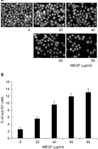
HepG2 인체간암세포의 증식에 미치는 MEGF의 영향을 조사하기 위하여 MEGF를 다양한 농도(20∼100 μg/ml) 로 72시간동안 처리한 후 hemocytometer counting을 실시 하였다. Fig. 1의 결과에서 알 수 있듯이 MEGF의 처리 농도 의존적으로 HepG2 세포의 생존율이 억제됨을 알 수 있었는데, 특히 40 μg/ml 처리군 이상에서 매우 낮은 생존율을 보이기 시작하여 100 μg/ml 처리군에서 세포
Fig. 1. Effect of methanol extracts of G. furcata (MEGF) on the cell viability of HepG2 human hepatocarcinoma cells. Cells were seeded as described in materials and methods, and the viable cells were counted after MEGF treatment for 72 h.
Results are expressed as averages +/ SD form separate experiments.
Fig. 2. Induction of apoptosis by MEGF in HepG2 cells. (A) After treated with MEGF for 72 h, the cells were fixed and stained with DAPI. The nuclear morphology was photographed with a fluorescence using blue filter. Magnification, X400. (B) Cells treated for 72 h with MEGF and then the cells were collected and stained with PI for flow cytometry analysis. The fraction of apoptotic sub-G1 cells is indicated. Results are expressed as averages +/ SD form separate experiments.
의 생존율이 20% 이하로 나타났으며, 이는 선행연구에 서 조사된 다른 암세포에서의 효과와 매우 유사한 결과 였다.13,14)
이러한 MEGF 처리에 따른 HepG2 세포의 생존율 저하 가 apoptosis 유발과 연관성이 있는지를 조사하기 위하여 MEGF 처리 후 암세포 핵의 형태변화를 조사하였다. Fig.
2A에 나타낸 바와 같이 정상 배지에서 배양된 세포의 경우 핵의 전체가 완전한 형태로 염색되는 양상을 보였 으나 MEGF의 처리 농도가 증가할수록 apoptosis가 일어 난 세포에서 전형적으로 관찰되는 chromatin이 응축에 의한 apoptotic body의 출현 정도가 증가되었다. 또한 Fig.
2B에서는 flow cytometry 분석을 이용하여 세포사멸의 증 거가 되고 있는 sub-G1기의 측정값을 조사해본 결과 sub-G1기의 값이 대조군에서 3.0% 미만이었으나, 80μg/
ml MEGF 처리군에서는 약 13%로 나타나 4.3배 증가되 었음을 알 수 있었다. 한편 선행연구에 의하면 MEGF 처 리에 의한 암세포의 증식억제는 세포주기 G2/M arrest와 연관성이 있는 것으로 나타나,13) MEGF의 처리에 따른 HepG2 세포의 생존율의 감소는 세포주기 G2/M arrest와 연관된 apoptosis 유발과 밀접한 연관성이 있음을 알 수 있었다.
2. Bcl-2 family 및 Fas/FasL system의 발현에 미치 는 MEGF의 영향
현재까지 apoptosis의 유발 경로는 death receptor에 의해 death signal을 전달하는 death receptor pathway와 mito-
chondria를 경유하는 mitochondrial pathway로 크게 나누어 지며, 두 경로에서의 주요 인자들 사이의 cross-talking 또 한 apoptosis 유발에 매우 중요한 역할을 한다.15,16) 그 중 death receptor pathway에서는 Fas (CD95/Apo1) 및 이와 결 합하는 ligand인 FasL가 널리 알려져 있다.17,18) 또한 apoptosis의 조절인자 중 Bcl-2 family에 속하는 몇 가지 중 요한 인자들은 apoptosis 유발 조절에 가장 대표적인 유전 자로 알려져 있는데, 그중 Bcl-2는 anti-apoptotic 분자로서 apoptosis의 유발을 억제하는 기능을 가지며, Bax는 pro-apoptotic 분자로 Bax 단백질 발현의 증가는 apoptosis 의 유발과 관계가 있다. 이들 두 유전자는 세포 내 소기 관 중 mitochondria로부터의 cytochrome c를 유리시켜 cys-
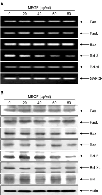
Fig. 3. Inhibition of anti-apoptotic Bcl-2 expression by MEGF treatment in HepG2 cells. HepG2 cells were incubated with or without MEGF for 72 h. (A) Total RNA was isolated and reverse-transcribed. The resulting cDNA was subjected to PCR with the indicated primers, and the reaction products were subjected to electrophoresis in 1.0% agarose gel and visualized by EtBr staining. GAPDH was used as an internal control. (B) The cells were lysed and then cellular proteins were separated by SDS-polyacrylamide gels and transferred onto nitrocellulose membranes. The membranes were probed with the indicated antibodies. Proteins were visualized using an ECL detection system. Actin was used as an internal control.
teine-related protease인 caspase, 종양억제 유전자인 p53, DNA의 단편화와 연관된 endonuclease 등의 활성을 조절 하며, 이들은 서로 dimer의 형태로 존재하며 그들의 발현
수준에 변화가 초래되면 apoptosis가 유발되는 것으로 알 려져 있다.6,19) 따라서 HepG2 세포에서 MEGF에 의한 apoptosis 유발에 이들 유전자가 관련되어 있는지의 여부 를 RT-PCR 및 Western blot 분석으로 조사하였다. Fig. 3 에 나타낸 바와 같이 Fas/FasL system에 속하는 대표적인 두 유전자인 Fas와 FasL의 발현은 전사 및 번역 수준 모 두에서 변화가 없었다. 그리고 Bcl-2 family에 속하는 다 양한 유전자들의 발현 변화에서는 대표적인 anti-apopto- tic 인자인 Bcl-2의 발현은 mRNA 및 단백질 수준에서 모 두 MEGF 처리 농도의 증가에 따라 발현의 정도가 감소 되었으나, 다른 인자들의 발현에는 큰 변화를 관찰할 수 없었다. 이는 인체백혈병세포에 나타난 MEGF 처리에 따 른 Fas 및 FasL의 발현 증가와는 상반된 결과로서 MEGF 의 처리에 의한 인체 간암세포 HepG2에서의 생존율 저 하에 따른 apoptosis 유발은 Fas/FasL death receptor를 경유 하는 apoptosis 유발과는 큰 상관관계가 없으며, 최소한 Bcl-2 family의 발현 변화는 관여할 것으로 추측된다.
3. IAP family의 발현 및 caspase의 활성에 미치는 WEPN의 영향
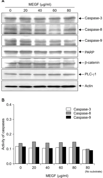
Apoptosis 조절에 관여하는 또 다른 인자 중 IAP family 에 속하는 여러 유전자 산물은 caspase와의 직접적인 결 합을 통하여 그들의 apoptotic 활성을 억제할 수 있을 것 으로 밝혀져 있다.20,21) 한편 caspase protease라는 효소 역 시 apoptosis 유발에 중요한 조절인자로서 작용하는데, 이 들 family에 속하는 단백질들은 세포에서 핵과 mitochon- dria의 외막에 불활성 상태로 존재하며, Bcl-2/Bax family 발현의 변화에 따라 이들의 활성도가 조절될 수 있다.22) 이들은 proenzyme 형태로 존재하다가 apoptosis 유도를 활 성화 시키는 신호에 의해 활성화된 protease로 전환되어 직접 또는 간접적으로 세포 내에 존재하는 많은 표적 단 백질의 분해에 관여하는 것으로 알려져 있으나, 많은 경 우에 caspase의 활성과는 무관하게 apoptosis가 유발될 수 도 있다.23,24) 따라서 본 연구에서는 MEGF 처리에 의한 HepG2 세포의 apoptosis 유발에 이들 IAP family 및 caspase 가 관여하는지의 여부를 조사하였다. 먼저 Fig. 4에 나타 난 바와 같이 MEGF 처리에 따라서 조사된 IAP family의 인자들은 전사 및 번역 수준에서 큰 변화를 보여 주지 않았으며, 지금까지 알려진 caspase 중 대부분의 apoptosis 가 유발된 세포에서 높은 활성 증가를 보여주는 caspase- 3, -8 및 -9의 발현 역시 MEGF 처리에 따라 큰 변화가 없었고, in vitro caspase activity assay를 통한 결과에서도 MEGF 처리에 따른 활성의 증가를 관찰할 수가 없었다.
또한 활성화된 caspase의 대표적인 기질 단백질에 해당되
Fig. 4. Effects of MEGF on IAP family members expression in HepG2 cells. (A) After incubation with MEGF for 72 h, the total RNA was isolated and reverse-transcribed. The resulting cDNA was subjected to PCR with indicated primers. The reaction products were subjected to electrophoresis in 1.0%
agarose gel and visualized by EtBr staining. GAPDH was used as the internal control. (B) The cells were lysed and the cellular proteins were then separated by SDS-polyacrylamide gels and transferred onto nitrocellulose membranes. The membranes were probed with the indicated antibodies. The proteins were visualized using an ECL detection system. Actin was used as the internal control.
Fig. 5. Effects of MEGF on the expression and activity of caspases in HepG2 cells. (A) After being treated with MEGF for 72 h, equal amounts of the cell lysates were resolved by SDS-polyacrylamide gels, transferred to nitrocellulose mem- branes and probed with the indicated antibodies. Proteins were then visualized using an ECL detection system. Actin was used as the internal control. (B) The cell lysates obtained from cells grown under the same conditions as (A) were assayed for the in vitro caspase-3, -8 and -9 activity using DEVD-pNA, IETD-pNA and LEHD-pNA, respectively, as substrates. The concentrations of the fluorescent products released were then measured. Data are means average of two separate experiments.
는 poly (ADP-ribose) polymerase (PARP) 및 phospholipase C- γ1 (PLC-γ1)의 단편화도25,26) 관찰되지 않았다(Fig. 5).
즉 MEGF 처리에 따른 HepG2 세포의 apoptosis 유발에는 IAP family가 크게 관여하지 않으며, 이에 따른 caspase 활 성의 증가도 관찰되지 않아 MEGF의 apoptosis 유도활성 은 caspase 비의존적임을 알 수 있었다.
결 론
본 연구에서는 인체간암세포인 HepG2 세포의 증식에 미치는 등불가사리 메탄올 추출물(MEGF)의 영향을 조사 하였다. MEGF의 처리 농도의 증가에 따라 농도의존적으
로 암세포의 생존율은 감소되었으며, 이러한 현상이 apoptosis 유발과 연관성이 있었음을 DAPI 염색에 의한 핵의 응축에 따른 apoptotic body의 출현 증가 및 flow cytometry 분석에 따른 sub-G1기 세포의 빈도 증가로 확 인을 하였다. MEGF 처리에 의한 apoptosis 유발에 관여하
는 유전자들의 발현 변화를 RT-PCR 및 Western blot 방법 으로 조사한 결과, Bcl-2 family에 속하는 anti-apoptotic 인 자인 Bcl-2의 발현만이 전사 및 번역 수준에서 감소되었 으며, 나머지 인자들의 발현은 큰 변화를 보이지 않았다.
특히 caspase의 발현 및 그들의 활성화에도 MEGF는 아무 런 영향을 미치지 않아 MEGF 처리에 의한 HepG2 세포 의 apoptosis 유발은 caspase 비의존적임을 알 수 있었다.
이상의 결과들은 MEGF 내 함유된 생리활성 물질의 분 리 및 그들의 항암작용 연구를 위한 기초 자료로서 활용 될 것이다.
감사의 글
김성윤은 2006년도 2단계 두뇌한국21사업의 박사후연 구원 지원을 받았으며, 본 연구는 해양수산부 마린바이오 21사업의 해양바이오프로세스연구단 연구비 지원(과제관 리번호 M2007-03)에 의해 수행된 결과의 일부입니다.
참 고 문 헌
1) Aneiros A, Garateix A. Bioactive peptides from marine sources:
pharmacological properties and isolation procedures. J Chromatogr B Analyt Technol Biomed Life Sci 803, 41-53, 2004.
2) Ara J, Sultana V, Qasim R, Ahmad VU. Hypolipidaemic activity of seaweed from Karachi coast. Phytother Res 16, 479- 483, 2002.
3) Heo SJ, Park EJ, Lee KW, Jeon YJ. Antioxidant activities of enzymatic extracts from brown seaweeds. Bioresour Technol 96, 1613-1623, 2005.
4) Yuan YV, Walsh NA. Antioxidant and antiproliferative activi- ties of extracts from a variety of edible seaweeds. Food Chem Toxicol 44, 1144-1150, 2006.
5) Barisic K, Petrik J, Rumora L. Biochemistry of apoptotic cell death. Acta Pharm 53, 151-164, 2003.
6) Liu S, Seidel-Dugan C. In search of p53 target genes for the therapeutic manipulation of cancer. Curr Opin Drug Discov Devel 9, 176-183, 2006.
7) Schultz DR, Harrington Jr WJ. Apoptosis: programmed cell death at a molecular level. Semin Arthritis Rheum 32, 345-369, 2003.
8) FitzGerald, G. A. COX-2 and beyond: Approaches to prosta- glandin inhibition in human disease. Nat Rev Drug Discov 2, 879-890, 2003.
9) Dempke W, Rie C, Grothey A, Schmoll HJ. Cyclooxy- genase-2: a novel target for cancer chemotherapy? J Cancer Res Clin Oncol 127, 411-417, 2001.
10) Ham SS, Lee SY, Choi M, HwangBo HJ. Antimutagenicity and cytotoxicity effect of Woorimil wheat flour extracts added
with wild herb and seaweed powder. J Korean Soc Food Sci Nutr 27, 1177-1182, 1998.
11) Tokudome S, Kuriki K, Moore MA. Seaweed and cancer prevention. Jpn J Cancer Res 92, 1008-1009, 2001.
12) Park SY, Jung BM, Choi YH, Bae SJ. Growth inhibition effects of cancer cell lines by Gloiopeltis furcata fractions in vitro.
J Korean Soc Food Sci Nutr 34, 771-775, 2005.
13) Bae SJ, Choi YH. Methanol extract of the seaweed Gloiopeltis furcata induces G2/M arrest and inhibits cyclooxygenase-2 activity in human hepatocarcinoma HepG2 cells. Phytother Res 21, 52-57, 2007.
14) Choi WY, Park C, Kim GY, Lee WH, Bae SJ, Choi YH.
Induction of apoptosis by methanol extract of Gloiopeltis furcata in human leukemia cell line U937. J Marine Biosci Biotechnol 1, 76-83, 2007.
15) Jin Z, El-Deiry WS. Overview of cell death signaling pathways. Cancer Biol Ther 4, 139-163, 2005.
16) Ghobrial IM, Witzig TE, Adjei AA. Targeting apoptosis pathways in cancer therapy. CA Cancer J Clin 55, 178-194, 2005.
17) Scholz M, Cinatl J. Fas/FasL interaction: a novel immune therapy approach with immobilized biologicals. Med Res Rev 25, 331-342, 2005.
18) Wajant H. CD95L/FasL and TRAIL in tumour surveillance and cancer therapy. Cancer Treat Res 130, 141-165, 2006.
19) Bettaieb A, Dubrez-Daloz L, Launay S, Plenchette S, Rebe C, Cathelin S, Solary E. Bcl-2 proteins: targets and tools for chemosensitisation of tumor cells. Curr Med Chem Anticancer Agents 3, 307-318, 2003.
20) de Graaf AO, de Witte T, Jansen JH. Inhibitor of apoptosis proteins: new therapeutic targets in hematological cancer?
Leukemia 18, 1751-1759, 2004.
21) Wrzesien-Kus A, Smolewski P, Sobczak-Pluta A, Wierzbowska A, Robak T. The inhibitor of apoptosis protein family and its antagonists in acute leukemias. Apoptosis 9, 705-715, 2004.
22) Philchenkov A, Zavelevich M, Kroczak TJ, Los M. Caspases and cancer: mechanisms of inactivation and new treatment modalities. Exp Oncol 26, 82-97, 2004.
23) Kolenko VM, Uzzo RG, Bukowski R, Finke JH. Caspase- dependent and -independent death pathways in cancer therapy. Apoptosis 5, 17-20, 2000.
24) Chang HY, Yang X. Proteases for cell suicide: functions and regulation of caspases. Microbiol Mol Biol Rev 64, 821-846, 2000.
25) Bae SS, Perry DK, Oh YS, Choi JH, Galadari SH, Ghayur T, Ryu SH, Hannun YA, Suh PG. Proteolytic cleavage of phospholipase C-gamma1 during apoptosis in Molt-4 cells.
FASEB J 14, 1083-1092, 2000.
26) Kaufmann SH, Desnoyers S, Ottaviano Y, Davidson NE, Poirier GG. Specific proteolytic cleavage of poly (ADP-ribose) polymerase: an early marker of chemotherapy-induced apopto- sis. Cancer Res 53, 3976-3985, 1993.